Slit-like jugular foramen due to abnormal bone growth at jugular fossa
Rakhi Rastogi* and Virendra Budhiraja
Department of Anatomy, Subharti Medical College, Meerut (U.P.), India
- *Corresponding Author:
- Dr. Rakhi Rastogi
Associate Professor, Department of Anatomy, Subharti Medical College, Delhi-Haridwar By Pass Road Meerut (U.P.), India
Tel: +91 989 7898515
E-mail: rakhirastogi1207@gmail.com
Date of Received: September 22nd, 2009
Date of Accepted: April 26th, 2010
Published Online: May 17th, 2010
© Int J Anat Var (IJAV). 2010; 3: 74–75.
[ft_below_content] =>Keywords
jugular foramen, jugular fossa, skull base, variation, Vernet’s syndrome
Introduction
The jugular foramen is a depression on the medial and inferior surface of the petrous pyramid formed by temporal and occipital bones [1,2]. It extends anteriorly, laterally and inferiorly from the endocranium to the exocranium between the temporal and occipital bones. The foramen is divided into two parts by a fibrous or osseous bridge that connects the jugular spine on petrous part of the temporal bone and the jugular process of the occipital bone. The foramen’s anteromedial compartment, the pars nervosa, contains the glossopharyngeal nerve and inferior petrosal sinus. The pars vascularis contains the jugular bulb and the vagus and spinal accessory nerves. The pars vascularis is usually larger on the right causing asymmetry of the jugular foramina [3]. The patients with lesions of the jugular foramen most commonly present with combinations of cranial nerve palsies (Vernet syndrome), which is characterized by loss of taste at the posterior third of the tongue (cranial nerve IX); vocal cord paralysis and dysphasia (cranial nerve X); and weakness of sternocleidomastoid and trapezius (cranial nerve XI) [4].
Case Report
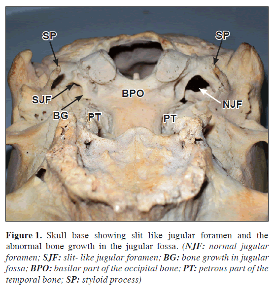
During osteology demonstration classes for medical undergraduates, an unusual bony projection was found in the left jugular fossa of a skull (Figure 1). On the right side the jugular fossa was as usual. Presence of the bony projection narrowed the jugular foramen into a slit. The right jugular foramen measured 13.61 mm in length fossaand 9.98 mm in width, on the other hand left jugular foramen measured 7.74 mm in length and 2.47 mm in width. The left jugular foramen was reduced less than 3/4 in its dimensions when compared with the right jugular foramen and on external observation it appeared like a slit. No other abnormality detected in the skull.
Figure 1: Skull base showing slit like jugular foramen and the abnormal bone growth in the jugular fossa. (NJF: normal jugular foramen; SJF: slit- like jugular foramen; BG: bone growth in jugular fossa; BPO: basilar part of the occipital bone; PT: petrous part of the temporal bone; SP: styloid process)
Discussion
The jugular foramen is indeed the most complex of the foramina through which the cranial nerves pass. It is traditionally a region of interest for neuro-otologists and neurosurgeons [5]. There are studies comparing the size of jugular foramen with anomalies of transverse and sigmoid sinuses [6]. There are also studies of variations in the structure of jugular foramen [7] and jugular bulb [8]. Hatiboglu and Anil studied 300 Anatolian skulls and found that in 61.6% the foramen was larger on the right side and in 26% it was larger on the left side [9]. Sturrock had also observed that in 68.6% cases jugular foramen was larger on right side and in 23.1% cases on left side and 8.3% of equal size [10].
Bony growth in relation to jugular foramen has been reported once [11]. In our case, the bony growth has converted the jugular foramen into a slit. It might cause the neurovascular symptoms which can mimic the symptoms caused by jugular meningiomas, glomus jugular tumors and even a nodule reducing size of jugular foramen in Varicella-Zoster virus infections [12]. The involvement of ninth, tenth and eleventh cranial nerves at jugular foramen is known as Vernet’s syndrome, which might occur in this case due to narrowing of jugular foramen.
This variation may lead to wrong clinical diagnosis. The knowledge of this variation may be important for neurologists, radiologists and anthropologists. The case is reported due to its rarity and clinically significance.
References
- Caldemeyer KS, Mathews VP, Azzarelli B, Smith RR. The jugular foramen: a review of anatomy, masses and imaging characteristics. Radiographics. 1997; 17: 1123–1139.
- Rhoton AL Jr, Buza R. Microsurgical anatomy of the jugular foramen. J Neurosurg.1975; 42: 541–550.
- Daniels DL, Williams AL, Haughton VM. Jugular foramen: anatomic and computed tomographic study. AJR Am J Roentgenol. 1984; 142: 153–158.
- Hakuba A, Hashi K, Fujitani K, Ikuno H, Nakamura T, Inoue Y. Jugular foramen neurinomas. Surg Neurol. 1979; 11: 83–94.
- Kanno T, Kiya N, Sunil MV. Microsurgical anatomy of the retroauricular, transcervicomastoid, infralabyrinthine approach to jugular foramen. Neuroanatomy. 2003; 2: 28–34.
- Manjunath KY. Anomalies of transverse and sigmoid sinuses associated with contracted jugular foramina. Indian J Otolaryngol Head Neck Surg. 2004; 56: 108–114.
- Patel MM, Singel TC. Variations in the structure of the jugular foramen of the human skull in Saurashtra region. J Anat Soc India. 2007; 56: 34–37.
- Sawyer DR, Kiely ML. Jugular foramen and mylohyoid bridging in an Asian Indian population. Am J Phys Anthropol.1987; 72: 473–477.
- Hatiboglu MT, Anil A. Structural variations in the jugular foramen of the human skull. J Anat. 1992; 180: 191–196.
- Sturrock RR. Variation in the structure of the jugular foramen of the human skull. J Anat. 1988; 160: 227–230.
- Nayak S. Partial obstruction of jugular foramen by abnormal bone growth at jugular fossa. Internet J Biol Anthropol. 2008; 1: 2.
- Kawabe K, Sekine T, Murata K, Sato R, Aoyagi J, Kawase Y, Ogura N, Kiyozuka T, Igarashi O, Iguchi H, Fujioka T, Iwasaki Y. A case of Vernet syndrome with varicella zoster virus infection. J Neurol Sci. 2008; 270: 209–210.
Rakhi Rastogi* and Virendra Budhiraja
Department of Anatomy, Subharti Medical College, Meerut (U.P.), India
- *Corresponding Author:
- Dr. Rakhi Rastogi
Associate Professor, Department of Anatomy, Subharti Medical College, Delhi-Haridwar By Pass Road Meerut (U.P.), India
Tel: +91 989 7898515
E-mail: rakhirastogi1207@gmail.com
Date of Received: September 22nd, 2009
Date of Accepted: April 26th, 2010
Published Online: May 17th, 2010
© Int J Anat Var (IJAV). 2010; 3: 74–75.
Abstract
An abnormal unilateral blockage of the jugular foramen by a bone growth converting it into a slit was noted in a skull during osteology demonstration classes for medical undergraduates. The left jugular foramen was narrowed by a thick bony projection filling the jugular fossa. This kind of narrowing of the foramen might results in neurovascular symptoms as it transmits important cranial nerves and internal jugular vein. Injury of ninth, tenth and eleventh cranial nerves can occur due to narrowing of jugular foramen know as Vernet’s syndrome is discussed along with case.
-Keywords
jugular foramen, jugular fossa, skull base, variation, Vernet’s syndrome
Introduction
The jugular foramen is a depression on the medial and inferior surface of the petrous pyramid formed by temporal and occipital bones [1,2]. It extends anteriorly, laterally and inferiorly from the endocranium to the exocranium between the temporal and occipital bones. The foramen is divided into two parts by a fibrous or osseous bridge that connects the jugular spine on petrous part of the temporal bone and the jugular process of the occipital bone. The foramen’s anteromedial compartment, the pars nervosa, contains the glossopharyngeal nerve and inferior petrosal sinus. The pars vascularis contains the jugular bulb and the vagus and spinal accessory nerves. The pars vascularis is usually larger on the right causing asymmetry of the jugular foramina [3]. The patients with lesions of the jugular foramen most commonly present with combinations of cranial nerve palsies (Vernet syndrome), which is characterized by loss of taste at the posterior third of the tongue (cranial nerve IX); vocal cord paralysis and dysphasia (cranial nerve X); and weakness of sternocleidomastoid and trapezius (cranial nerve XI) [4].
Case Report
During osteology demonstration classes for medical undergraduates, an unusual bony projection was found in the left jugular fossa of a skull (Figure 1). On the right side the jugular fossa was as usual. Presence of the bony projection narrowed the jugular foramen into a slit. The right jugular foramen measured 13.61 mm in length fossaand 9.98 mm in width, on the other hand left jugular foramen measured 7.74 mm in length and 2.47 mm in width. The left jugular foramen was reduced less than 3/4 in its dimensions when compared with the right jugular foramen and on external observation it appeared like a slit. No other abnormality detected in the skull.
Figure 1: Skull base showing slit like jugular foramen and the abnormal bone growth in the jugular fossa. (NJF: normal jugular foramen; SJF: slit- like jugular foramen; BG: bone growth in jugular fossa; BPO: basilar part of the occipital bone; PT: petrous part of the temporal bone; SP: styloid process)
Discussion
The jugular foramen is indeed the most complex of the foramina through which the cranial nerves pass. It is traditionally a region of interest for neuro-otologists and neurosurgeons [5]. There are studies comparing the size of jugular foramen with anomalies of transverse and sigmoid sinuses [6]. There are also studies of variations in the structure of jugular foramen [7] and jugular bulb [8]. Hatiboglu and Anil studied 300 Anatolian skulls and found that in 61.6% the foramen was larger on the right side and in 26% it was larger on the left side [9]. Sturrock had also observed that in 68.6% cases jugular foramen was larger on right side and in 23.1% cases on left side and 8.3% of equal size [10].
Bony growth in relation to jugular foramen has been reported once [11]. In our case, the bony growth has converted the jugular foramen into a slit. It might cause the neurovascular symptoms which can mimic the symptoms caused by jugular meningiomas, glomus jugular tumors and even a nodule reducing size of jugular foramen in Varicella-Zoster virus infections [12]. The involvement of ninth, tenth and eleventh cranial nerves at jugular foramen is known as Vernet’s syndrome, which might occur in this case due to narrowing of jugular foramen.
This variation may lead to wrong clinical diagnosis. The knowledge of this variation may be important for neurologists, radiologists and anthropologists. The case is reported due to its rarity and clinically significance.
References
- Caldemeyer KS, Mathews VP, Azzarelli B, Smith RR. The jugular foramen: a review of anatomy, masses and imaging characteristics. Radiographics. 1997; 17: 1123–1139.
- Rhoton AL Jr, Buza R. Microsurgical anatomy of the jugular foramen. J Neurosurg.1975; 42: 541–550.
- Daniels DL, Williams AL, Haughton VM. Jugular foramen: anatomic and computed tomographic study. AJR Am J Roentgenol. 1984; 142: 153–158.
- Hakuba A, Hashi K, Fujitani K, Ikuno H, Nakamura T, Inoue Y. Jugular foramen neurinomas. Surg Neurol. 1979; 11: 83–94.
- Kanno T, Kiya N, Sunil MV. Microsurgical anatomy of the retroauricular, transcervicomastoid, infralabyrinthine approach to jugular foramen. Neuroanatomy. 2003; 2: 28–34.
- Manjunath KY. Anomalies of transverse and sigmoid sinuses associated with contracted jugular foramina. Indian J Otolaryngol Head Neck Surg. 2004; 56: 108–114.
- Patel MM, Singel TC. Variations in the structure of the jugular foramen of the human skull in Saurashtra region. J Anat Soc India. 2007; 56: 34–37.
- Sawyer DR, Kiely ML. Jugular foramen and mylohyoid bridging in an Asian Indian population. Am J Phys Anthropol.1987; 72: 473–477.
- Hatiboglu MT, Anil A. Structural variations in the jugular foramen of the human skull. J Anat. 1992; 180: 191–196.
- Sturrock RR. Variation in the structure of the jugular foramen of the human skull. J Anat. 1988; 160: 227–230.
- Nayak S. Partial obstruction of jugular foramen by abnormal bone growth at jugular fossa. Internet J Biol Anthropol. 2008; 1: 2.
- Kawabe K, Sekine T, Murata K, Sato R, Aoyagi J, Kawase Y, Ogura N, Kiyozuka T, Igarashi O, Iguchi H, Fujioka T, Iwasaki Y. A case of Vernet syndrome with varicella zoster virus infection. J Neurol Sci. 2008; 270: 209–210.


