Superficial course of brachial and ulnar arteries and high origin of common interosseous artery
Saju Binu Cherian*, Satheesha Nayak B, Nagabhooshana Somayaji
Department of Anatomy, Melaka Manipal Medical College (Manipal Campus), International Centre for Health Sciences, Manipal, Karnataka State, India.
- *Corresponding Author:
- Saju Binu Cherian, MSc
Senior Grade Lecturer of Anatomy, Melaka Manipal Medical College (Manipal Campus), International Centre for Health Sciences, Madhav Nagar, Manipal, Udupi District, Karnataka State, 576 104, India.
Tel: +91 820 2922519
Fax: +91 820 2571905
E-mail: sajubinu@gmail.com
Date of Received: September 17th, 2008
Date of Accepted: December 26th, 2008
Published Online: January 7th, 2009
© IJAV. 2009; 2: 4–6.
[ft_below_content] =>Keywords
brachial artery, ulnar artery, common interosseous artery, variation
Introduction
Brachial artery is the major artery of the upper limb. It is the direct continuation of the axillary artery. It extends from the lower border of the teres major muscle to the neck of the radius where it terminates by dividing into radial and ulnar arteries. In the upper part of the arm, it is very superficial but in the lower part of the arm, it is overlapped by the biceps brachii muscle. It gives profunda brachii artery, superior and inferior ulnar collateral arteries, nutrient artery to the humerus and muscular branches in the arm.
The ulnar artery is one of the terminal branches of the brachial artery. It starts at the cubital fossa and leaves the cubital fossa by passing deep to both heads of pronator teres muscle. It runs along the medial border of the forearm along with the ulnar nerve and enters the palm by passing superficial to the flexor retinaculum.
The common interosseous artery is normally a branch of the ulnar artery. It divides into anterior and posterior interosseous arteries, which supply anterior and posterior compartments of the forearm respectively.
Case Report
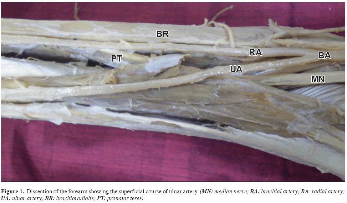
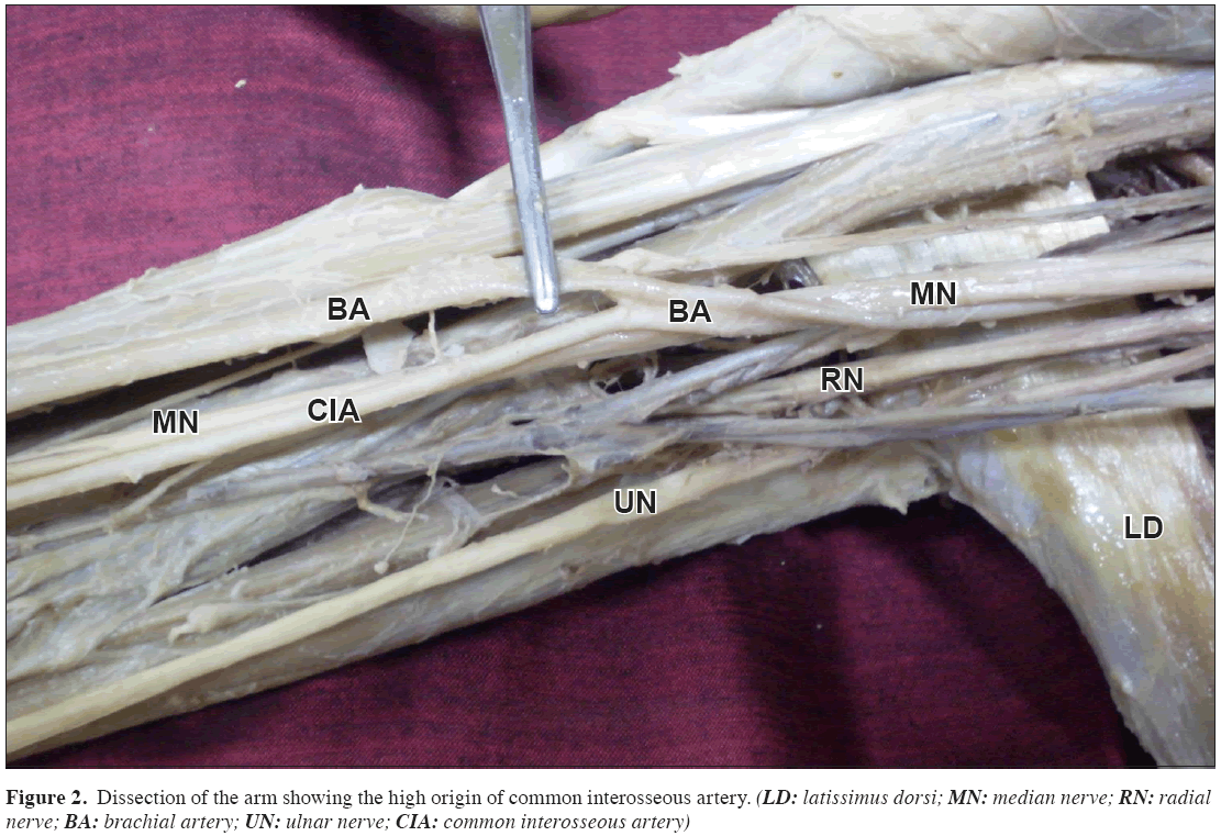
During regular dissection classes for undergraduate medical students, we observed variations of ulnar and common interosseous arteries in an adult male cadaver. The variations were on the right upper limb and were unilateral. The brachial artery was very superficial throughout its course. It divided into radial and ulnar arteries in the cubital fossa. The ulnar artery passed superficial to the pronator teres, flexor carpi radialis, palmaris longus and flexor digitorum superficialis muscles (Figure 1). The course, relations and distribution of the ulnar artery were normal in the distal part of the forearm. The radial artery had a normal course and distribution. The common interosseous artery was large and originated from the brachial artery approximately 5 cm below the lower border of the teres major muscle (Figure 2). It replaced the brachial artery’s normal course in the arm. The median nerve was lateral to it at first, and in the lower part of the arm it crossed the common interosseous artery from lateral to medial direction. In the cubital fossa also the median nerve was medial to the common interosseous artery. The brachial artery was just anterior to the common interosseous artery in the cubital fossa. The common interosseous artery divided into anterior and posterior interosseous arteries that had their normal course and distribution in the forearm.
Discussion
The ulnar artery normally arises from brachial artery in the cubital fossa. But it may arise from the brachial artery above the usual point of division or from the axillary artery. The superficial ulnar artery is a rare variation of the upper limb arterial system that arises from the brachial or axillary artery and runs superficial to the muscles arising from the medial epicondyle. The incidence is about 0.7 to 7% [1]. Dartnell et al., have reported the presence of superficial ulnar artery in 4.2% of cases [2]. The superficial ulnar artery may replace the ulnar artery altogether or it may supplement it. In a study by Mannan et al., the superficial ulnar artery terminated by joining the normal artery in the distal part of the forearm [3]. Origin of superficial ulnar artery from the brachial artery, in the middle of the arm has been reported [4]. In this case, the common interosseous artery arose from brachial artery as one of its terminal branches. The common interosseous artery may present variations in its origin. It may originate from axillary artery or brachial artery. Previous studies have shown high origin of radial and common interosseous arteries [5,6].
The knowledge of variations reported here is important for surgeons, orthopedic surgeons, plastic surgeons, radiologists and nurses. The superficial ulnar artery might get damaged in the superficial injuries. The needle for intravenous injections might enter such a superficial ulnar artery instead of median cubital vein. But it may be advantageous in case of catheterizations. The superficial ulnar artery may be useful in arterial grafts and plastic surgeries.
References
- Senanayake KJ, Salgado S, Rathnayake MJ, Fernando R, Somarathne K. A rare variant of the superficial ulnar artery, and its clinical implications: a case report. J. Med. Case Reports. 2007; 1: 128.
- Dartnell J, Sekaran P, Ellis H. The superficial ulnar artery: incidence and calibre in 95 cadaveric specimens. Clin. Anat. 2007; 20: 929–932.
- Mannan A, Sarikcioglu L, Ghani S, Hunter A. Superficial ulnar artery terminating in a normal ulnar artery. Clin. Anat. 2005; 18: 602–605.
- Krishnamurthy A, Kumar M, Nayak Sr, Prabhu LV. High origin and superficial course of ulnar artery: a case report. Fırat Tip Dergisi. 2006; 11: 66–67.
- Bergman RA, Afifi AK, Miyauchi R. Common Interosseous Artery. Illustrated Encyclopedia of Human Anatomic Variation: Opus II: Cardiovascular System: Arteries: Upper Limb. http://www.anatomyatlases.org/AnatomicVariants/Cardiovascular/Text/Arteries/InterosseousCommon.shtml (accessed September 2008).
- Sargon M, Celik HH. Proximal origins of radial and common interosseous arteries. Kaibogaku Zasshi. 1994; 69: 406–409.. Arch. Med. Sci. 2007; 3: 284–286.
Saju Binu Cherian*, Satheesha Nayak B, Nagabhooshana Somayaji
Department of Anatomy, Melaka Manipal Medical College (Manipal Campus), International Centre for Health Sciences, Manipal, Karnataka State, India.
- *Corresponding Author:
- Saju Binu Cherian, MSc
Senior Grade Lecturer of Anatomy, Melaka Manipal Medical College (Manipal Campus), International Centre for Health Sciences, Madhav Nagar, Manipal, Udupi District, Karnataka State, 576 104, India.
Tel: +91 820 2922519
Fax: +91 820 2571905
E-mail: sajubinu@gmail.com
Date of Received: September 17th, 2008
Date of Accepted: December 26th, 2008
Published Online: January 7th, 2009
© IJAV. 2009; 2: 4–6.
Abstract
Knowledge of variations in the course and branching pattern of the arteries of upper limb is important for clinicians. We report the variations of the branches of brachial artery. The brachial artery was superficial throughout its course. It divided into radial and ulnar arteries in the cubital fossa. The ulnar artery passed superficial to the flexor muscles of the forearm as it leaved the cubital fossa. The common interosseous artery was large in size and it was a direct branch of brachial artery. It took its origin from brachial artery approximately 5 cm below the lower border of teres major muscle and followed the median nerve till the cubital fossa and divided into anterior and posterior interosseous branches
-Keywords
brachial artery, ulnar artery, common interosseous artery, variation
Introduction
Brachial artery is the major artery of the upper limb. It is the direct continuation of the axillary artery. It extends from the lower border of the teres major muscle to the neck of the radius where it terminates by dividing into radial and ulnar arteries. In the upper part of the arm, it is very superficial but in the lower part of the arm, it is overlapped by the biceps brachii muscle. It gives profunda brachii artery, superior and inferior ulnar collateral arteries, nutrient artery to the humerus and muscular branches in the arm.
The ulnar artery is one of the terminal branches of the brachial artery. It starts at the cubital fossa and leaves the cubital fossa by passing deep to both heads of pronator teres muscle. It runs along the medial border of the forearm along with the ulnar nerve and enters the palm by passing superficial to the flexor retinaculum.
The common interosseous artery is normally a branch of the ulnar artery. It divides into anterior and posterior interosseous arteries, which supply anterior and posterior compartments of the forearm respectively.
Case Report
During regular dissection classes for undergraduate medical students, we observed variations of ulnar and common interosseous arteries in an adult male cadaver. The variations were on the right upper limb and were unilateral. The brachial artery was very superficial throughout its course. It divided into radial and ulnar arteries in the cubital fossa. The ulnar artery passed superficial to the pronator teres, flexor carpi radialis, palmaris longus and flexor digitorum superficialis muscles (Figure 1). The course, relations and distribution of the ulnar artery were normal in the distal part of the forearm. The radial artery had a normal course and distribution. The common interosseous artery was large and originated from the brachial artery approximately 5 cm below the lower border of the teres major muscle (Figure 2). It replaced the brachial artery’s normal course in the arm. The median nerve was lateral to it at first, and in the lower part of the arm it crossed the common interosseous artery from lateral to medial direction. In the cubital fossa also the median nerve was medial to the common interosseous artery. The brachial artery was just anterior to the common interosseous artery in the cubital fossa. The common interosseous artery divided into anterior and posterior interosseous arteries that had their normal course and distribution in the forearm.
Discussion
The ulnar artery normally arises from brachial artery in the cubital fossa. But it may arise from the brachial artery above the usual point of division or from the axillary artery. The superficial ulnar artery is a rare variation of the upper limb arterial system that arises from the brachial or axillary artery and runs superficial to the muscles arising from the medial epicondyle. The incidence is about 0.7 to 7% [1]. Dartnell et al., have reported the presence of superficial ulnar artery in 4.2% of cases [2]. The superficial ulnar artery may replace the ulnar artery altogether or it may supplement it. In a study by Mannan et al., the superficial ulnar artery terminated by joining the normal artery in the distal part of the forearm [3]. Origin of superficial ulnar artery from the brachial artery, in the middle of the arm has been reported [4]. In this case, the common interosseous artery arose from brachial artery as one of its terminal branches. The common interosseous artery may present variations in its origin. It may originate from axillary artery or brachial artery. Previous studies have shown high origin of radial and common interosseous arteries [5,6].
The knowledge of variations reported here is important for surgeons, orthopedic surgeons, plastic surgeons, radiologists and nurses. The superficial ulnar artery might get damaged in the superficial injuries. The needle for intravenous injections might enter such a superficial ulnar artery instead of median cubital vein. But it may be advantageous in case of catheterizations. The superficial ulnar artery may be useful in arterial grafts and plastic surgeries.
References
- Senanayake KJ, Salgado S, Rathnayake MJ, Fernando R, Somarathne K. A rare variant of the superficial ulnar artery, and its clinical implications: a case report. J. Med. Case Reports. 2007; 1: 128.
- Dartnell J, Sekaran P, Ellis H. The superficial ulnar artery: incidence and calibre in 95 cadaveric specimens. Clin. Anat. 2007; 20: 929–932.
- Mannan A, Sarikcioglu L, Ghani S, Hunter A. Superficial ulnar artery terminating in a normal ulnar artery. Clin. Anat. 2005; 18: 602–605.
- Krishnamurthy A, Kumar M, Nayak Sr, Prabhu LV. High origin and superficial course of ulnar artery: a case report. Fırat Tip Dergisi. 2006; 11: 66–67.
- Bergman RA, Afifi AK, Miyauchi R. Common Interosseous Artery. Illustrated Encyclopedia of Human Anatomic Variation: Opus II: Cardiovascular System: Arteries: Upper Limb. http://www.anatomyatlases.org/AnatomicVariants/Cardiovascular/Text/Arteries/InterosseousCommon.shtml (accessed September 2008).
- Sargon M, Celik HH. Proximal origins of radial and common interosseous arteries. Kaibogaku Zasshi. 1994; 69: 406–409.. Arch. Med. Sci. 2007; 3: 284–286.


