Superior sagittal sinus draining only to the left transverse sinus
Received: 31-Jan-2023, Manuscript No. ijav-23-6139; Editor assigned: 01-Feb-2023, Pre QC No. ijav-23-6139 (PQ); Accepted Date: Feb 21, 2023; Reviewed: 14-Feb-2023 QC No. ijav-23-6139; Revised: 21-Feb-2023, Manuscript No. ijav-23-6139 (R); Published: 28-Feb-2023, DOI: 10.37532/1308-4038.16(2).240
Citation: Mulugeta F, Bogale A, Sharew M, Assefa S, Tadesse Y. Superior Sagittal Sinus Draining Only to the Left Transverse Sinus. Int J Anat Var. 2023;16(2):253-254.
This open-access article is distributed under the terms of the Creative Commons Attribution Non-Commercial License (CC BY-NC) (http://creativecommons.org/licenses/by-nc/4.0/), which permits reuse, distribution and reproduction of the article, provided that the original work is properly cited and the reuse is restricted to noncommercial purposes. For commercial reuse, contact reprints@pulsus.com
Abstract
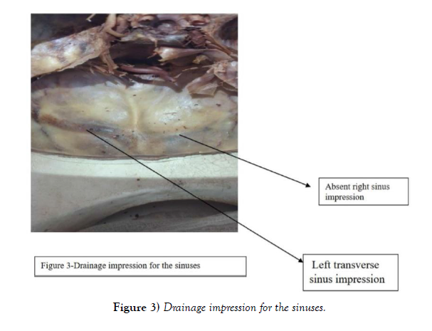
In anatomical studies things expected to be studied and remembered by students are structural formats that are statistically dominant i.e. structural formats that are found in most of the population. But anatomical locations of the body organs and structures are not always found in the same form in all the people of the world which gives rise to features known as anatomical variations. Knowing about variations has great clinical implications especially diagnosis based on imaging because unawareness of the variation may lead to diagnosing normal structures as pathological. In our routine dissection room session at Myungsung Medical College, Addis Ababa, we observed a drainage pattern of the superior sagittal sinus that is statistically not dominant. In our case the superior sagittal sinus was draining to the left transverse sinus with no observable confluence of sinus. We also observed there the right transverse sinus and its impression on the bone was absent.
Keywords
Anatomy; Variations; Superior sagittal sinus; Left transverse sinus; Right transverse sinus
CASE REPORT
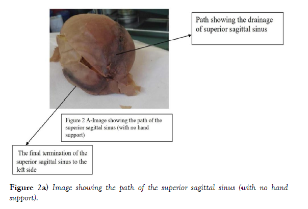
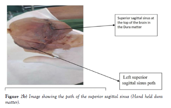
During our dissection session which was focusing on the brain and its related structures we started by observing the drainage of the sinus as it is the most superficial part next to the scalp. When we followed the superior sagittal sinus it was not placed in the place where it is found in most of the population rather it was going to the left with no observable right transverse sinus.
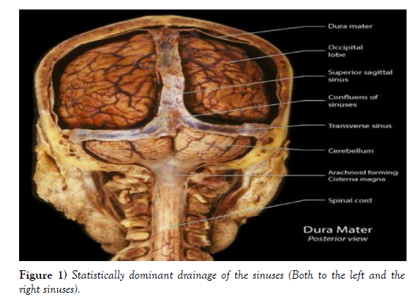
Our cadaver is 1.63 m in length with no observable gross deformity in its outer structure, has gray hair with an estimated age of 50-60 years. The superior sagittal sinus after going to the left transverse sinus it will be changed to sigmoid sinus then to the left internal jugular vein. Below there is a figure showing the statistically dominant feature superior sagittal sinus (Figure 1) and the next figure shows the variation we observed (Figure 2) and Figure 3 shows Drainage impression for the sinuses.
DISCUSSION
The brain as an organ that requires a high amount of energy and oxygen takes 15%-20% of the total cardiac output per minute which is approximately 750 milliliters per minute [1]. After this amount of blood is used and gives off the nutrients and oxygen that are needed for the normal functioning of the brain it will be metabolized into different substances and they will be drained by the venous system to the heart. The inner surface of the skull bone has several jugular veins. The venous sinuses are found at the attachment of the dural venous septa and their walls are formed by the dura mater and periosteum of the skull. This makes them in close contact with the skull and their course, existence and anatomical variation can be implicated by the impressions they make on the skull bone. Determining the normal impression and anatomical variation will help pathologists and radiologists make an appropriate diagnosis. Particularly on the occipital bone we can observe the impression of the right and the left transverse sinus. This impression is formed by its respective transverse sinus. Impression size varies in size among individuals. The average width of the right transverse sinus is 0.80 ± 0.044 and of left transverse sinus is 0.82 ± 0.035 [2].
There are six types of drainage variations that can be observed in drainage of the superior sagittal sinuses [3-5].
These are:-
Type I- The superior sagittal sinus continuing as the right transverse sinus and two occipital sinuses open to the confluence of sinuses (30.1% of cases).
Type II- The superior sagittal sinus draining as the right transverse sinus and the confluence of sinuses draining more than two occipital sinuses (21.2% of cases)
Type III- The superior sagittal sinus draining to the left transverse sinus without showing any interruptions and the two occipital veins drain to the confluence of sinuses (12.1% of cases).
Type IV- The superior sagittal sinus diverted to the left side and continuing as the corresponding transverse sinus and multiple small occipital sinuses joining it (6.1% of cases).
Type V- Superior sagittal sinus showing anatomical continuity through transverse sinus of each side by joining in the confluence of sinuses and two occipital veins joining in the confluence of sinuses (9.1% of cases)
Type VI- The superior sagittal sinus bifurcating symmetrically before joining the transverse sinus and multiple occipital sinuses will drain to the confluence of sinuses (21.2% of cases) From this type of variations the case we are reporting can be grouped as the type IV as the superior sagittal sinus diverted to the left side and continued as the left transverse sinus without major occipital sinus joining it.
ACKNOWLEDGEMENT:
None.
CONFLICTS OF INTEREST:
None.
REFERENCES
- Kay LW. Some anthropologic investigations of interest to oral surgeons. Int J Oral Surg. 1974; 3(6):363-79.
- Agthong S, Huanmanop T, Chentanez V. Anatomical variation of the supraorbital, infraorbital, and mental foramina related to gender and side. J Oral Maxillofac Surg. 2005; 63(6):800-804.
- Katakami K, Mishima A, Shiozaki K, Shimoda S, et al. Characteristics of accessory mental foramina observed on limited cone-beam computed tomography images. J Endod. 2008; 34(12):1441-1445.
- Sonick M, Abrahams J, Faiella RA. A comparison of the accuracy of periapical, panoramic, and computerized tomographic radiographs in locating the mandibular canal. Int J Oral Maxil-lofac Implants. 1994; 9:455-468.
- Naitoh M, Hiraiwa Y, Aimiya H, Gotoh K, et al. Accessory mental foramen assessment using cone-beam computed tomography. Oral Surg Oral Med Oral Pathol Oral Radiol Endod. 2009; 107(2):289-294.
Indexed at, Google Scholar, Crossref
Indexed at, Google Scholar, Crossref
Indexed at, Google Scholar, Crossref


