44 2033180199
All submissions of the EM system will be redirected to Online Manuscript Submission System. Authors are requested to submit articles directly to Online Manuscript Submission System of respective journal.
Current Research: Cardiology

Sign up for email alert when new content gets added: Sign up

Raj Kumar MD and Rahul Mehrotra MD*
 
1 Non-Invasive Cardiology, Max Super Speciality hospital, Saket, New Delhi, India, Email: Mehrotra@maxhealthcare.com
 
*Correspondence: Dr. Rahul Mehrotra, MD, Non-Invasive Cardiology, Max Super Speciality hospital, Saket, New Delhi, India, Tel: 011 2651 5050, Email: Rahul.Mehrotra@maxhealthcare.comm

Received: 21-Aug-2017 Accepted Date: Aug 22, 2017; Published: 24-Aug-2017, DOI: 10.4172/2368-0512.1000088

Citation: Kumar R, Mehrotra R. Three-dimensional echocardiography of tricuspid valve for visualizing all the three leaflets. Curr Res Cardiol 2017;4(3):35-35

This open-access article is distributed under the terms of the Creative Commons Attribution Non-Commercial License (CC BY-NC) (http://creativecommons.org/licenses/by-nc/4.0/), which permits reuse, distribution and reproduction of the article, provided that the original work is properly cited and the reuse is restricted to noncommercial purposes. For commercial reuse, contact reprints@pulsus.com

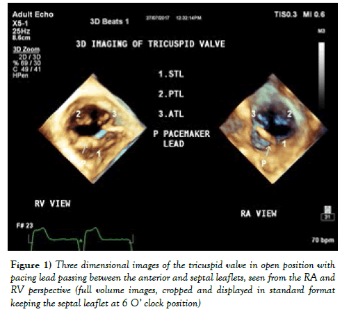
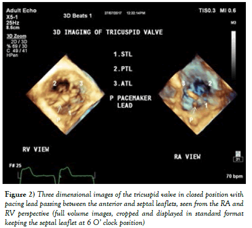
Three-dimensional echocardiography offers a unique opportunity of visualizing all the three leaflets of tricuspid valve in en-face view from transthoracic windows. It is very useful in localizing the position of the pacing leads and for planimetry (Figures 1 and 2).

current-research-cardiology-anterior-septal-leaflets

Figure 1: Three dimensional images of the tricuspid valve in open position with pacing lead passing between the anterior and septal leaflets, seen from the RA and RV perspective (full volume images, cropped and displayed in standard format keeping the septal leaflet at 6 o' clock position)

current-research-cardiology-tricuspid

Figure 2: Three dimensional images of the tricuspid valve in closed position with pacing lead passing between the anterior and septal leaflets, seen from the RA and RV perspective (full volume images, cropped and displayed in standard format keeping the septal leaflet at 6 o' clock position)

 
Google Scholar citation report
Citations : 177

Current Research: Cardiology received 177 citations as per Google Scholar report

Current Research: Cardiology peer review process verified at publons
Current Research: Cardiology
Top