Thrombosis Of Giant Right Coronary Aneurysm: 3-D Reconstruction
2 Department of Heart and Vessels, Cardiac Surgery Unit and Heart Transplantation Center , “S. Camillo-Forlanini” Hospital, 00149 Rome, Italy
Received: 08-Aug-2017 Accepted Date: Oct 24, 2017; Published: 26-Oct-2017
Citation: Buffa V, Cottini M, Madau M, et al. Thrombosis of giant right coronary aneurysm: A 3-D reconstruction. Surg Case Rep 2017;1(1):11.
This open-access article is distributed under the terms of the Creative Commons Attribution Non-Commercial License (CC BY-NC) (http://creativecommons.org/licenses/by-nc/4.0/), which permits reuse, distribution and reproduction of the article, provided that the original work is properly cited and the reuse is restricted to noncommercial purposes. For commercial reuse, contact reprints@pulsus.com
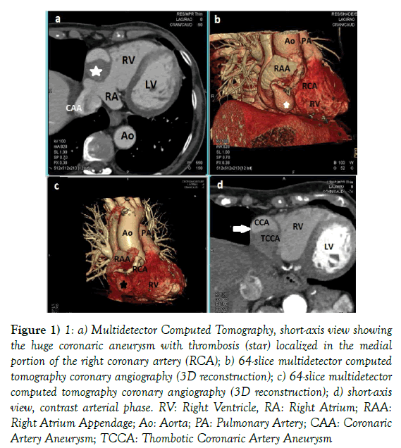
The Coronary artery aneurysm (CAA) is defined as the dilatation of the coronary artery exceeding 50% of the reference vessel diameter, is uncommon and occurs in <5% of coronary angiographic series. CAAs are termed giant if their diameter exceeds the reference vessel diameter by >4 times or if they are >8 mm in diameter. Up to one third of CAAs are associated with obstructive coronary artery disease and have been associated with myocardial infarction, arrhythmias, or sudden cardiac death. We reported an 84-year-old female with hypertension and promyelocytic leukemia (PML), presented to our emergency room for acute prolonged thoracic pain spreading out left arm, neck and back. Because of the suspicion of aortic root and arch dissection, she was immediately undergone to 64-slice Multidetector Computed Tomography (MDCT-64) documented an acute extensive giant coronaric aneurysm (2.01 × 2.51 cm, area 3.97 cmq) of the medial segment of the right coronary artery (RCA, Figure 1 (a-d). The ECG showed ST-segment elevation in leads of the inferior-posterior wall and the cardiac troponin T was 3.78 ng/ml. The patient was successfully managed to standard STEMI-management guideline: antiplatelet medication and emergency conventional coronary angiography (CCA). The CCA demonstrated an extensive aneurysm of right coronary artery that was occluded proximally, hence three second-generation drug-eluting stents were implanted (Endeavor Resolute stent, Medtronic, stent diameter 4.5-5.0-4.5, length 15-18-12 respectively). No IVUS control was done because the optimal results of PTCA and because the patient was immediately admitted to the intensive cardiology unit to stabilize and monitor the hemodynamic. The postoperative period was free of complication and the patient was discharge eight days, the CCA and she was prescribed aspirin and clopidogrel as a lifelong therapy. At the six months follow up, the patient referred a good quality of life; the echocardiogram documented a mild hypokinesis of infero-lateral sides and akinesis of infero-basal segment.
Figure 1: 1: a) Multidetector Computed Tomography, short-axis view showing the huge coronaric aneurysm with thrombosis (star) localized in the medial portion of the right coronary artery (RCA); b) 64-slice multidetector computed tomography coronary angiography (3D reconstruction); c) 64-slice multidetector computed tomography coronary angiography (3D reconstruction); d) short-axis view, contrast arterial phase. RV: Right Ventricle, RA: Right Atrium; RAA: Right Atrium Appendage; Ao: Aorta; PA: Pulmonary Artery; CAA: Coronaric Artery Aneurysm; TCCA: Thombotic Coronaric Artery Aneurysm
What is the peculiarity of this clinical case? The CT image has demonstrated the aneurysm and occlusion of RCA but the etio-pathogenesis of them were so interesting and curious. Indeed, we could not have detected if the aneurysm was grown independently and had occluded RCA or the aneurysm were grown because of a kink in the coronary artery that induced by it.
Unfortunately, there were no specific data that could explain the origin of aneurysm in this reported clinical case but in scientific context it was very interesting and educational for the peculiarities of the case and doubtful origin.
Acknowledgement
None.
Conflict of Interest
None.


