Toxicogenic symptoms provoke in isolated histological and histopathological cells is an inherent property of small size silver nanoparticles dispersed in particle colloidal silver
2 NYTIMS Karjat New Panvel, Navi-Mumbai, India
Received: 07-May-2018 Accepted Date: May 23, 2018; Published: 30-May-2018
Citation: Jyoti P, Royana S, Sankarsan P. Toxicogenic symptoms provoke in isolated histological and histopathological cells is an inherent property of small size silver nanoparticles dispersed in particle colloidal silver. J Histol Histopathol Res 2018;2(1): 8-14.
This open-access article is distributed under the terms of the Creative Commons Attribution Non-Commercial License (CC BY-NC) (http://creativecommons.org/licenses/by-nc/4.0/), which permits reuse, distribution and reproduction of the article, provided that the original work is properly cited and the reuse is restricted to noncommercial purposes. For commercial reuse, contact reprints@pulsus.com
Abstract
Toxicogenic risk evaluation for small size (2.75 to 20.11 nm range). nano silver metalloid particulate is still under explorative search by new mode of nano experiment due to some of the doubtful, undiagnosed and hidden facts. Some of the inherent mechanical properties of small size nano silver are yet to be discovered. Toxicogenic sign and symptom arousal in histological and histopathological isolated cells of vital organs primate vertebrate tissues is an inherent property of small size nano silver metallic particulate. The objective and intension behind this small size nanosilver experimental study is to prove that; small size nanosilver particles are toxicogenic arousal agents in most of the histological and histopathological cells isolated from primate vertebrate tissue and through this mode of new small size nanosilver repeated oral gavages experiment the objective and intension is proved also novelty of this study is addressed. This query also needs more and more new mode of nano experiment. Whether toxicogenic sequels provoke is due to sharp irregular edges of small size nanosilver metallic particulate while swift penetration into deep core of histological and histopathological cells through cell membrane or hazardous chemical reaction produced by coated and stabilized chemicals in colloidal aqua is a question mark yet to be diagnosed needs this new mode of experiment.
Keywords
AgNps colloidal solution; Magnetic stirring and cooling method; Toxicity; Pregnant Swiss Albino mice and fetuses vital organs tissues and cells; Colloidal solution matrix; Repeated oral gavages experiment; Gestational age; Dose; Experimental anionic double distilled water vehicle sham control and small size AgNps colloidal aqua treated groups
Introduction
Small size silver nano particles (AgNps). with diameters of less than 50 nm or 2.75 to 20.11 nm range have inherent toxicogenic arousal properties. New mode of nano research involving the synthesis and experimental applications of small size silver NPs and tiny nano dimension metalloid particulate has evoked a large margin of interest on behalf of nano scientists since the mechanical toxicogenic properties of these particles are size and dose-dependent. Physical and chemical properties of such nano metal are unique. Examples of such properties include optical interference and magnetism attractive properties, specific heats conduction, melting points and surface reactivity but toxicogenic arousal is a unique mechanochemical property executed by small size nano silver crystals. Observable and macroscopic size and dose-dependent toxicogenic arousal mechanochemical properties are apprehended to be consequences of the high ratio of irregular and spiny surface area of smaller size silver nanoparticle and volume. [1]. The potential applications of small size silver NPs in nano-biotechnology have been exhibited by a wide variety of emerging and developing laboratory uses in the study of histological and histopathological cells and inter with subcellular processes of fundamental importance in biology which arouses toxicogenic consequences. Examples include the use of colloidal silver small size nano-crystals due to their toxicogenic catalysis features (high irregular and spiny surface area with controlled crystal transparent surfaces)., and light wave refractory properties which is quite beneficial in fluorescent biological labels, bio-detection with evaluation of bacterial and viral pathogens, probing of Deoxy ribo nucleic acid structure, tumor destruction through temperature conduction (hyperthermia)., magnetic resonance image contrast enhancement and sun screen. Small size silver nano particles also have vast range of supportive applications in tissue engineering, synthesis, characterization, separation and purification of biological molecules and cells, drug and gene delivery [2-4]. but the negative repurcation arises in form of toxicogenic consequences arousal after such applications. Many of these applications result from the unique mechanochemical properties of noblemetal nano crystals. Small size silver nano particles magnetic, electromagnetic (optical). and catalytic properties are shape and size dependant, which helps in discovery of new synthesis and characterization techniques for a better control of small size silver nano particles shape and size so small size nano silver have maintain its identity in nano world. [5]. In spite of the vast utilization of small size silver nano particles (AgNps). in diagnostic medicine there is a lack of information concerning their impact on human health and the environment? Small size silver nanoparticle is toxic to a wide range of microorganisms in its various chemical forms. [6]. However, very little is known about the toxicity of small size nano silver particles. Yet, researchers only know that size and surface area are thought to be important factors of toxicogenic arousal. [7]. Bigger size silver nano particles, always produces teratogenic effects after passing through vital organs of the body even it also produces teratogenic effects in F1 generation fetuses after passing through pregnant mother, including teratogenicity on the central nervous system (CNS). and other vital systems of the body. Smaller size silver nano particles migrate to the systemic circulation and the central nervous system in a unique manner due to their small size and large surface area but irregular and spiny surface area. Smaller size silver nano particles can easily cross the blood-brain barrier (BBB). [8]., also the smallest size silver nano particles can easily penetrate the central nervous system (CNS). if it is exposed to primate vertebrate, experimental pregnant Swiss Albino mice and reach up to fresh delivered fetuses through placenta into their newly developing central nervous system in dose and size dependent manner and in predominant rate produces toxicogenic sequels which also inculcate in dose and size dependent application in both mother and fetuses. [9-12]. Recent researches [13,14]. have demonstrated that through vein approach (32 mg/kg)., through intra peritoneal approach (53 mg/kg). or through intra cerebral approach (23 μg in 13 μl). by injecting small size nano silver nano particulate (AgNps)., copper nano particulate (CuNps). or aluminum nanoparticulate in colloidal form (AlNps). such nano particles (~20-50 nm). disrupt the blood brain barrier also arises multi organ toxic genomic sequels in peak manner in Evans blue albumenized serum proteins of pregnant rats and mice and their fetuses in a highly selective, specific and segregated manner. Apart from that, a new study of recent era [15]. reported that the experimental exposure and application of small size AgNps (24 nm). to pregnant rats and their fetuses for continuous 20 to 30 days significantly changed the activity of various organs, including the brain also exhibited toxic genomic sign and symptoms in peak manner correspond to size and dose. The plasma 1/2-period of small size silver in the primate vertebrate central nervous system which includes brain and spinal cord is lengthier than its 1/2-period in rest vital organs and its isolated histological and histopathological cells, which suggests that exposure to small size silver nano particles may result in severe pathological consequences to the mammalian vital organs as well as histological and histopathological cells. [16]. Some of the recent researches have also point out that small size silver nanoparticle can destruct normal histological cell functions and can induce cell apoptosis in certain group of histological and histopathological cells, such as hippocampal neurons, hepatocytes, splenocytes, nephroblast cells, cardiomyocytes, etc. [16-19]. A world famous nanosciencist in year 2006 found that small size AgNps (6×10⁻⁶ g/ml). depletes dopamine concentration and that they are highly toxicogenic to multi vital organ histological and histopathological cells than other metalloid nano particles in neuroendocrine cells tracts. [20]. So all in one, these scientific reports indicates that small size AgNps must have toxicogenic and ambiguatory effects on the primate vertebrate and their F1 generation fetuses vital organs histological and histopathological cells. The multi vital organs histological and histopathological cells are predominantly irretentive to singlet oxygen misbalanced stress due to its peak cell metabolic rate. Small size silver nanoparticle also bears minimal capacity for regenerating histological and histopathological cells through normal cell mitotic division and aggravates oxidative stress compatibility in those oxidative stress chemical agents which is ultimately secreted from mature and developed histological and histopathological cells, and the presence of various histological and histopathological cellular oxidative stress targets. [11,12,21]. Reactive oxygen species (ROS). and oxidative stress have been implicated in the pathogenesis of histological and histopathological neural cells which is evoked from neurodegenerative diseases. [22]. Toxicogenic sign and symptoms are linked term for oxidative stress which results from an increased generation of reactive oxygen species or from a weak antioxidant defense system. A nano scientific study of recent era discovered that the generation of reactive oxygen species increased after application of small size AgNps (15 and 25 nm). through serial exposure in developing rat liver histological and histopathological isolated cells and neuroendocrine cells. [20]. Small size silver nanoparticle (5-18 nm size range). can cause toxicogenic sign and symptoms along with high peak oxidative stress resulting in a decrease of glutathione (GSH). content and increased GSH and GSSG ratio, decreased superoxide dismutase (SOD). activity and an significant increase in Malondialdehyde (MDA). levels in histological and histopathological cells isolated from multi vital organs of primate vertebrate. [23]. Apart from all these, small size nano silver of 20 to 30 nm size ranges produces central nervous system toxicity through generating free radical and scavenger cells induced singlet oxygen stress and by deviating the expression of normal genes related to singlet oxygen stress. [24]. Small size silver nano particles of 7 to 13 nm size ranges also introduces the over secretion of temperature anaphylaxes protein 75, singlet oxygen stress as well as programmed cell death in fruit fly. [25]. Also produces apoptosis in the liver cells of the mature zebra fish. [26]. Toxicity evaluations of small size silver nano particles of different small sizes and surface area by means of chondroblast and cell with plasma membrane penetration along with reactive oxygen species assay suggest that the cytotoxicity of small size silver nano particles is probably mediated through oxidative stress. [27,28]. Versatile results from these various cumbersome researches suggest that the impact of small size silver nano materials on multi vital organs histological and histopathological cells is predominant and grievous in terms of toxic genesis. To gain a new discovery insights on the deleterious effects of small size silver nanoparticle on CNS functions, some of the internationally recognized nano scientists inoculated electro synthesized small size nano silver of 25 to 30 nm ranges (AgNps I). and small size nano silver of 20 to 26 nm ranges (AgNps II). with a complicated chemical capping agent in pregnant rats and their fetuses to investigate the effects of these small size nano silver particles on cognitive processes and oxidative stress generation in the temporal cortical lobe of brain. They observed arousal of toxicogenic consequences in histological and histopathological neural cells, where this cortical area after this cumbersome experiment found most vulnerable to oxidative stress effects [29].
Materials and Methods
Permission from animal ethical committee and selection of animals
Permission for animal experiment has been approved from Central Animal Ethical Committee Institute of Medical Sciences, Banaras Hindu University (no./Dean/2014/CAEC/614- Dated 30.05.2014). Pregnant Swiss Albino mice from different breeding colonies acquired from Departmental Animal house Anatomy Department IMS BHU. 10 pregnant mice were taken into sphere of experiment from each control and treated group (Figure 1).
Synthesis and characterization of small size colloidal nanosilver
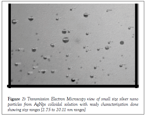
Small size 2.75 to 20.11 nm size ranges silver nano (AgNO3). crystal, PVP crystal, fresh sodium borohydried crystal, 1.5 molar sodium chloride powder purchased from Trimurti Scientific Dealer Varanasi Uttarpradesh India. AgNps I and II (0.017 g of AgNO3 was dissolved into 100 ml distilled water). (Ag I). 0.002 M NaBH4 (0.0189 g of NaBH4 was dissolved into 250 ml distilled water). (This solution must be made fresh and was fresh before the experiment). 0.3% PVP solution (0.1 g of PVP was dissolved into 33 ml distilled water). Colloidal solution is prepared by specific synthesis method called stirring on magnetic stirrer of 380Hz to 860Hz range with 1” magnetic stirrer bead inoculation and circumferential ice berg cooling method covering Erlenmeyer flask. Size of nano silver from cool stirring solution was characterized by Transmission Electron Microscopy (Indian Institute of Toxicological Research; Lucknow under esteemed guidance of Dr. L K Chauhan)., Dynamic Light Scattering, Zeta potential (Pharmaceutical Division IIT, BHU under esteemed guidance of Dr Sanjay Singh). and Image J Analysis [30-32].
Repeated oral gavages animal feeding, dissection, tissue and cell preparation protocol
Nano filtered colloidal solution inoculated orally in form of repeated gavages to pregnant Swiss Albino mice from 0 to 19 gestational ages once daily every day at 9 AM morning in 24°C aseptic air conditioned Animal house. Different doses (negotiable and higher dose). of small size AgNps colloidal solution were selected viz. 0.65, 1.3 mg/kg b.w. (negotiable dose).; 6, 12, 18 and 24 (higher dose). mg/kg b.w.). to each pregnant mouse for repeated oral gavages schedule. On 20 gestational day morning pregnant mice were killed by deep ether/chloroform anesthesia. Ventral laparotomy done for collection of abdominal and pelvic vital organ and periorbital chipping done for brain and spinal cord removal following collection. Organs were preserved in 10% formalin for 7 to 10 days duration for proper irrigation of formalin into deep core of tissue. Tissues were further processed by microtome cutting, trimming, molten liquid paraffin embedding section (2, 5, 8 and 30 μm). preparation for histological (Haematoxylin and Eosin stain). and histopathological (Immunology and Camello Golgi stain). view. Photo view (Downloaded SIS Image view). and Analysis of dry colloidal and processed tissue sections (60 to 80μm size range). was done under FEI Tecnai G2 spirit twin transmission electron microscope equipped with Gatan digital CCD camera (Netherland). at 60 or 80 KV. Gross, histological and histopathological Photographic view of various histological and histopathological cells isolated from anionic double distilled water vehicle treated sham control and small size AgNps treated and fresh dissected 10% formalin preserved multi vital organ tissues of pregnant Swiss Albino mice and their fresh delivered fetuses were taken by Digital camera (only Japan). Nikon 30 camera of Department of Anatomy IMS, BHU for histology and histopathology cell view purpose for apoptosis and necrosis toxicity and TEM from Indian Institute of toxicological research for TEM view purpose.
Results
Characterization of small size AgNps
Nano filtered colloidal stock nano silver solutions of small size AgNps at a AgNO3 concentration of 0.001M AgNO3 (0.017 g of AgNO3 was dissolved into 100 ml distilled water). (Ag I). 0.002M NaBH4 (0.0189 g of NaBH4 was dissolved into 250 ml distilled water).(This solution must be made fresh and was fresh before the experiment). 0.3% PVP solution (0.1 g of PVP was dissolved into 33 ml distilled water). (PVP/NaBH4I). were obtained by magnetic stirring and cooling electro synthesis as described in Experimental Procedures. From the nano filtered solutions, In accordance with X-Rays Diffraction analyses, Transmission Electron Microscopy photographs from small size AgNps revealed that the small size silver nano particles are irregularly spiny shaped but bears large surface area. The mean diameter of small size AgNps I was found 2.75 nm minimum and AgNps II comparatively large among smallest found 20.11 nm. Shows that the size range was 2.75 to 20.11nm. And the small size AgNps are aggregates of poly vinyl pyrollidone crystallites. From the spectroscopy size distribution diagram, we observe that the small size AgNps dimensions are mild larger than those obtained from the Transmission Electron Microscopy analysis, so which confirms the fact that the small size AgNps are coated with deaggregator PVP/ Stabilizer NaBH4 I. Zeta potential vs. intensity (a.u.). for AgNps I was found -17.52mV which proves that the solution was thin and colloidal in nature. Dynamic Light Scattering and Image-J Analysis also showed more or less similar result (Figure 2).
Pregnant mouse and fetuses’ liver, kidney, brain, placenta and spleen gross morphological and histopathological observation
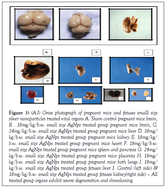
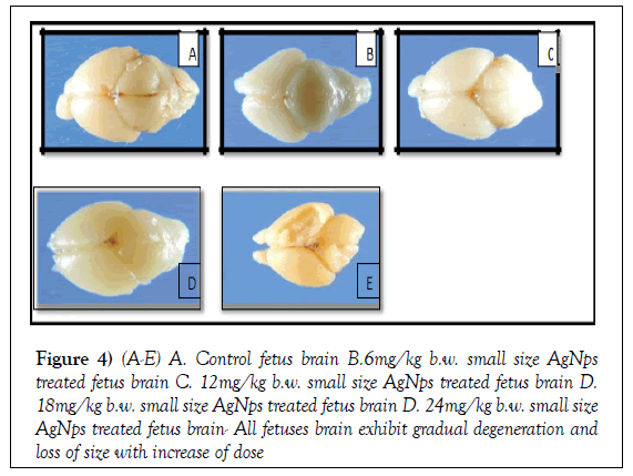
Histopathological and gross morphological assessment of toxicity in pregnant mice and their fresh born fetuses from control and treated groups brain tissue revealed significant histopathological and gross morphological changes (liver, kidney, brain, placenta and spleen tissue damage which includes insignificant decrease in anteroposterior length and breadth). in all small size Ag-NPexposed groups. In lower dose (0.65, 1.3 mg/kg b.w.). found very negotiable and minimal depletion whereas in higher dose (6, 12, 18, 24 mg/kg b.w.). found insignificant depletion of liver, kidney, brain, placenta and spleen gross morphological and histopathological parameters in pregnant mice and their fresh born fetuses. AgNps I and II (PVP/NaBH4I aggregated and stabilized). higher and lower dose treated groups insignificantly compared to the sham control (p<0.006 pregnant mother/ p<0.0052 fetuses). (Figures 3 and 4 ). (Tables 1 and 2).
Figure 3: (A-J) Gross photograph of pregnant mice and fetuses smalll size silver nanoparticles treated vital organs A. Sham control pregnant mice brain, B . 18mg/kg/b.w. small size AgNps treated group pregnant mice brain, C. 24mg/kg/b.w. small size AgNps treated group pregnant mice liver D. 18mg/ kg/b.w. small size AgNps treated group pregnant mice kidney E. 18mg/kg/ b.w. small size AgNps treated group pregnant mice heart F. 18mg/kg/b.w. small size AgNps treated group pregnant mice spleen and pancreas G. 24mg/ kg/b.w. small size AgNps treated group pregnant mice placenta H. 18mg/ kg/b.w. small size AgNps treated group pregnant mice both lungs I. 18mg/ kg/b.w. small size AgNps treated group fetuses liver J. Control (left side) & 18mg/kg/b.w. small size AgNps treated group fetuses kidney(right side) – All treated group organs exhibit severe degeneration and shrunkening
Figure 4: (A-E) A. Control fetus brain B.6mg/kg b.w. small size AgNps treated fetus brain C. 12mg/kg b.w. small size AgNps treated fetus brain D. 18mg/kg b.w. small size AgNps treated fetus brain D. 24mg/kg b.w. small size AgNps treated fetus brain- All fetuses brain exhibit gradual degeneration and loss of size with increase of dose
Table 1: Different anthropometrical measures of liver, kidney, brain and placentas from control and treated group pregnant mother parameters taken length and breadth in cm ( liver), length and breadth in cm ( kidney), ant. to post. Length and breadth in cm ( brain) and breadth distance in cm ( placenta).
| Groups | Pregnant mice Liver Ap Length in cm. | Pregnant mice liver Breadth in cm. | Pregnant mice liver Breadth in cm. | Pregnant mice kidney Breadth in cm. | Pregnant mice brain Ap Length in cm. | Pregnant mice brain Breadth in cm. | Pregnant mice placenta Breadth in cm |
|---|---|---|---|---|---|---|---|
| Control | 4.12 ± 0.12 | 3.01 ± 0.09 | 1.32 ± 0.006 | 0.75 ± 0.004 | 3.25 ± 0.06 | 2.02 ± 0.06 | 1.81 ± 0.06 |
| 0.65 mg | 4.09 ± 0.11 | 2.97 ± 0.06 | 1.29 ± 0.006 | 0.74 ± 0.004 | 3.22 ± 0.05 | 1.99 ± 0.05 | 1.80 ± 0.06 |
| 1.3 mg | 4.04 ± 0.10 | 2.96 ± 0.06 | 1.28 ± 0.006 | 0.73 ± 0.003 | 3.20 ± 0.05 | 1.96 ± 0.04 | 1.79 ± 0.05 |
| 6 mg | 3.92 ± 0.08 | 2.91 ± 0.03 | 1.26 ± 0.005 | 0.71 ± 0.003 | 3.17 ± 0.04 | 1.93 ± 0.04 | 1.78 ± 0.04 |
| 12 mg | 3.90 ± 0.06 | 2.88 ± 0.03 | 1.25 ± 0.005 | 0.69 ± 0.003 | 3.15 ± 0.04 | 1.92 ± 0.04 | 1.73 ± 0.04 |
| 18 mg | 3.59 ± 0.04 | 2.55 ± 0.06 | 1.13 ± 0.004 | 0.57 ± 0.003 | 3.03 ± 0.03 | 1.85 ± 0.03 | 1.53 ± 0.03 |
| 24 mg | 3.89 ± 0.02 | 2.83 ± 0.06 | 1.22 ± 0.002 | 0.66 ± 0.003 | 3.11 ± 0.03 | 1.86 ± 0.03 | 1.69 ± 0.03 |
Table 2: Mean ± S.D. of distance of length and breadth of fetus liver, Mean ± S.D. of of length and breadth of fetus kidney , Mean ± S.D. of distance of length and breadth fetus brain from control to treated group given below in table 2.( Marked reduction in lengths are underlined).
| Groups | Fetus liver Ap Length in cm | Fetus liver Breadth in cm | Fetus kidney Ap Length in cm | Fetus kidney Breadth in cm | Fetus brain Ap length in cm | Fetus brain Breadth in cm |
|---|---|---|---|---|---|---|
| Control | 1.12 ± 0.02 | 1.01 ± 0.03 | 0.3 ± 0.002 | 0.25 ± 0.002 | 1.25 ± 0.03 | 1.02 ± 0.02 |
| 0.65 mgtr.gr. | 1.11 ± 0.02 | 1.00 ± 0.02 | 0.29 ± 0.002 | 0.24 ± 0.002 | 1.24 ± 0.03 | 1.01 ± 0.02 |
| 1.3 mg tr. gr. | 1.10 ± 0.02 | 0.99 ± 0.02 | 0.28 ± 0.002 | 0.23 ± 0.002 | 1.23 ± 0.02 | 1.00 ± 0.02 |
| 6 mg tr. gr. | 0.84 ± 0.01 | 0.83 ± 0.01 | 0.26 ± 0.002 | 0.22 ± 0.001 | 1.21 ± 0.02 | 0.99 ± 0.02 |
| 12 mgtr.gr. | 0.79 ± 0.01 | 0.78 ± 0.01 | 0.25 ± 0.002 | 0.21 ± 0.001 | 1.19 ± 0.02 | 0.97 ± 0.02 |
| 18 mgtr.gr. | 1.09 ± 0.02 | 0.99 ± 0.02 | 0.18 ± 0.001 | 0.15 ± 0.001 | 1.17 ± 0.01 | 0.95 ± 0.01 |
| 24 mgtr.gr. | 1.08 ± 0.02 | 0.98 ± 0.02 | 0.24 ± 0.002 | 0.21 ± 0.001 | 0.97 ± 0.01 | 0.77 ± 0.01 |
Histological toxicogenic effects of small size AgNps on central nervous system and rest of the vital organs
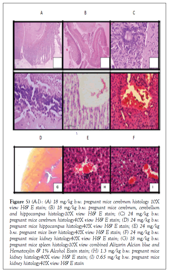
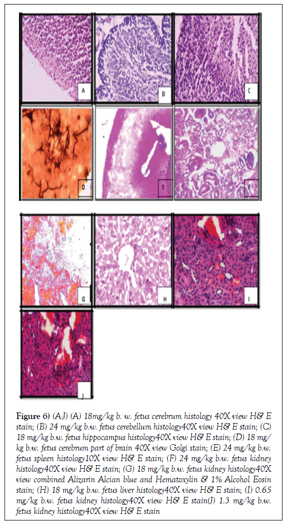
In the histological view trial, we observed a significant degeneration and impairment of nervous system histology which includes cerebrum, cerebellum and hippocampus of pregnant mother and their fetuses. In lower treated groups exposed to low dose (0.65/1.3 mg/kg b.w.). small size AgNps we observed very minimal and insignificant histological deviation cerebrum, cerebellum and hippocampus organs of mother and fetuses which is quite nearly comparable to control (p>1.03). whereas at higher to highest doses we observed significant histological deviation. (p< 0.003). (6, 12, 18 and 24 mg/ kg b.w. small size AgNps treated group). (Figures 5 and 6). This histological view trend was reflected by a decrease in the spontaneous alternation percentage compared to the control group, suggesting effects on short term memory. In lower dose group we observed very negotiable and minimal cortico medullary histological deviation such as degeneration and dystrophy of granular layer, molecular layer, pyramidal cell layer, Pyknotic cells, Honey comb deformity, degeneration of Lugaro cells, Purkinje cell layer and Betz cell layer which is found quite nearly comparable to normal sham control group of both pregnant mother and fetuses. Whereas in higher dose treated group we observed significant and higher to highest intensity histological deviation of central nervous system which includes degeneration and dystrophy of granular layer, molecular layer, pyramidal cell layer, Pyknotic cells, Honey comb deformity, degeneration of Lugaro cells, Purkinje cell layer and Betz cell layer in same experimental animals (Figures 5 and 6). As a histological toxicogenic focus we observed same variation or alteration in view of cortical part as well as medullary part of all vital internal organs of pregnant mice and fetuses. In liver histology of pregnant mice and fetuses as toxicogenic focus in principal form we observed dilatation of central vein, discontinuity of endothelial parenchyma of central vein or multiple tears in between, degeneration and atrophy with dystrophy of hepatocytes and portal triad. In kidney histology we observed degeneration, atrophy with dystrophy of Glomerulus or Malphigian corpuscles or even absence, degeneration or dystrophy with severe congestion and atrophy of proximal convoluted tubules, distal convoluted tubules and loop of Henley’s in both pregnant mice and fetuses. In spleen histology we observed degeneration, atrophy with dystrophy of splenic corpuscles or even absence, degeneration or dystrophy with severe atrophy of red blood corpuscles, white blood corpuscles and severe congestion in medullary sinuses in both pregnant mice and fetuses. In lung, trachea and bronchus histology we observed degeneration, atrophy with dystrophy of alveolus and atrium or even absence, degeneration or dystrophy with severe atrophy of cortical and medullary pneumocytes, and severe congestion in lumen of alveolus with tear of internal regularity of alveolar lumen or parenchyma we also observed severe loss of squamous cell pattern of lumen of alveolus in both pregnant mice and fetuses and at last in cardiac histology we observed degeneration, atrophy with dystrophy of cardiomyocytes or even absence, degeneration or dystrophy with severe atrophy of endocardial parenchyma , and severe congestion in lumen of valves in horizontal view in both pregnant mice and fetuses. In Golgi staining view we also observed degeneration dystrophy of multipolar neurons, loss of axons and dendrites from matrix, loss of depthness of neural tree branching pattern and loss of Basket cells and Hoffbauer cells with increased intensities of dose of small size AgNps in fetuses brain bright field microscopic view. (Figures 5 and 6). In lower dose treated group the intensities of such was found lower almost nearby or equal to sham control group whereas in higher and highest dose treated group the intensities was found higher and highest. Post hoc analyses indicated that the small size AgNps I lower and higher dosetreated groups made more spontaneous alternation errors than the control group (P<0.0001). (Figures 5 and 6)., but no statistically significant difference was found between the small size AgNps I lower dose treated groups and sham control. But statistically significant difference was found between the small size AgNps I higher to highest dose treated groups and sham control.
Figure 5: (A-I)-: (A) 18 mg/kg b.w. pregnant mice cerebrum histology 10X view H& E stain; (B) 18 mg/kg b.w. pregnant mice cerebrum, cerebellum and hippocampus histology10X view H& E stain; (C) 24 mg/kg b.w. pregnant mice cerebrum histology40X view H& E stain; (D) 24 mg/kg b.w. pregnant mice hippocampus histology40X view H& E stain; (E) 24 mg/kg b.w. pregnant mice liver histology40X view H& E stain; (F) 24 mg/kg b.w. pregnant mice kidney histology40X view H& E stain; (G) 18 mg/kg b.w. pregnant mice spleen histology10X view combined Alizarin Alcian blue and Hematoxylin & 1% Alcohol Eosin stain; (H) 1.3 mg/kg b.w. pregnant mice kidney histology40X view H& E stain; (I) 0.65 mg/kg b.w. pregnant mice kidney histology40X view H& E stain
Figure 6: (A-J) (A) 18mg/kg b. w. fetus cerebrum histology 40X view H& E stain; (B) 24 mg/kg b.w. fetus cerebellum histology40X view H& E stain; (C) 18 mg/kg b.w. fetus hippocampus histology40X view H& E stain; (D) 18 mg/ kg b.w. fetus cerebrum part of brain 40X view Golgi stain; (E) 24 mg/kg b.w. fetus spleen histology10X view H& E stain; (F) 24 mg/kg b.w. fetus kidney histology40X view H& E stain; (G) 18 mg/kg b.w. fetus kidney histology40X view combined Alizarin Alcian blue and Hematoxylin & 1% Alcohol Eosin stain; (H) 18 mg/kg b.w. fetus liver histology40X view H& E stain; (I) 0.65 mg/kg b.w. fetus kidney histology40X view H& E stain(J) 1.3 mg/kg b.w. fetus kidney histology40X view H& E stain
Discussion
Results after synthesis and characterization of small size nanosilver dispersed in fresh particle colloidal silver
The cause of poly vinyl pyrollidone coated and sodium borohydried stabilized small size AgNps colloidal solution prepared by magnetic stirring and cooling synthesizing method used in this study was to ensure a visualized toxicity. Small size AgNps were found to have a hydrodynamic radius measured 2.75 to 20.11nm size range that the radius obtained by transmission electron microscopy analysis. Based on the DLS, TEM and Image-J analysis measurements, our data suggest some degree of poly vinyl pyrollidone coated and sodium borohydried stabilized small size AgNps crystallites agglomeration in colloidal suspension.
Selection of animals, groups which corresponds to doses and oral gavages schedules and maintain of temperature inside animal house
The agglomerated small size nanosilver particles arises toxicogenic sign and symptoms in primate vertebrate experimental pregnant Swiss Albino mice and their fresh born fetuses from sham control, 0.65, 1.3, 6, 12, 18 and 24 mg/kg b.w. small size AgNps colloidal solution treated group which is been introduced into pregnant mice body in repeated oral gavages schedule from 0 to 19 gestational days once daily in morning inside animal house in suitable maintenance of temperature (20-24°C).
Results of analysis of zeta potential and TEM of small size nanosilver particles in terms of toxicity
This limited matrix bottom agglomeration supports the dependence of Zeta potential for AgNpsI stock solutions, as the colloidal solution AgNps I showed reasonable stability (AZP =-17.52 mV). After synthesis processes significant aggregation of small size AgNps in suspension was observed by TEM under relevant physiological condition. These findings suggest that the small size silver nano particles colloidal solution experimented in this existing study are positively demonstrate toxicity if tested in vivo or in vitro.
Utility of small size nanosilver in medical and consumer field and toxic hazards are its normal behavior in human and primate vertebrate
In the treatment and medical field, nanosilver is used as burn and chronic wounds ointpaint more over it is strongly used as dermal cream and it is a dermal tissue inhaler. [33]. Small size nanosilver made new consumer products are recently being developed and introduced in market. Small size AgNps has received considerable attention because of their attractive mechanochemical properties when compared to other metal nanomaterials. The strong toxicogenic properties of nanosilver to a vast range of species is well-known [34,35]. and small size AgNps has currently been shown to be a promising antimicrobial and antibacterial material. (36). However, some of the useful properties of small size silver nano materials are also pose a toxicogenic hazard to humans, primate vertebrate and the surrounding environment. [37,38]. place in the xylene. Repeat the treatment.
Corrosive effects of small size nanosilver in histological and histopathological cells with vital organs histopathological and gross morphological deviations as toxicogenic symptoms provoke Hydration: Due to their small size, NPs can enter plant or animal tissues and can pass through cell membranes such as the blood-brain barrier into critical areas within the brain or into other organs or tissues and due to its large surface area and irregular spiny surface it produces severe toxicogenic dilemma. [39]. Recent researches have reported various toxicogenic effects of small size silver NPs, including detrimental effects on multi vital internal organ functions which also include central nervous system. [15,22]. In our study, treated groups exposed to low dose (0.65 and 1.3 mg/kg b.w.). and high doses (6, 12, 18 and 24 mg/kg b.w.). of AgNps I exhibited a significant toxicity inform of degeneration dystrophy and severe atrophy or even absence of histological and histopathological cells and severe congestion of luminal structures which are macroscopic and microscopic both. Histopathological and gross morphological assessment of toxicity in pregnant mice and their fresh born fetuses from control and treated groups brain tissue revealed significant histopathological and gross morphological changes (liver, kidney, brain, placenta and spleen tissue damage which includes insignificant decrease in anteroposterior length and breadth). in all small size Ag-NP-exposed groups. In lower dose (0.65, 1.3 mg/ kg b.w.). found very negotiable and minimal depletion whereas in higher dose (6, 12, 18, 24 mg/kg b.w.). found insignificant depletion of liver, kidney, brain, placenta and spleen gross morphological and histopathological parameters in pregnant mice and their fresh born fetuses. AgNps I and II (PVP/NaBH4I aggregated and stabilized). higher and lower dose treated groups insignificantly compared to the sham control (p<0.006 pregnant mother/p<0.0052 fetuses). (Figures 4 and 5). (Tables 1 and 2).
Results of histological trials
In the histological view trial, we observed a significant degeneration and impairment of nervous system histology which includes cerebrum, cerebellum and hippocampus of pregnant mother and their fetuses. In lower treated groups exposed to low dose (0.65/1.3 mg/kg b.w.). small size AgNps we observed very minimal and insignificant histological deviation cerebrum, cerebellum and hippocampus organs of mother and fetuses which is quite nearly comparable to control (p>1.03). whereas at higher to highest doses we observed significant histological deviation. (p< 0.003). (6, 12, 18 and 24 mg/ kg b.w. small size AgNps treated group). (Figures 5 and 6). This histological view trend was reflected by a decrease in the spontaneous alternation percentage compared to the control group, suggesting effects on short term memory. In lower dose group we observed very negotiable and minimal corticomedullary histological deviation such as degeneration and dystrophy of granular layer, molecular layer, pyramidal cell layer, Pyknotic cells, Honey comb deformity, degeneration of Lugaro cells, Purkinje cell layer and Betz cell layer which is found quite nearly comparable to normal sham control group of both pregnant mother and fetuses. Whereas in higher dose treated group we observed significant and higher to highest intensity histological deviation of central nervous system which includes degeneration and dystrophy of granular layer, molecular layer, pyramidal cell layer, Pyknotic cells, Honey comb deformity, degeneration of Lugaro cells, Purkinje cell layer and Betz cell layer in same experimental animals (Figures 5 and 6). As a histological toxicogenic focus we observed same variation or alteration in view of cortical part as well as medullary part of all vital internal organs of pregnant mice and fetuses. In liver histology of pregnant mice and fetuses as toxicogenic focus in principal form we observed dilatation of central vein, discontinuity of endothelial parenchyma of central vein or multiple tears in between, degeneration and atrophy with dystrophy of hepatocytes and portal triad. In kidney histology we observed degeneration, atrophy with dystrophy of Glomerulus or Malphigian renal corpuscles or even absence, degeneration or dystrophy with severe congestion and atrophy of proximal convoluted tubules, distal convoluted tubules and loop of Henley’s in both pregnant mice and fetuses. In spleen histology we observed degeneration, atrophy with dystrophy of splenic corpuscles or even absence, degeneration or dystrophy with severe atrophy of red blood corpuscles, white blood corpuscles and severe congestion in medullary sinuses in both pregnant mice and fetuses. In lung, trachea and bronchus histology we observed degeneration, atrophy with dystrophy of alveolus and atrium or even absence, degeneration or dystrophy with severe atrophy of cortical and medullary pneumocytes, and severe congestion in lumen of alveolus with tear of internal regularity of alveolar lumen or parenchyma we also observed severe loss of squamous cell pattern of lumen of alveolus in both pregnant mice and fetuses and at last in cardiac histology we observed degeneration, atrophy with dystrophy of cardiomyocytes or even absence, degeneration or dystrophy with severe atrophy of endocardial parenchyma , and severe congestion in lumen of valves in horizontal view in both pregnant mice and fetuses.
Results of special Golgi stain trials for fetal CNS toxicity
In Golgi staining view we also observed degeneration dystrophy of multi polar neurons, loss of axons and dendrites from matrix, loss of depthness of neural tree branching pattern and loss of Basket cells and Hoffbauer cells with increased intensities of dose of small size AgNps in fetuses brain bright field microscopic view. (Figures 5 and 6). In lower dose treated group the intensities of such was found lower almost nearby or equal to sham control group whereas in higher and highest dose treated group the intensities was found higher and highest. Post hoc analyses indicated that the small size AgNps I lower and higher dose-treated groups made more spontaneous alternation errors than the control group (P<0.0001). (Figures 5 and 6)., but no statistically significant difference was found between the small size AgNps I lower dose treated groups and sham control. But statistically significant difference was found between the small size AgNps I higher to highest dose treated groups and sham control.
Overall small size AgNps CNS toxicity which stimulates neurodegenerative diseases and oxidative stress However we found no differences in the working and cognitive memory errors between both groups of Pregnant Swiss Albino mice and fetuses exposed to different small sized silver nano particles (AgNps I-:2.75 nm size and AgNps II-:20.11 nm size ranges). While the body weight (BW). of pregnant mice and fetuses that were treated with small size AgNps (AgNps I and AgNps II). did not change significantly, their brain to body weight ratio decreased significantly compared to the control pregnant mice and fetuses. We also observed significant histological and histopathological changes also changes in the brains and other vital internal organs of AgNps (AgNps I-:2.75 nm size and AgNps II-:20.11 nm size ranges). exposed groups. Therefore, we conclude that AgNps I-:2.75 nm size and AgNps II-:20.11 nm size ranges treatment might cause brain damage along with other vital internal organs damage and execute toxicogenic sequels. Current, reports have indicated that the small silver nano sized components of particulate matter can reach the brain and other internal vital organs and may be associated with histological and histopathological changes and diseases with neurodegenerative disease and neurological deficit. [40,41]. Small size AgNps have been found to increase oxidative stress [24,25,42].
Conclusion
These findings support the hypothesis that neuronal cells and other histological and histopathological cells which are exposed from all small size AgNps (2.75 to 20.11 nm size range). in pregnant mice and fetuses’ and other primate vertebrate vital internal organs and brain undergo oxidative stress, which could ultimately lead to the observed histological and histopathological cells significant toxicogenic response and central nervous system toxicity. Nanotechnology allows the manipulation and application of engineered small size silver nanoparticles or systems that have at least one dimension less than 100 nm in length or 2.75 to 20.11 nm ranges. New industries in collaboration with university research departments are being formed and are finding so many investors eager to back their ideas and products. There is no doubt that governments and major industrial companies are committing significant resources for research into the expansion of nanometer scale processes, materials and products. Growing exploration of nanotechnology has resulted in the discovery of many distinctive properties of silver nanomaterials such as superior catalytic, optical, magnetic, mechanical and electrical properties when compared to conventional formulations of the same material. Nanomaterials are progressively more being used for commercial purposes and in consumer products leading to increased direct and indirect exposure in humans which definitely leads to human toxicity and drastically affects public health. In the field of medicine, small size silver nanoparticles are purposefully injected into the human body. For drug delivery and imaging nanomaterials are often deliberately coated with biomolecules such as protein, DNA and monoclonal antibodies to aim particular cells and deaggregator and stabilizer chemicals. The novel physicochemical properties of these engineered nanomaterials may initiate new mechanisms of injury and toxicological effects due to destructive interactions of small size silver nanomaterials with biological systems and the environment. Even though less studied than human health, there is also basis for concern regarding environmental impacts and areas such as ecotoxicology, environmental chemistry, behavior and fate are areas of concentrated current research. Nanomaterials may potentially impact the environment in three possible ways: [1] direct effect on invertebrates, microorganisms, fish and other species; [2] interaction with other pollutants, that may transform the bioavailability of toxic compounds and nutrients; and [3] changes to nonliving environmental structures. Understanding the environmental and human health impacts of silver nanomaterials is a crucial stage for the responsible development of nanotechnology and to gain full benefit of its applications. As nanotechnology matures, questions are being aroused regarding whether the products or materials of nanotechnology will provide hazards to human health or the environment and whether the manufacturing of these materials will spawn new hazards or waste streams. Considerate the environmental and human health impact of nanomaterials is a fundamental stage for the liable development nanotechnology and to gain full advantage of its applications. Alternatively, green nanoscience has been described as the progress of clean technologies to reduce possible environmental and human health risks coupled with the production and use of nanomaterials and to promote replacement of existing products with new nano-products that are further environmentally friendly all over their life cycle. Green nanoscience is the successful integration of green chemistry and nanoscience that brings the collective approaches and methods to build up greener products, processes, and applications. Therefore, green chemistry and nanoscience are promising fields that take advantage of molecular level design and have massive potential to overcome the adverse impacts of small size silver nanoparticles and nanomaterials on health and environment. If the global research community can take advantage of green nanoscience then we can surely look forward to the advent of safe nanotechnologies. In this review we have summarized the adverse effects of different small size silver a nanoparticle (2.75 to 20.11 nm ranges). which evokes human toxicity and affects public health and environment along with alternative strategies to overcome ill impacts. Our results implies small size silver nanoparticles have definitely stimulates human toxicity and public health also affects environment. So more and more nano business tycoons of this recent world must be very careful in production of consumer utility goods.
Conflict of Interest
The present manuscript 1st author, corresponding author and co-authors declare no conflict of interest.
Acknowledgements
This work was supported by minor division 5012 scheme of Department of Anatomy; Central Banaras Hindu University Uttarpradesh and University Grant Commission New Delhi, India.
REFERENCES
- Toshima N. Nano scale Materials. In: M Liz Marzan L, Kamat PV, Eds. Kluwer Academic Pub. London 2003:79-96.
- Nam JM, Thaxton CS, Mirkin CA. Nanoparticle based bio-bar codes for the ultrasensitive detection of proteins. Science 2003;301:1884-6.
- Tkachenko AG, Xie H, Coleman D, et al. Multifunctional gold nanoparticle peptide complexes for nuclear targeting. J Am Chem Soc 2003;125:4700-1.
- Hirsch LR, Stafford RJ, Bankson JA, et al. Nano shell mediated near-infrared thermal therapy of tumors under magnetic resonance guidance. Proc Natl Acad Sci 2003;100:13549-54.
- Burda C, Chen X, Narayanan R, et al. Chemistry and properties of nano crystals of different shapes. Chem Rev 2005;105:1025-102.
- Gupta A, Silver S. Silver as biocide: Will resistance become a problem?. Nat Biotechnol 1998;16: 888.
- Ji JH, Jung JH, Kim SS, et al. Twenty-eight-day inhalation toxicity study of silver nanoparticles in Sprague Dawley rats. Inhal Toxicol 2007;19:857-71.
- Lockman PR, Koziara JM, Mumper RJ, et al. Nanoparticle surface charges alter blood brain barrier integrity and permeability. J Drug Target 2004;12:635-41.
- Kreyling WG, Semmler M, Erbe F, et al. Translocation of ultrafine insoluble iridium particles from lung epithelium to extra pulmonary organs is size Brought to you by | Banaras Hindu University Authenticated Download Date | 11/1/15 10:43 AM Exposure to silver nano particles induces oxidative stress and memory deficits in laboratory rats dependent but very low. J Toxicol Environ Health Part A 2002;65:1513-30.
- Oberdorster G, Sharp Z, Atudorei V, et al. Translocation of inhaled ultrafine particles to the brain, Inhal. Toxicol 2004; 16:437-45.
- Ma L, Liu J, Li N, et al. Oxidative stress in the brain of mice caused by translocated nano particulate TiO2 delivered to the abdominal cavity, Biomaterials, 2010;31:99-105.
- Hu R, Gong X, Duan Y, et al. Neuro toxicological effects and the impairment of spatial recognition memory in mice caused by exposure to TiO2 nano particles. Biomaterials 2010;31:8043-50.
- Sharma HS. Nano neuroscience: Emerging concepts on nano neurotoxicity and nano neuro protection. Nano medicine 2007;2:753-8.
- Sharma HS, Sharma A. Nano particles aggravate heat stress induced cognitive deficits, blood–brain barrier disruption, edema formation and brain pathology. Prog Brain Res 2007;162:245-73.
- Kim YS, Kim JS, Cho HS, et al. Twenty-eight-day oral toxicity, genotoxicity, and gender related tissue distribution of silver nano particles in Sprague-Dawley rats. Inhal Toxicol 2008;20:575-83.
- Liu Z, Ren G, Zhang T, et al. Action potential changes associated with the inhibitory effects on voltage-gated sodium current of hippocampal CA1 neurons by silver nano particles. Toxicology 2009;264:179-84.
- Cha K, Hong HW, Choi YG, et al. Comparison of acute responses of mice livers to short term exposure to nano-sized or micro-sized silver particles, Biotechnol. Lett 2008;30:1893-9.
- Shin SH, Ye MK, Kim HS, et al. The effects of nano-silver on the proliferation and cytokine expression by peripheral blood mononuclear cells. Int Immunopharmacol 2007;7:1813-8.
- Tang M, Xing T, Zeng J, et al. Unmodifed CdSe quantum dots induce elevation of cytoplasmic calcium levels and impairment of functional properties of sodium channels in rat primary cultured hippocampal neurons. Environ Health Perspect 2008;116:915-22.
- Hussain SM, Javorina AK, Schrand AM, et al. The interaction of manganese nanoparticles with PC-12 cells induces dopamine depletion. Toxicol Sci 2006;92:456-63.
- Cui K, Luo X, Xu K, et al. Role of oxidative stress in neurodegeneration:recent developments in assay methods for oxidative stress and nutraceutical antioxidants. Prog Neuro Psychopharmacol Biol Psychiatry 2004;28:771- 99.
- Curtin JF, Donovan M, Cotter TG. Regulation and measurement of oxidative stress in apoptosis. J Immunol Methods 2002;265:49-72.
- Arora S, Jain J, Rajwade JM, et al. Cellular responses induced by silver nano particles: In vitro studies. Toxicol Lett 2008;179:93-100.
- Rahman MF, Wang J, Patterson TA, et al. Expression of genes related to oxidative stress in the mouse brain after exposure to silver-25 nanoparticles. Toxicol Lett 2009;187:15-21.
- Ahamed M, Posgai R, Gorey TJ, et al. Silver nanoparticles induced heat shock protein 70, oxidative stress and apoptosis in Drosophila melanogaster. Toxicol Appl Pharmacol 2010;242:263-9.
- Choi JE, Kim S, Ahn JH, et al. Induction of oxidative stress and apoptosis by silver nanoparticles in the liver of adult zebrafish. Aquat Toxicol 2010;100:151-9.
- Sun L, Singh AK, Vig K, et al. Silver nano particles inhibit replication of respiratory syncytial virus. J Biomed Biotechnol 2008;4:149-58.
- Duran N, Marcato PD, De Conti R, et al. Potential use of silver nanoparticles on pathogenic bacteria, their toxicity and possible mechanisms of action. J Braz Chem Soc 2010;21:949-959.
- Karelson E, Bogdanovic N, Garlind A, et al. The cerebrocortical areas in normal brain aging and in Alzheimer’s disease:noticeable differences in the lipid peroxidation level and in antioxidant defense. Neurochem Res 2001;26:353-361.
- Berne BJ, Pecora R. Dynamic light scattering. Courier Dover Publications 2000 ISBN 48641155-9.
- Schneider CA, Rasband WS, Eliceiri KW. NIH Image to Image J: A 25 years of image analysis. Nat Methods 2012;9 (7).:671–675.
- Reimer L. Transmission electron microscopy: Image formation and microanalysis (4th edn). Berlin: Springer 1997.
- Liau SY, Read DC, Pugh WJ, et al Interaction of silver nitrate with readily identifiable groups: Relationship to the antibacterial action of silver ions. Lett Appl Microbiol 1997;25:279-83.
- Hritcu L. Nabeshima T. Kainic acid lesion-induced spatial memory deficits of rats. Cent Eur J Biol 2009;4:179-85.
- Hritcu L, Clicinschi M, Nabeshima T. Brain serotonin depletion impairs short-term memory, but not long-term memory in rats. Physiol Behav 2007;91:652-57.
- Sondi I, Salopek-Sondi B. Silver nano particles as antimicrobial agent:A case study on E. coli as a model for Gram-negative bacteria. J Colloid Interface Sci 2004;275:177-82.
- Brown DM, Wilson MR, MacNee W, et al. Size-dependent pro-inflammatory effects of ultrafine polystyrene particles: A role for surface area and oxidative stress in the enhanced activity of ultra-fines. Toxicol Appl Pharmacol 2001;175:191-99.
- Muller M, Mackeben S, Muller-Goymann CC. Physicochemical characterization of liposomes with encapsulated local anaesthetics. Int J Pharm 2004;274:139-48.
- Foley S, Crowley C, Smaihi M, et al. Cellular localization of a water-soluble fullerene derivative. Biochem Biophys Res Commun 2002;294:116-9.
- Block ML, Wu X, Pei Z, et al. Nanometer size diesel exhaust particles are selectively toxic to dopaminergic neurons: The role of microglia, phagocytosis, and NADPH oxidase. FASEB J 2004;18:1618-20
- Peters A, Veronesi B, Calderon-Garciduenas L, et al. Translocation and potential neurological effects of fine and ultrafine particles a critical update. Part Fibre Toxicol 2006;3:1-13.
- Carlson C, Hussain SM, Schrand AM, et al. Unique cellular interaction of silver nano particles: Size dependent generation of reactive oxygen species. J Phys Chem B 2008;112:13608-19.


