Unilateral Large Mylohyoid Bridging the Dry Human Mandible.
Received: 01-Sep-2022, Manuscript No. ijav-22-5289; Editor assigned: 05-Sep-2022, Pre QC No. ijav-22-5289 (PQ); Accepted Date: Sep 23, 2022; Reviewed: 19-Sep-2022 QC No. ijav-22-5289; Revised: 23-Sep-2022, Manuscript No. ijav-22-5289 (R); Published: 30-Sep-2022, DOI: 10.37532/1308-4038.15(9).216
Citation: Parlak M. Unilateral Large Mylohyoid Bridging the Dry Human Mandible. Int J Anat Var. 2022;15(9):216-217.
This open-access article is distributed under the terms of the Creative Commons Attribution Non-Commercial License (CC BY-NC) (http://creativecommons.org/licenses/by-nc/4.0/), which permits reuse, distribution and reproduction of the article, provided that the original work is properly cited and the reuse is restricted to noncommercial purposes. For commercial reuse, contact reprints@pulsus.com
Abstract
45 mandibles used in education in Bezmialem Vakıf, Hitit and Marmara University Medical Faculty Anatomy Departments were examined macroscopically. In some cases, the proximal part of the sulcus mylohyoideus may appear as a canal through a bony bridge (arcus mylohyoideus = mylohyoid bridge). Knowing the frequency of Mylohyoid Bridge variations will contribute to the literature studies on this subject and will guide oral surgery and dentistry practices in order to prevent possible complications in clinical applications.
Keywords
Mandibula; Arcus mylohyoideus; Sulcus mylohyoideus; Canalis mylohyoidus; Foramen mandibula
INTRODUTION
Mandibula is a strong mobile bone that constitutes the lower jaw skeleton, consisting of a corpus and two rami. On the inner surface of the ramus of the mandible, there is the mandibular foramen with a protrusion on its anterior side, called lingula, which is formed by the fusion of the medial and lateral laminae of the compact bone. Mylohyoid groove, which courses inferiorly arising from the mandibular foramen, is also observed inferior to this bony protrusion. The mylohyoid nerve passes through mylohyoid Groove and innervates the mylohyoid muscle and the anterior belly of the digastric muscle [1-2]. Histological studies have shown that the mylohyoid nerve is a mixed nerve containing both motor and sensory fibers [3]. In some cases, the proximal part of the mylohyoid groove may appear as a canal due to a bony bridge (Mylohyoid Bridge or arch) which is a hyperostotic derivation of Meckel’s cartilage on the mylohyoid Groove [4]. In cases with the Mylohyoid Bridge, the vessels and nerves coursing inside the bridge may be entrapped or the bridge structure may act as a barrier preventing anesthetic injections [5]. In addition to collecting valuable anthropometric data, the investigation of anatomical variations has always been important since these variations may bring significant contributions to the clinical applications. The aim of the current study is to determine the frequency rates of the Mylohyoid Bridge and to discuss the clinical significance of the subject.
METHOD
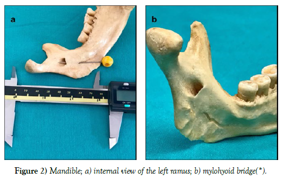
45 human mandibles which are used in regular educational sessions in Bezmialem Vakıf University, Hitit University and Marmara University Medical Schools, Departments of Anatomy, were macroscopically examined and the frequency of Mylohyoid Bridge was investigated. Vertical lengths of mylohyoid grooves and mylohyoid bridges were measured with digital calipers in the mylohyoid bridge variation presenting cases.
RESULTS
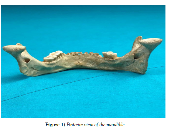
In one of the total 45 mandibles (90 mylohyoid grooves) examined in the current study, a large mylohyoid bridge extending towards the mandibular foramen was determined on the proximal part of the left mylohyoid groove (1.1%). The vertical length of this bridge was 12.41mm and the length of the mylohyoid groove was 20, 21mm. The length of the right mylohyoid groove, in this case, was measured 22.7mm (Figure 1-2).
CONCLUSION
The knowledge about the frequency of mylohyoid bridge variations will contribute to the literature on this subject and will guide oral surgical and dental practices to prevent possible complications in the clinical practice.
ACKNOWLEDGEMENT
None
CONFLICTS OF INTEREST
None
REFERENCES
- Arifoğlu Y. Her Yönüyle Anatomi, 3. Baskı, 2021 İstanbul Tıp Kitapevleri. Bezmi Vakıf Uni Res Info Sys. 77-79.
- Standring S, Ellis H, Healy J, Johnson D et al. Gray's anatomy: the anatomical basis of clinical practice. AJNR Am J Neuroradiol. 2005; 26(10), 2703.
- Bennett S, Townsend G. Distribution of the mylohyoid nerve: anatomical variability and clinical implications. Aust Endod J. 2001; 27(3):109-111.
- Rusu MC, Săndulescu M, Bichir C, Muntianu et al. Combined anatomical variations: The mylohyoid bridge, retromolar canal and accessory palatine canals branched from the canalis sinuosus. Ann Anat. 2017; 214:75-79.
- Balcioglu HA, Kose TE, Uyanikgil Y. Multiple morphological variations in a human mandible. Int J Morphol. 2015; 33(3):1023-1026.
Indexed at, Google Scholar, Crossref
Indexed at, Google Scholar, Crossref
Indexed at, Google Scholar, Crossref


