Unusual branching pattern of common hepatic artery – a case report
Satheesha Nayak B*, Sudarshan S, Venu Madhav N and Srinivasa Rao Sirasanagandla
Department of Anatomy, Melaka Manipal Medical College (Manipal Campus), International Centre for Health Sciences, Manipal University, Madhav Nagar, Manipal, Karnataka, India
- *Corresponding Author:
- Dr. Satheesha Nayak B.
Professor and Head, Department of Anatomy, MMMC, Int. Centre for Health Sci. Manipal University, Madhav Nagar, Manipal, Udupi District, Karnataka, 576 104, India
Tel: +91 820 2922519
E-mail: nayaksathish@yahoo.com
Date of Received: November 16th, 2011
Date of Accepted: September 22nd, 2012
Published Online: December 24th, 2012
© Int J Anat Var (IJAV). 2012; 5: 126–127.
[ft_below_content] =>Keywords
common hepatic artery, right gastric artery, gastroduodenal artery, hepatic artery, superior pancreaticoduodenal artery, coeliac trunk
Introduction
The common hepatic artery is normally a branch of coeliac trunk. It runs forwards and to the right and divides into a hepatic artery proper and a gastroduodenal artery. The hepatic artery proper terminates by dividing into right and left hepatic arteries and the gastroduodenal artery terminates by dividing into superior pancreaticoduodenal artery and right gastroepiploic artery. The right gastric artery arises either from common hepatic artery or the hepatic artery proper. The variations in the branching pattern of common hepatic artery are rare and the knowledge of the variations may be very useful for radiologists and surgeons. We report one of the very rare variations of common hepatic artery and its branches.
Case Report
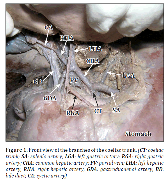
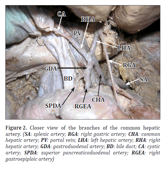
During the dissection classes for first year medical students, we observed the variation in the branching pattern of the hepatic arteries. This variation was found in an adult male cadaver of Indian origin. The coeliac trunk divided into its three terminal branches as usual: the left gastric, splenic and common hepatic arteries (Figure 1). The common hepatic artery coursed forwards and to the right and entered the lesser omentum. In the lesser omentum, it terminated by dividing into three branches; the right hepatic artery, the left hepatic artery and the gastroduodenal artery. The gastroduodenal artery, after a course of about 2.5 cm, terminated by dividing into superior pancreaticoduodenal and right gastroepiploic arteries. This division occurred in the lesser omentum above the pyloric part of the stomach. Both the branches of the gastroduodenal artery descended down behind the gastro-duodenal junction (Figure 2). The right gastric artery took its origin from the left hepatic artery within the porta hepatis and descended down in the lesser omentum. It passed in front of the trifurcation of the common hepatic artery and reached the lesser curvature of the stomach (Figure 2).
Figure 1. Front view of the branches of the coeliac trunk. (CT: coeliac trunk; SA: splenic artery; LGA: left gastric artery; RGA: right gastric artery; CHA: common hepatic artery; PV: portal vein; LHA: left hepatic artery; RHA: right hepatic artery; GDA: gastroduodenal artery; BD: bile duct; CA: cystic artery)
Figure 2. Closer view of the branches of the common hepatic artery. (SA: splenic artery; RGA: right gastric artery; CHA: common hepatic artery; PV: portal vein; LHA: left hepatic artery; RHA: right hepatic artery; GDA: gastroduodenal artery; BD: bile duct; CA: cystic artery; SPDA: superior pancreaticoduodenal artery; RGEA: right gastroepiploic artery)
Discussion
Though the variations in the branching pattern of the coeliac trunk are very common, variations of the course and branches of the common hepatic artery are very rare. Song et al., did an extensive study (on 5002 patients) on the common hepatic artery and found variations in only 3.71% of cases [1]. In their study, the common hepatic artery arose from the left gastric artery in 0.16% cases and passed into the liver through the fissure for ligamentum venosum. It arose from superior mesenteric artery in 3% of cases and from the abdominal aorta in 0.40% of cases. Okada et al., have also reported the origin of common hepatic artery from left gastric artery in 3% of cases [2]. A case of right hepatic artery forming a caterpillar hump has been reported by Priti and Lakshmi [3].
Knowledge of variations in the branches of the coeliac trunk is of importance for surgeons performing upper abdominal surgery and to radiologists performing any procedures in this area. In the upper abdominal surgeries, the blood flow in the hepatic arteries has to be maintained to minimise serious hepatic ischemic complications. This can be best done by a preoperative three dimensional imaging [1,2,4–6]. Recently the conventional angiography is replaced owing to recent advances in spiral and multidetector computed tomography (CT) technology [2,7–10]. Therefore, diagnostic as well as interventional radiologists should be familiar with the spectrum and cross-sectional and 3D appearances of variations in the celiac axis and hepatic artery anatomy. In the current case, we observed a trifurcation of the common hepatic artery; early division of gastroduodenal artery and origin of right gastric artery from the left hepatic artery. These variations are extremely rare and even if they are detected multidetector computed tomography, the varied branching pattern can make the hepatic arterial infusion chemotherapy and microcatheter embolizations techniques difficult. A case of catheterization and embolization of a replaced left hepatic artery through the right gastric artery has been reported [11]. The current variation of the origin of the right gastric artery from the left hepatic artery might be of additional advantage to pass a catheter into left hepatic artery through it to embolize the left hepatic artery.
Trifurcation of common hepatic artery and origin of right gastric artery from the left hepatic artery are very rare variations. Since the trifurcation of the common hepatic artery was in the lesser omentum, it makes the case one of the rarest variations.
References
- Song SY, Chung JW, Yin YH, Jae HJ, Kim HC, Jeon UB, Cho BH, So YH, Park JH. Celiac axis and common hepatic artery variations in 5002 patients: systematic analysis with spiral CT and DSA. Radiology. 2010; 255: 278–288.
- Okada Y, Nishi N, Matsuo Y, Watadani T, Kimura F. The common hepatic artery arising from the left gastric artery. Surg Radiol Anat. 2010; 32: 703–705.
- Mishall PL, Rajgopal L. Variant right hepatic artery forming Moynihan’s hump – clinical relevance. Int J Anat Var (IJAV). 2010; 3: 144–145.
- Koops A, Wojciechowski B, Broering DC, Adam G, Krupski-Berdien G. Anatomic variations of the hepatic arteries in 604 selective celiac and superior mesenteric angiographies. Surg Radiol Anat. 2004; 26: 239–244.
- Nghiem HV, Dimas CT, McVicar JP, Perkins JD, Luna JA, Winter TC 3rd, Harris A, Freeny PC. Impact of double helical CT and three-dimensional CT arteriography on surgical planning for hepatic transplantation. Abdom Imaging. 1999; 24: 278–284.
- Rygaard H, Forrest M, Mygind T, Baden H. Anatomic variants of the hepatic arteries. Acta Radiol Diagn (Stockh). 1986; 27: 425–427.
- Suzuki T, Nakayasu A, Kawabe K, Takeda H, Honjo I. Surgical significance of anatomic variations of the hepatic artery. Am J Surg. 1971; 122: 505–512.
- Curley SA, Chase JL, Roh MS, Hohn DC. Technical considerations and complications associated with the placement of 180 implantable hepatic arterial infusion devices. Surgery. 1993; 114: 928–935.
- Winter TC 3rd, Nghiem HV, Freeny PC, Hommeyer SC, Mack LA. Hepatic arterial anatomy: demonstration of normal supply and vascular variants with three-dimensional CT angiography. Radiographics. 1995; 15: 771–780.
- Takahashi S, Murakami T, Takamura M, Kim T, Hori M, Narumi Y, Nakamura H, Kudo M. Multi-detector row helical CT angiography of hepatic vessels: depiction with dual-arterial phase acquisition during single breath hold. Radiology. 2002; 222: 81–88.
- Miyazaki M, Shibuya K, Tsushima Y, Endo K. Catheterization and embolization of a replaced left hepatic artery via the right gastric artery through the anastomosis: a case report. J Med Case Rep. 2011; 5: 346.
Satheesha Nayak B*, Sudarshan S, Venu Madhav N and Srinivasa Rao Sirasanagandla
Department of Anatomy, Melaka Manipal Medical College (Manipal Campus), International Centre for Health Sciences, Manipal University, Madhav Nagar, Manipal, Karnataka, India
- *Corresponding Author:
- Dr. Satheesha Nayak B.
Professor and Head, Department of Anatomy, MMMC, Int. Centre for Health Sci. Manipal University, Madhav Nagar, Manipal, Udupi District, Karnataka, 576 104, India
Tel: +91 820 2922519
E-mail: nayaksathish@yahoo.com
Date of Received: November 16th, 2011
Date of Accepted: September 22nd, 2012
Published Online: December 24th, 2012
© Int J Anat Var (IJAV). 2012; 5: 126–127.
Abstract
Common hepatic artery is a branch of coeliac trunk. It normally terminates by dividing into hepatic artery proper and gastroduodenal artery. We report here the trifurcation of the common hepatic artery. The common hepatic artery terminated in the lesser omentum by dividing into three branches; the left hepatic artery, right hepatic artery and gastroduodenal artery. This termination was at a higher level than the usual termination. The gastroduodenal artery terminated by dividing into superior pancreaticoduodenal and right gastroepiploic arteries within the lesser omentum, above the pyloric part of the stomach. The right gastric artery arose from the left hepatic artery in the porta hepatis and descended down through the lesser omentum to reach the lesser curvature of the stomach.
-Keywords
common hepatic artery, right gastric artery, gastroduodenal artery, hepatic artery, superior pancreaticoduodenal artery, coeliac trunk
Introduction
The common hepatic artery is normally a branch of coeliac trunk. It runs forwards and to the right and divides into a hepatic artery proper and a gastroduodenal artery. The hepatic artery proper terminates by dividing into right and left hepatic arteries and the gastroduodenal artery terminates by dividing into superior pancreaticoduodenal artery and right gastroepiploic artery. The right gastric artery arises either from common hepatic artery or the hepatic artery proper. The variations in the branching pattern of common hepatic artery are rare and the knowledge of the variations may be very useful for radiologists and surgeons. We report one of the very rare variations of common hepatic artery and its branches.
Case Report
During the dissection classes for first year medical students, we observed the variation in the branching pattern of the hepatic arteries. This variation was found in an adult male cadaver of Indian origin. The coeliac trunk divided into its three terminal branches as usual: the left gastric, splenic and common hepatic arteries (Figure 1). The common hepatic artery coursed forwards and to the right and entered the lesser omentum. In the lesser omentum, it terminated by dividing into three branches; the right hepatic artery, the left hepatic artery and the gastroduodenal artery. The gastroduodenal artery, after a course of about 2.5 cm, terminated by dividing into superior pancreaticoduodenal and right gastroepiploic arteries. This division occurred in the lesser omentum above the pyloric part of the stomach. Both the branches of the gastroduodenal artery descended down behind the gastro-duodenal junction (Figure 2). The right gastric artery took its origin from the left hepatic artery within the porta hepatis and descended down in the lesser omentum. It passed in front of the trifurcation of the common hepatic artery and reached the lesser curvature of the stomach (Figure 2).
Figure 1. Front view of the branches of the coeliac trunk. (CT: coeliac trunk; SA: splenic artery; LGA: left gastric artery; RGA: right gastric artery; CHA: common hepatic artery; PV: portal vein; LHA: left hepatic artery; RHA: right hepatic artery; GDA: gastroduodenal artery; BD: bile duct; CA: cystic artery)
Figure 2. Closer view of the branches of the common hepatic artery. (SA: splenic artery; RGA: right gastric artery; CHA: common hepatic artery; PV: portal vein; LHA: left hepatic artery; RHA: right hepatic artery; GDA: gastroduodenal artery; BD: bile duct; CA: cystic artery; SPDA: superior pancreaticoduodenal artery; RGEA: right gastroepiploic artery)
Discussion
Though the variations in the branching pattern of the coeliac trunk are very common, variations of the course and branches of the common hepatic artery are very rare. Song et al., did an extensive study (on 5002 patients) on the common hepatic artery and found variations in only 3.71% of cases [1]. In their study, the common hepatic artery arose from the left gastric artery in 0.16% cases and passed into the liver through the fissure for ligamentum venosum. It arose from superior mesenteric artery in 3% of cases and from the abdominal aorta in 0.40% of cases. Okada et al., have also reported the origin of common hepatic artery from left gastric artery in 3% of cases [2]. A case of right hepatic artery forming a caterpillar hump has been reported by Priti and Lakshmi [3].
Knowledge of variations in the branches of the coeliac trunk is of importance for surgeons performing upper abdominal surgery and to radiologists performing any procedures in this area. In the upper abdominal surgeries, the blood flow in the hepatic arteries has to be maintained to minimise serious hepatic ischemic complications. This can be best done by a preoperative three dimensional imaging [1,2,4–6]. Recently the conventional angiography is replaced owing to recent advances in spiral and multidetector computed tomography (CT) technology [2,7–10]. Therefore, diagnostic as well as interventional radiologists should be familiar with the spectrum and cross-sectional and 3D appearances of variations in the celiac axis and hepatic artery anatomy. In the current case, we observed a trifurcation of the common hepatic artery; early division of gastroduodenal artery and origin of right gastric artery from the left hepatic artery. These variations are extremely rare and even if they are detected multidetector computed tomography, the varied branching pattern can make the hepatic arterial infusion chemotherapy and microcatheter embolizations techniques difficult. A case of catheterization and embolization of a replaced left hepatic artery through the right gastric artery has been reported [11]. The current variation of the origin of the right gastric artery from the left hepatic artery might be of additional advantage to pass a catheter into left hepatic artery through it to embolize the left hepatic artery.
Trifurcation of common hepatic artery and origin of right gastric artery from the left hepatic artery are very rare variations. Since the trifurcation of the common hepatic artery was in the lesser omentum, it makes the case one of the rarest variations.
References
- Song SY, Chung JW, Yin YH, Jae HJ, Kim HC, Jeon UB, Cho BH, So YH, Park JH. Celiac axis and common hepatic artery variations in 5002 patients: systematic analysis with spiral CT and DSA. Radiology. 2010; 255: 278–288.
- Okada Y, Nishi N, Matsuo Y, Watadani T, Kimura F. The common hepatic artery arising from the left gastric artery. Surg Radiol Anat. 2010; 32: 703–705.
- Mishall PL, Rajgopal L. Variant right hepatic artery forming Moynihan’s hump – clinical relevance. Int J Anat Var (IJAV). 2010; 3: 144–145.
- Koops A, Wojciechowski B, Broering DC, Adam G, Krupski-Berdien G. Anatomic variations of the hepatic arteries in 604 selective celiac and superior mesenteric angiographies. Surg Radiol Anat. 2004; 26: 239–244.
- Nghiem HV, Dimas CT, McVicar JP, Perkins JD, Luna JA, Winter TC 3rd, Harris A, Freeny PC. Impact of double helical CT and three-dimensional CT arteriography on surgical planning for hepatic transplantation. Abdom Imaging. 1999; 24: 278–284.
- Rygaard H, Forrest M, Mygind T, Baden H. Anatomic variants of the hepatic arteries. Acta Radiol Diagn (Stockh). 1986; 27: 425–427.
- Suzuki T, Nakayasu A, Kawabe K, Takeda H, Honjo I. Surgical significance of anatomic variations of the hepatic artery. Am J Surg. 1971; 122: 505–512.
- Curley SA, Chase JL, Roh MS, Hohn DC. Technical considerations and complications associated with the placement of 180 implantable hepatic arterial infusion devices. Surgery. 1993; 114: 928–935.
- Winter TC 3rd, Nghiem HV, Freeny PC, Hommeyer SC, Mack LA. Hepatic arterial anatomy: demonstration of normal supply and vascular variants with three-dimensional CT angiography. Radiographics. 1995; 15: 771–780.
- Takahashi S, Murakami T, Takamura M, Kim T, Hori M, Narumi Y, Nakamura H, Kudo M. Multi-detector row helical CT angiography of hepatic vessels: depiction with dual-arterial phase acquisition during single breath hold. Radiology. 2002; 222: 81–88.
- Miyazaki M, Shibuya K, Tsushima Y, Endo K. Catheterization and embolization of a replaced left hepatic artery via the right gastric artery through the anastomosis: a case report. J Med Case Rep. 2011; 5: 346.


