Unusual concurrence of intermediate pectoralis muscle and variant insertion of pectoralis minor with its clinical aspects.
2 Department of Anatomy, Mahatma Gandhi Medical College & Research Institute, SBV University, Puducherry, India
Received: 29-Jun-2018 Accepted Date: Jul 17, 2018; Published: 27-Jul-2018
Citation: Vani PC, Anbalagan J, Rajasekar SS. Unusual concurrence of intermediate pectoralis muscle and variant insertion of pectoralis minor with its clinical aspects. Int J Anat Var. 2018;11(3):81-83.
This open-access article is distributed under the terms of the Creative Commons Attribution Non-Commercial License (CC BY-NC) (http://creativecommons.org/licenses/by-nc/4.0/), which permits reuse, distribution and reproduction of the article, provided that the original work is properly cited and the reuse is restricted to noncommercial purposes. For commercial reuse, contact reprints@pulsus.com
Abstract
Pectoralis minor is a triangular muscle situated in upper chest deep to pectoralis major. Intermediate pectoralis muscle is an accessory muscle that intervenes between pectoralis major and minor. We report a rare case of bilateral variant insertion of pectoralis minor associated with intermediate pectoralis muscle in a female cadaver. On right side the origin of intermediate pectoralis muscle was from 4th rib, 5th rib and external oblique aponeurosis. The right pectoralis minor originated from 3rd to 5th ribs. Both intermediate pectoralis muscle and pectoralis minor inserted onto coracoid process as well as capsule of the glenohumeral joint. On the left side the intermediate pectoralis muscle originated from 6th rib, external oblique aponeurosis, fascia over rectus sheath. The pectoralis minor originated from 2nd to 5th ribs. Both inserted onto the fascia over coracobrachialis. The knowledge of these variants is important to clinicians in procedures involving pectoral and axillary region.
Keywords
Accessory muscle; Pectoralis minor; Pectoralis quartus; Pectoralis tertius; Variant pectoral muscle.
Introduction
Accessory muscle of pectoral region intervening between the pectoralis major and minor have been reported in humans. The variants of these accessory muscles previously documented include pectoralis minimus [1], pectoralis intermedius [2], pectoralis tertius [3] and pectoralis quartus [4-8]. Pectoralis minimus originated from first or second rib and inserted onto coracoid process [1,9]. Pectoralis intermedius originated from third and fourth ribs between pectoralis major and minor, and inserted onto coracoid process [2]. Pectoralis tertius originated from the lower ribs and inserted into humerus or coracoid process [3,9]. Pectoralis quartus originated from lower ribs, lateral border of pectoralis major or rectus sheath and inserted into bicipital groove or fascia of upper arm [2,9]. The literature also states that the pectoralis intermedius and pectoralis quartus described by many authors are in fact none other than the pectoralis tertius making classification of these muscles complicated [9]. Hence the accessory muscle of pectoral region in the present study intervening between pectoralis major and minor was referred to as intermediate pectoralis muscle (IPM).
Various studies described the insertional variations of pectoralis minor to the capsule of glenohumeral joint [10], supraspinatus tendon [11] and the coracobrachialis [12]. We report a rare case of bilateral coexistence of IPM with variant insertion of pectoralis minor. Such coexistence has not been reported in the literature so far to the best of our knowledge.
Case Report
The variation was observed bilaterally during routine dissection in a 48-year-old female cadaver obtained from the Department of Anatomy at Mahatma Gandhi Medical College and Research Institute, SBV University, Puducherry. On the right side the IPM originated as muscular fibers from the outer surface of the 5th to 6th ribs and aponeurosis of external oblique muscle. It then coursed deep to the lower border of pectoralis major in relation to anterior wall of axilla. Close to its insertion fibers became aponeurotic and fused with fibers of pectoralis minor. Majority of the fibers inserted onto the capsule of gleno-humeral joint. Some fibers inserted on the coracoid process and few fibers fused with fascia over coracobrachialis and short head of biceps brachii. This accessory muscle was 19 cm in length and 1.5 cm in width at its midpoint. It was supplied by the branch from 3rd intercostal nerve. Pectoralis minor of the same side originated from the outer surface of 2nd to 4th ribs close to its insertion fused with fibers of IPM and attached to coracoid process as well as to the capsule of gleno-humeral joint (Figure 1).
Figure 1) Representative photograph of the left axilla and pectoral region after reflection of pectoralis major (Insert: Anterior view of left axilla) showing the insertion of intermediate pectoralis muscle to fascia over coracobrachialis (↙A), along with fibers of pectoralis minor to the capsule of gleno-humeral joint (↙B) and coracoid process (↙C). (1-Pectoralis minor, 2-Intermediate Pectoralis muscle, 3-Capsule of glenohumeral joint).
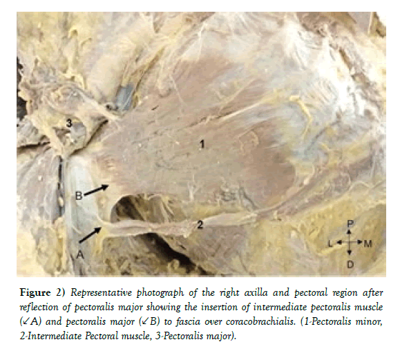
On the left side also IPM was associated with variant insertion of pectoralis minor. The IPM originated from the outer surface of 6th rib, external oblique aponeurosis and fascia over rectus sheath. It extended along the inferior margin of pectoralis major in a deeper plane forming a part of anterior wall of axilla. At insertion fused with the fascia over coracobrachialis. It measured about 17.8 cm in length and 0.9 cm in width at its midpoint. It was supplied by 3rd intercostal nerve. Pectoralis minor on this side originated from the outer surface of 2nd to 5th rib. The insertion was onto the coracoid process as well as the fascia over coracobrachialis (Figure 2).
Figure 2) Representative photograph of the right axilla and pectoral region after reflection of pectoralis major showing the insertion of intermediate pectoralis muscle (↙A) and pectoralis major (↙B) to fascia over coracobrachialis. (1-Pectoralis minor, 2-Intermediate Pectoral muscle, 3-Pectoralis major).
Discussion
The origin of IPM in present study is from 5th rib, 6th rib and aponeurosis of external oblique. This is similar to various other studies reported in the literature on pectoralis quartus and tertius. Pectoralis quartus has been reported to originate from 4th to 7th ribs and its costal cartilages [2,6-8] and rectus sheath [5]. Pectoralis tertius was found to arise from external surface of 6th,7th ribs and external oblique aponeurosis [3]. But the insertion of IPM reported in present study differed from the previous studies.
Previous reports cited that the accessory muscle inserted onto pectoralis major [6], fascia over coracobrachialis [7], fascia over short head of biceps and coracobrachialis [8], lateral lip of bicipital groove and tendon of short head of biceps [4,7,8] and axillary arch [5]. But in the present study the majority of IPM fibers inserted mainly onto the capsule of gleno-humeral joint. This type of variation in insertion of IPM has not been reported so far in the literature to the best of our knowledge.
Earlier studies showed that pectoralis quartus received its innervations from branches of medial pectoral nerve [4,7,8] and 4th intercostal nerve [2]. In the present study the IPM was innervated by the 3rd intercostal nerve thus differing from the earlier reports.
The pectoralis minor in majority is attached to the superior surface of coracoid process. Le double classified the ectopic insertion of pectoralis minor onto supraspinatus, coracoacromial ligament, tubercle of humerus and glenoid labrum into three different types [13]. In present study the right side pectoralis minor after fusing with fibers of IPM inserted onto coracoid process as well as the capsule of gleno-humeral joint which is similar to type 2 variant of Le Double. On the left side few fibers of pectoralis minor blended with fascia over coracobrachialis similar to that observed in literatures [12]. But the co-existence of anomalous insertion of pectoralis minor along with the IPM has not been reported previously.
Pectoralis quartus is normal finding in gorilla and mandrill [3]. Pectoralis minor is found to be inserted at the gleno-humeral capsule in orangutans and chimpanzees [14]. The pectoral muscle mass of present-day tetrapods has evolved from the abductor superficialis muscle of the lobe-finned fish. With evolution this undivided muscle mass separated into superficial and deep layers [14].The previous studies proposed pectoralis quartus as a segmented portion of pectoralis major [4]. Literature also described quartus as a remnant of the ventral part of the subcutaneous trunk muscle in lower mammals. So with evolution the insertions of minor and major had shifted cranially and hence the accessory muscles are considered to be the atavistic remnants present in lower mammal quadrupeds [14]. De sol et al. states the pectoralis quartus muscle described by some authors is none other than the pectoralis tertius muscle [3].
Developmentally when the embryo is about 4 mm in length the arm bud appears opposite to the ventral ends of fifth to eighth cervical and first thoracic segments. The muscles arising from the arm bud and later spreading out to the trunk include the pectoralis major and minor. The pectoral muscles develop from the pectoral premuscle mass which appears first in a 9 mm embryo anterior to first rib. Initially the premuscle mass is continuous with arm premuscle sheath and remains attached to humerus, coracoid process and clavicular rudiment. As the mass differentiates further it extends both caudally and ventrally. As it reaches level of 5th rib in a 14 mm embryo the proximal portion of the mass splits into pectoralis major and minor, one being attached by tendon to humerus and other to coracoid process, with fusion still persisting towards the costal attachments. The two muscles become quite distinct in a 16 mm embryo. With further migration and differentiation the adult bilaminar pattern of pectoralis major is established only in a 40 mm embryo [15]. Hence any defects in migration and differentiation of the pectoral premuscle mass during the development from 4 mm stage to 40 mm stage of embryo could lead to the occurrence of the IPM.
Thus the pectoral muscles develop from a common muscle mass during the embryonic development, forming the upper limb and subsequently extend to the thorax. Hence it’s proposed that IPM is also derived from the same muscle mass as that of the intercostal muscles [2]. This explains the nerve supply of IPM by branches of intercostal nerve in the present study.
The presence of accessory muscles in pectoral region has been associated with complications during axillary lymphadenectomy due to it reducing the area of surgical field [5,8]. Ectopic insertion of pectoralis minor has been associated with shoulder stiffness [11]. Hence the knowledge of co-existence of intermediate pectoralis muscle with variant insertion of pectoralis minor is noteworthy to the clinicians.
Conclusion
The anatomical knowledge of these muscle variations involving pectoral region and axilla is important for surgeons as well as orthopaedicians during chest wall surgeries and shoulder arthroscopic procedures. Prior knowledge of these variations also might help the radiologists with diagnostic imaging methods.
REFERENCES
- Rai R, Ranade AV, Prabhu LV, et al. Unilateral pectoralis minimus muscle: a case report. Int J Morphol. 2008;26:27-30.
- Arican RY, Coskun N, Sarikcioglu L, et al. Co-existence of the pectoralis quartus and pectoralis intermedius muscles. Morphologie. 2006;90:157- 9.
- Del Sol M, Vasquez B. Anatomical and clinical considerations of the pectoralis tertius muscle in man. Int J Morphol. 2009;27:715-8.
- Birmingham A. Homology and innervation of the achselbogen and pectoralis quartus, and the nature of the lateral cutaneous nerve of the thorax. J Anat Physiol. 1889;23:206.
- Bonastre V, Rodríguez-Niedenführ M, Choi D, et al. Coexistence of a pectoralis quartus muscle and an unusual axillary arch: case report and review. Clin Anat. 2002;15:366-70.
- Hunt JD. Bilateral pectoralis major and pectoralis quartus variants: A conjoined tendon passing through the inter tubercular groove. Int J Anat Var. 2017;10:88-90.
- Michelle AH. An accessory muscle of the thoracic wall. Int J Anat Var. 2009;2:93-5.
- Suman V, Sulochana S. Concomitant pectoralis minor, pectoralis quartus and axillary arch. Eur J Pharm Med Sci. 2017;4:570-2.
- Tubbs RS, Shoja MM, Loukas M. Bergman’s Comprehensive Encyclopedia of Human Anatomic Variation. John Wiley & Sons; 2016;pp:339-42.
- Tubbs RS, Oakes WJ, Salter EG. Unusual attachment of the pectoralis minor muscle. Clin Anat. 2005;18:302-4.
- Moineau G, Cikes A, Trojani C, et al. Ectopic insertion of the pectoralis minor: implication in the arthroscopic treatment of shoulder stiffness. Knee Surg Sports Traumatol Arthrosc. 2008; 16:869-71.
- Anil Kumar D. An unusual variation of pectoralis minor muscle and its clinical significance. Int J Biomed Res. 2016;7:613-8.
- Le Double. Traite des variations du systeme musculaire de l’home et leur signification au point de vue du l’anthropologie. Paris; 1897;p: 516.
- Potau JM, Arias-Martorell J, Bello-Hellegouarch G, et al. Inter and intraspecific variations in the pectoral muscles of common chimpanzees (pan troglodytes), bonobos (pan paniscus) and humans (homo sapiens). BioMed Res Int. 2018; 1-12.
- Lewis WH. The development of the muscular system. In: Keibel F, Mall FP (ed) Manual of Human Embryology. 1910; pp:454-522.


