Unusual innervation of thenar muscles
Kuntal Vashishtha*, Subhash Kaushal and Usha Chhabra
Department of Anatomy, Government Medical College, Chandigarh, India
- *Corresponding Author:
- Kuntal Vashishtha, MS Anatomy
Demonstrator, Department of Anatomy, Government Medical College Chandigarh, India
Tel: +91 987 6398056
E-mail: kuntalvashishtha@ymail.com
Date of Received: November 11th, 2010
Date of Accepted: August 4th, 2011
Published Online: August 18th, 2011
© Int J Anat Var (IJAV). 2011; 4: 149–151.
[ft_below_content] =>Keywords
multiple thenar nerves, subligamentous, extraligamentous, median nerve
Introduction
Thenar muscles are innervated by motor branch or recurrent branch of median nerve, also known as thenar nerve. This branch is usually given off at the level of the distal margin of flexor retinaculum. There are several anatomical variations in origin, course and number of thenar branch [1]. One very unusual observation, which to the best of our knowledge has not been published so far, is being reported in this case report.
Case Report
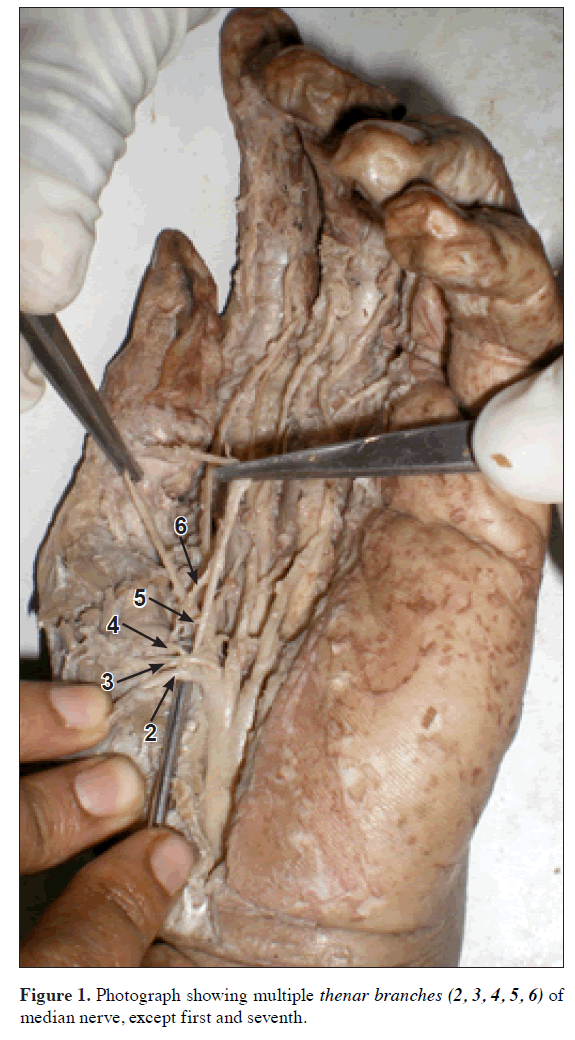
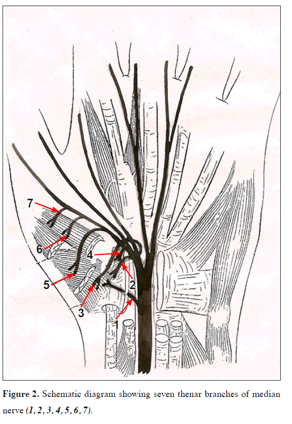
During dissection of the left hand of an adult human cadaver we observed seven thenar branches supplying the thenar musculature. The pattern of origin was as follows: First branch was arising from the posterolateral aspect of main trunk of median nerve beneath the flexor retinaculum just before the division of median nerve (subligamentous origin). Second branch was arising from the lateral division of median nerve having a recurrent course to flexor pollicis brevis. Third branch was a twig from the medial side of lateral division of median nerve. Fourth branch was arising slightly distal to second branch just at origin of the proper palmar digital nerve to the lateral side of thumb. Fifth branch was a branch from proper palmar digital nerve to the lateral side of thumb. This was joining the fourth branch and forming a loop. Sixth and seventh branches were arising from proper palmar digital nerve to the lateral side of thumb distal to fifth branch. Seventh branch was accidentally broken during dissection (Figures 1, 2).
Except for the first branch all other branches arose distal to the lower border of the flexor retinaculum (extraligamentous). All these branches were entering flexor pollicis brevis proximally, three branches ended by supplying it and four supplied rest of the thenar muscles.
Discussion
The present report is unique in that we found seven thenar branches supplying thenar muscles. Earlier, Lanz observed variations in the course of motor branch specially relating to its origin which was either subligamentous, extraligamentous or transligamentous [1]. In addition, Lanz reported accessory branches at the distal portion of the carpal tunnel in 18 out of 246 hands. In present specimen we found one subligamentous branch while six others were extraligamentous. Falconer and Spinner studied ten specimens [2]. Transligamentous passage of the recurrent motor branch of the median nerve was noted in six dissections. In two specimens, out of six transligamentous thenar branches, one more extraligamentous motor branch to the flexor pollicis brevis was observed. Akio studied variations of the branching pattern of median nerve and their variant course in a series of 147 hands in which the carpal tunnel was explored during surgery. One hundred and fourteen hands had one thenar branch, 23 hands two and 10 hands had three branches [3]. Alp et al. studied ramification pattern of the thenar branch in 144 hands of 74 cadavers and found that thenar branch was a single trunk in 84% cases, two branches were observed in 13.2%, three in 2.1% and four branches in 0.7% cases [4].
Mumford et al. studied thenar nerve in 20 fresh-frozen cadaveric hands. The traditionally described pattern of one main thenar trunk with three terminal branches, one each to the abductor pollicis brevis, opponens pollicis and flexor pollicis brevis muscles, was observed in nine specimens (45%). One main trunk with two terminal branches (one branch to the abductor and one to the opponens) was seen in six specimens (30%). The remaining five specimens (25%) exhibited four other terminal patterns with two, three, or four branches off the main trunk. In 15 specimens (75%), accessory thenar nerve arose from either the first common digital nerve (25%) or the radial proper digital nerve to the thumb (50%) and innervated the flexor pollicis brevis [5]. Thenar loop has been described between thenar, accessory thenar and deep branch of ulnar nerve [6]. We observed a similar loop formation by union of fourth and fifth branch in our specimen. Knowledge of the branching patterns and unusual variations helps in diagnosis and proper treatment of the disorders of the median nerve. During surgery unusual branching patterns could lead to injury of these branches causing permanent disability if one is not aware of such branching patterns. Anticipating and understanding unexpected anatomic differences during surgery enhances the surgeon’s ability to perform a safer dissection.
References
- Lanz U. Anatomical variations of the median nerve in the carpal tunnel. J Hand Surg Am. 1977; 2: 44–53.
- Falconer D, Spinner M. Anatomic variations in the motor and sensory supply of the thumb. Clin Orthop Relat Res. 1985; 195: 83–96.
- Akio Matsuzaki. Variations and anomalies of the branching of the median nerve observed on carpal tunnel release. J Jpn Soc Surg Hand. 1998; 15: 452–456.
- Alp M, Marur T, Akkin SM, Yalcin L, Demirci S. Ramification pattern of the thenar branch of the median nerve entering the thenar fascia and the distribution of the terminal branches in the thenar musculature: Anatomic cadaver study in 144 hands. Clin Anat. 2005; 18: 195–199.
- Mumford J, Morecraft R, Blair WF. Anatomy of the thenar branch of the median nerve. J Hand Surg Am. 1987; 12: 361–365.
- Homma T, Sakai T. Thenar and hypothenar muscles and their innervation by the ulnar and median nerves in the human hand. Acta Anat (Basel). 1992; 145: 44–49.
Kuntal Vashishtha*, Subhash Kaushal and Usha Chhabra
Department of Anatomy, Government Medical College, Chandigarh, India
- *Corresponding Author:
- Kuntal Vashishtha, MS Anatomy
Demonstrator, Department of Anatomy, Government Medical College Chandigarh, India
Tel: +91 987 6398056
E-mail: kuntalvashishtha@ymail.com
Date of Received: November 11th, 2010
Date of Accepted: August 4th, 2011
Published Online: August 18th, 2011
© Int J Anat Var (IJAV). 2011; 4: 149–151.
Abstract
One very unusual observation has been reported in this case report. We found seven thenar branches supplying the thenar musculature. One branch was arising from the main trunk of median nerve, second from the lateral aspect of lateral division of median nerve, third from the medial aspect of lateral division of median nerve, fourth from the lateral aspect of lateral division of median nerve slightly distal to second branch. Fifth, sixth and seventh were arising from the proper palmar digital nerve to the lateral aspect of thumb. Fifth branch was joining the fourth after forming a loop. Except for the first branch, which was subligamentous, all other branches were extraligamentous. All these branches were entering flexor pollicis brevis muscle proximally, three branches ended by supplying it and four supplied rest of the thenar muscles. To the best of our knowledge the seven thenar branches have not been described in published literature.
-Keywords
multiple thenar nerves, subligamentous, extraligamentous, median nerve
Introduction
Thenar muscles are innervated by motor branch or recurrent branch of median nerve, also known as thenar nerve. This branch is usually given off at the level of the distal margin of flexor retinaculum. There are several anatomical variations in origin, course and number of thenar branch [1]. One very unusual observation, which to the best of our knowledge has not been published so far, is being reported in this case report.
Case Report
During dissection of the left hand of an adult human cadaver we observed seven thenar branches supplying the thenar musculature. The pattern of origin was as follows: First branch was arising from the posterolateral aspect of main trunk of median nerve beneath the flexor retinaculum just before the division of median nerve (subligamentous origin). Second branch was arising from the lateral division of median nerve having a recurrent course to flexor pollicis brevis. Third branch was a twig from the medial side of lateral division of median nerve. Fourth branch was arising slightly distal to second branch just at origin of the proper palmar digital nerve to the lateral side of thumb. Fifth branch was a branch from proper palmar digital nerve to the lateral side of thumb. This was joining the fourth branch and forming a loop. Sixth and seventh branches were arising from proper palmar digital nerve to the lateral side of thumb distal to fifth branch. Seventh branch was accidentally broken during dissection (Figures 1, 2).
Except for the first branch all other branches arose distal to the lower border of the flexor retinaculum (extraligamentous). All these branches were entering flexor pollicis brevis proximally, three branches ended by supplying it and four supplied rest of the thenar muscles.
Discussion
The present report is unique in that we found seven thenar branches supplying thenar muscles. Earlier, Lanz observed variations in the course of motor branch specially relating to its origin which was either subligamentous, extraligamentous or transligamentous [1]. In addition, Lanz reported accessory branches at the distal portion of the carpal tunnel in 18 out of 246 hands. In present specimen we found one subligamentous branch while six others were extraligamentous. Falconer and Spinner studied ten specimens [2]. Transligamentous passage of the recurrent motor branch of the median nerve was noted in six dissections. In two specimens, out of six transligamentous thenar branches, one more extraligamentous motor branch to the flexor pollicis brevis was observed. Akio studied variations of the branching pattern of median nerve and their variant course in a series of 147 hands in which the carpal tunnel was explored during surgery. One hundred and fourteen hands had one thenar branch, 23 hands two and 10 hands had three branches [3]. Alp et al. studied ramification pattern of the thenar branch in 144 hands of 74 cadavers and found that thenar branch was a single trunk in 84% cases, two branches were observed in 13.2%, three in 2.1% and four branches in 0.7% cases [4].
Mumford et al. studied thenar nerve in 20 fresh-frozen cadaveric hands. The traditionally described pattern of one main thenar trunk with three terminal branches, one each to the abductor pollicis brevis, opponens pollicis and flexor pollicis brevis muscles, was observed in nine specimens (45%). One main trunk with two terminal branches (one branch to the abductor and one to the opponens) was seen in six specimens (30%). The remaining five specimens (25%) exhibited four other terminal patterns with two, three, or four branches off the main trunk. In 15 specimens (75%), accessory thenar nerve arose from either the first common digital nerve (25%) or the radial proper digital nerve to the thumb (50%) and innervated the flexor pollicis brevis [5]. Thenar loop has been described between thenar, accessory thenar and deep branch of ulnar nerve [6]. We observed a similar loop formation by union of fourth and fifth branch in our specimen. Knowledge of the branching patterns and unusual variations helps in diagnosis and proper treatment of the disorders of the median nerve. During surgery unusual branching patterns could lead to injury of these branches causing permanent disability if one is not aware of such branching patterns. Anticipating and understanding unexpected anatomic differences during surgery enhances the surgeon’s ability to perform a safer dissection.
References
- Lanz U. Anatomical variations of the median nerve in the carpal tunnel. J Hand Surg Am. 1977; 2: 44–53.
- Falconer D, Spinner M. Anatomic variations in the motor and sensory supply of the thumb. Clin Orthop Relat Res. 1985; 195: 83–96.
- Akio Matsuzaki. Variations and anomalies of the branching of the median nerve observed on carpal tunnel release. J Jpn Soc Surg Hand. 1998; 15: 452–456.
- Alp M, Marur T, Akkin SM, Yalcin L, Demirci S. Ramification pattern of the thenar branch of the median nerve entering the thenar fascia and the distribution of the terminal branches in the thenar musculature: Anatomic cadaver study in 144 hands. Clin Anat. 2005; 18: 195–199.
- Mumford J, Morecraft R, Blair WF. Anatomy of the thenar branch of the median nerve. J Hand Surg Am. 1987; 12: 361–365.
- Homma T, Sakai T. Thenar and hypothenar muscles and their innervation by the ulnar and median nerves in the human hand. Acta Anat (Basel). 1992; 145: 44–49.


