Unusual ligament of the scapula
Philip A. Fabrizio*
Department of Physical Therapy, College of Pharmacy and Health Sciences, Mercer University, Atlanta, Georgia, USA
- *Corresponding Author:
- Philip A. Fabrizio, PT, DPT, MS
Department of Physical Therapy, College of Pharmacy and Health Sciences, Mercer University, 3001 Mercer University Drive, Suite 111 Davis Building, Atlanta, Georgia, 30341, USA
Tel: +1 (678) 547-6178
E-mail: fabrizio_pa@mercer.edu
Date of Received: March 1st, 2012
Date of Accepted: July 6th, 2012
Published Online: October 21st, 2012
© Int J Anat Var (IJAV). 2012; 5: 54–55.
[ft_below_content] =>Keywords
scapular ligament, suprascapular nerve, suprascapular artery
Introduction
Customary dissection in the Physical Therapy Anatomy Laboratory yielded a unilateral left-sided scapular ligament variation in a single cadaver. The embalmed specimen was a 58-year-old Caucasian male. The primary finding was a variant ligament which spanned across the supraspinous fossa in inferior to superior orientation. The suprascapular nerve and artery were seen passing under the variant ligament. Although variations in the transverse scapular ligament and spinoglenoid ligament have been documented, the author is not aware of any description which matches the variant ligament described in the current case.
Case Report
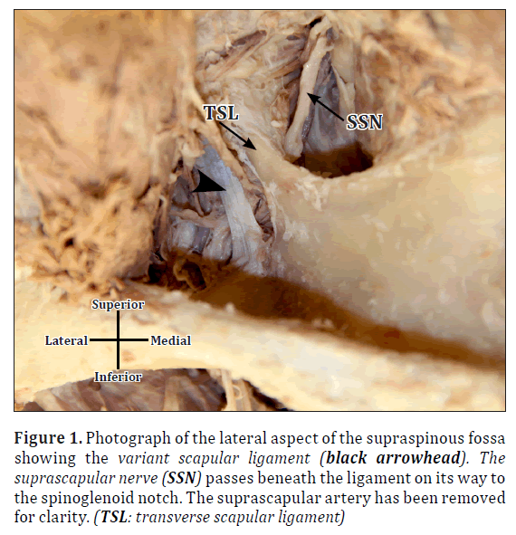
Routine dissection, performed to expose the course of the suprascapular nerve on the posterior aspect of the scapula, revealed an unusual scapular ligament distal to the superior transverse scapular ligament but proximal to the spinoglenoid notch. The ligament spanned the surface of the supraspinous fossa from a proximal attachment at the superior border of the base of the spine of the scapula vertically to the distal attachment at the posterior aspect of the base of the coracoid process (Figure 1). The dimensions of the ligament were 16.7 mm in length and 5.1 mm in width. The suprascapular nerve and artery coursed through the scapular notch, under an ossified transverse scapular ligament and laterally across the supraspinous fossa providing innervation and blood supply to the supraspinatus. The nerve and artery then passed beneath the variant scapular ligament before entering the spinoglenoid notch (Figure 1).
Figure 1. Photograph of the lateral aspect of the supraspinous fossa showing the variant scapular ligament (black arrowhead). The suprascapular nerve (SSN) passes beneath the ligament on its way to the spinoglenoid notch. The suprascapular artery has been removed for clarity. (TSL: transverse scapular ligament)
Discussion
The current finding may have clinical implications in suprascapular nerve impingement and suprascapular artery compression [1,2]. Typically two scapular ligaments are described related to the course of the suprascapular nerve in the supraspinatus and infraspinatus fossae. The superior transverse scapular ligament overlies the suprascapular notch creating an osseous fibrous foramen through which the suprascapular nerve typically passes at the beginning of its course on the scapula [1,3,4]. Yang et al. demonstrated the suprascapular nerve running through the suprascapular notch and under the superior transverse scapular ligament in 101 out of 103 shoulders with a suprascapular notch being absent in two shoulders [4]. Duparc et al. found the suprascapular nerve passing through the suprascapular notch under the superior transverse scapular ligament in 30 shoulders and noted suprascapular nerve compression in 50% of cases with concomitant atrophy of the supraspinatus and infraspinatus muscles [1]. The inferior transverse scapular ligament, sometimes referred to as the spinoglenoid ligament, has been inconsistently reported in the literature. The ligament when present has been described as a weak band, overlying the suprascapular artery and nerve, stretching from the lateral aspect of the spine of the scapula to the neck of the scapula or to the glenoid margin [3,5,6]. Demaio et al. examined 75 shoulders from 39 embalmed cadavers for the presence of the inferior transverse scapular ligament [7]. The inferior transverse scapular ligament was demonstrated in two shoulders with concomitant infraspinatus muscle atrophy. A thickened aponeurosis in the typical location of the inferior transverse scapular ligament was demonstrated in 10 shoulders [7]. Demirkan et al. demonstrated the inferior transverse scapular ligament in 19 out of 27 shoulders [8]. The authors described the proximal attachments of the ligaments as the lateral border of the base of the spine of the scapula. The distal attachments varied with 14 attaching to the neck of the scapula, five attaching to the shoulder joint capsule, and eight attaching to the neck of the scapula and posterior aspect of the shoulder joint capsule via two separate bundles. However, atrophy of the infraspinatus muscle was not noted [8]. In the present case, the variant ligament may have caused compression of the suprascapular nerve and limited function of the infraspinatus muscle.
References
- Duparc F, Coquerel D, Ozeel J, Noyon M, Gerometta A, Michot C. Anatomical basis of suprascapular nerve entrapment, and clinical relevance of the supraspinatus fascia. Surg Radiol Anat. 2010; 32: 277–284.
- Moore TP, Hunter RE. Suprascapular nerve entrapment. Oper Tech Sports Med. 1996; 4: 8–14.
- Standring S, ed. Gray’s Anatomy. 40th Ed., New York, Churchill Livingstone. 2008; 796.
- Yang HJ, Gil YC, Jin JD, Ahn SV, Lee HY. Topographical anatomy of the suprascapular nerve and vessels at the suprascapular notch. Clin Anat. 2012; 25: 359–365.
- Plancher KD, Peterson RK, Johnston JC, Luke TA. The spinoglenoid ligament. Anatomy, morphology and histological findings. J Bone Joint Surg Am. 2005; 87: 361–365.
- Williams PL, Warwick R, Dyson M, Bannister LH, eds. Gray’s Anatomy. 37th Ed., New York, Churchill Livingstone. 1989; 501.
- Demaio M, Drez D Jr, Mullins RC. The inferior transverse scapular ligament as a possible cause of entrapment neuropathy of the nerve to the infraspinatus. A brief note. J Bone Joint Surg Am. 1991; 73: 1061–1063.
- Demirkan AF, Sargon MF, Erkula G, Kiter E. The spinoglenoid ligament: An anatomic study. Clin Anat. 2003; 16: 511–513.
Philip A. Fabrizio*
Department of Physical Therapy, College of Pharmacy and Health Sciences, Mercer University, Atlanta, Georgia, USA
- *Corresponding Author:
- Philip A. Fabrizio, PT, DPT, MS
Department of Physical Therapy, College of Pharmacy and Health Sciences, Mercer University, 3001 Mercer University Drive, Suite 111 Davis Building, Atlanta, Georgia, 30341, USA
Tel: +1 (678) 547-6178
E-mail: fabrizio_pa@mercer.edu
Date of Received: March 1st, 2012
Date of Accepted: July 6th, 2012
Published Online: October 21st, 2012
© Int J Anat Var (IJAV). 2012; 5: 54–55.
Abstract
Routine dissection has identified a previously unrecorded scapular ligament found unilaterally in 58-year-old male cadaver. The ligament was found coursing in a superior inferior direction, distal to the transverse scapular ligament and proximal to the spinoglenoid notch. The suprascapular nerve and artery were found passing beneath the unusual ligament. The current finding and clinical significance are discussed.
-Keywords
scapular ligament, suprascapular nerve, suprascapular artery
Introduction
Customary dissection in the Physical Therapy Anatomy Laboratory yielded a unilateral left-sided scapular ligament variation in a single cadaver. The embalmed specimen was a 58-year-old Caucasian male. The primary finding was a variant ligament which spanned across the supraspinous fossa in inferior to superior orientation. The suprascapular nerve and artery were seen passing under the variant ligament. Although variations in the transverse scapular ligament and spinoglenoid ligament have been documented, the author is not aware of any description which matches the variant ligament described in the current case.
Case Report
Routine dissection, performed to expose the course of the suprascapular nerve on the posterior aspect of the scapula, revealed an unusual scapular ligament distal to the superior transverse scapular ligament but proximal to the spinoglenoid notch. The ligament spanned the surface of the supraspinous fossa from a proximal attachment at the superior border of the base of the spine of the scapula vertically to the distal attachment at the posterior aspect of the base of the coracoid process (Figure 1). The dimensions of the ligament were 16.7 mm in length and 5.1 mm in width. The suprascapular nerve and artery coursed through the scapular notch, under an ossified transverse scapular ligament and laterally across the supraspinous fossa providing innervation and blood supply to the supraspinatus. The nerve and artery then passed beneath the variant scapular ligament before entering the spinoglenoid notch (Figure 1).
Figure 1. Photograph of the lateral aspect of the supraspinous fossa showing the variant scapular ligament (black arrowhead). The suprascapular nerve (SSN) passes beneath the ligament on its way to the spinoglenoid notch. The suprascapular artery has been removed for clarity. (TSL: transverse scapular ligament)
Discussion
The current finding may have clinical implications in suprascapular nerve impingement and suprascapular artery compression [1,2]. Typically two scapular ligaments are described related to the course of the suprascapular nerve in the supraspinatus and infraspinatus fossae. The superior transverse scapular ligament overlies the suprascapular notch creating an osseous fibrous foramen through which the suprascapular nerve typically passes at the beginning of its course on the scapula [1,3,4]. Yang et al. demonstrated the suprascapular nerve running through the suprascapular notch and under the superior transverse scapular ligament in 101 out of 103 shoulders with a suprascapular notch being absent in two shoulders [4]. Duparc et al. found the suprascapular nerve passing through the suprascapular notch under the superior transverse scapular ligament in 30 shoulders and noted suprascapular nerve compression in 50% of cases with concomitant atrophy of the supraspinatus and infraspinatus muscles [1]. The inferior transverse scapular ligament, sometimes referred to as the spinoglenoid ligament, has been inconsistently reported in the literature. The ligament when present has been described as a weak band, overlying the suprascapular artery and nerve, stretching from the lateral aspect of the spine of the scapula to the neck of the scapula or to the glenoid margin [3,5,6]. Demaio et al. examined 75 shoulders from 39 embalmed cadavers for the presence of the inferior transverse scapular ligament [7]. The inferior transverse scapular ligament was demonstrated in two shoulders with concomitant infraspinatus muscle atrophy. A thickened aponeurosis in the typical location of the inferior transverse scapular ligament was demonstrated in 10 shoulders [7]. Demirkan et al. demonstrated the inferior transverse scapular ligament in 19 out of 27 shoulders [8]. The authors described the proximal attachments of the ligaments as the lateral border of the base of the spine of the scapula. The distal attachments varied with 14 attaching to the neck of the scapula, five attaching to the shoulder joint capsule, and eight attaching to the neck of the scapula and posterior aspect of the shoulder joint capsule via two separate bundles. However, atrophy of the infraspinatus muscle was not noted [8]. In the present case, the variant ligament may have caused compression of the suprascapular nerve and limited function of the infraspinatus muscle.
References
- Duparc F, Coquerel D, Ozeel J, Noyon M, Gerometta A, Michot C. Anatomical basis of suprascapular nerve entrapment, and clinical relevance of the supraspinatus fascia. Surg Radiol Anat. 2010; 32: 277–284.
- Moore TP, Hunter RE. Suprascapular nerve entrapment. Oper Tech Sports Med. 1996; 4: 8–14.
- Standring S, ed. Gray’s Anatomy. 40th Ed., New York, Churchill Livingstone. 2008; 796.
- Yang HJ, Gil YC, Jin JD, Ahn SV, Lee HY. Topographical anatomy of the suprascapular nerve and vessels at the suprascapular notch. Clin Anat. 2012; 25: 359–365.
- Plancher KD, Peterson RK, Johnston JC, Luke TA. The spinoglenoid ligament. Anatomy, morphology and histological findings. J Bone Joint Surg Am. 2005; 87: 361–365.
- Williams PL, Warwick R, Dyson M, Bannister LH, eds. Gray’s Anatomy. 37th Ed., New York, Churchill Livingstone. 1989; 501.
- Demaio M, Drez D Jr, Mullins RC. The inferior transverse scapular ligament as a possible cause of entrapment neuropathy of the nerve to the infraspinatus. A brief note. J Bone Joint Surg Am. 1991; 73: 1061–1063.
- Demirkan AF, Sargon MF, Erkula G, Kiter E. The spinoglenoid ligament: An anatomic study. Clin Anat. 2003; 16: 511–513.


