Unusual origin and potentially hazardous course of the major blood vessels in neck – A clinically relevant rare casein front
Virendra Budhiraja* and Rakhi Rastogi
Department of Anatomy, Subharti Medical College, Meerut (U.P.), India
- *Corresponding Author:
- Dr. Virendra Budhiraja
Associate Professor of Anatomy
Subharti Medical College
Delhi-Haridwar By Pass Road
Meerut (U.P.), INDIA.
Tel: +91 989 7244802
E-mail: budhirajasimran46@gmail.com
Date of Received: September 8th, 2009
Date of Accepted: April 15th, 2010
Published Online: April 27th, 2010
© Int J Anat Var (IJAV). 2010; 3: 61–62.
[ft_below_content] =>Keywords
left brachiocephalic vein, brachiocephalic trunk, tracheostomy, percutaneous dilatational tracheostomy
Introduction
Left brachiocephalic vein begins from junction of left subclavian and left internal jugular vein posterior to medial end of left clavicle. It crosses to the right moving slightly in inferior direction and joins with right brachiocephalic vein to form superior vena cava. During its oblique course it crosses the left subclavian artery, left common carotid artery and brachiocephalic trunk in the superior mediastinum.
Brachiocephalic trunk is described as the largest branch of the arch of the aorta, 4 to 5 cm in length, and arises from the arch’s convexity, posterior to the center of the manubrium sterni. It ascends posterolaterally to the right, at first anterior to trachea, then on its right. At the level of right sternoclavicular joint’s upper border it divides into right common carotid and right subclavian arteries [1]. At the lower part of neck the two common carotid arteries are separated from each other by a very narrow interval, which contains the trachea (Figure 1).
Case Report
During routine educational dissection for undergraduate students in our department, this unusual origin of left brachiocephalic vein and brachiocephalic artery was observed in a male cadaver aged about 60 years.
In the region of anterior triangle of the neck after removal of skin, superficial fascia, deep fascia and the infrahyoid muscles, it was found that the left brachiocephalic vein and brachiocephalic artery both were crossing obliquely casein front of trachea just below the thyroid gland (Figure 2).
Discussion
There are few reports of anomalies in left brachiocephalic vein like anomalous sub-aortic brachiocephalic vein [2], rare retro-aortic brachiocephalic vein [3]. Muhammad et al. observed that high brachiocephalic vein was source of bleeding during percutaneous dilatational tracheostomy (PDT), in one case out of 6 bleeding patients in a series of 497 PDT procedures [4]. We also found high left brachiocephalic vein, crossing from left to right in front of trachea and just below thyroid gland.
There are also reports of variations of brachiocephalic artery [5] and subclavian artery [6], which may lead to problems during tracheostomy. Comert et al. [7] found an innominate artery crossing 4th and 5th tracheal ring while Iterezote et al. [8] observed brachiocephalic trunk originating from the aortic arch in front of the trachea. Gupta and Mehta also reported a case of brachiocephalic artery crossing in front of trachea [9]. In present case, the brachiocephalic artery and left brachiocephalic vein following an unusual course and lying in front of trachea such as occluding space for tracheostomy. The anatomy of aberrant great vessels is relevant in surgeries of the anterior neck, especially tracheostomy and thyroidectomy [10]. This case is a rare variant where the left brachiocephalic vein and brachiocephalic artery lie directly in the line of incision for tracheostomy or in the path of guide wire that is inserted in PDT.
Such a variant is of vital significance during surgeries and even more important in PDT, which has gained wide acceptance due to its relative speed, simplicity and the ability to perform it at the bedside but the major disadvantage is the increased risk of peri-operative complication of severe bleeding.
Acknowledgement
The Authors are thankful and grateful to Dr. Satyam Khare Professor and head, Department of Anatomy and Dr. A. K. Asthana, Dean of the institute for their advice and support.
References
- Giorgio Gabella. Angiology, The Arterial System. In: Williams PL, ed. Gray’s Anatomy. 38th Ed., Edinburgh, Churchill Livingstone. 2000; 1513.
- Fujimoto K, Abe T, Kumabe T, Hayabuchi N, Nozaki Y. Anomalous left brachiocephalic (innominate) vein: MR demonstration. AJR Am J Roentgenol. 1992; 159: 479–480.
- Yilmaz M, Sargon MF, Dogan OF, Pasaoglu I. A very rare anatomic variation of the left brachiocephalic vein: left retro-aortic brachiocephalic vein with tetralogy of Fallot. Surg Radiol Anat. 2003; 25: 158–160.
- Muhammad JK, Major E, Wood A, Patton DW. Percutaneous dilatational tracheostomy: haemorrhagic complications and the vascular anatomy of the anterior neck. A review based on 497 cases. Int J Oral Maxillofac Surg. 2000; 29: 217–222.
- Maldjian PD, Saric M, Tsai SC. High brachiocephalic artery: CT Appearance and clinical implications. J Thorac Imaging. 2007; 22: 192–194.
- Chadha NK, Chiti-Batelli S. Tracheostomy reveals a rare aberrant right subclavian artery; a case report. BMC Ear Nose Throat Disord. 2004; 4: 1.
- Comert A, Comert E, Ozlugedik S, Kendir S, Tekdemir I. High-located aberrant innominate artery: an unusual cause of serious hemorrhage of percutaneous tracheostomy. Am J Otolaryngol. 2004; 25: 368–369.
- Iterezote AM, de Medeiros AD, Filho RCCB, Petrella S, de Andrade Junior LC, Marques SR, Prates JC. Anatomical variation of the brachiocephalic trunk and common carotid artery in neck dissection. Int J Morphol. 2009; 27: 601–603.
- Kumar GR, Mehta CD. Anomalous origin and potentially hazardous course of the brachiocephalic artery. J Anat Soc India. 2007; 56: 38–41.
- Upadhyaya PK, Bertellotti R, Laeeq A, Sugimoto J. Beware of the aberrant innominate artery. Ann Thorac Surg. 2008; 85: 653–654.
Virendra Budhiraja* and Rakhi Rastogi
Department of Anatomy, Subharti Medical College, Meerut (U.P.), India
- *Corresponding Author:
- Dr. Virendra Budhiraja
Associate Professor of Anatomy
Subharti Medical College
Delhi-Haridwar By Pass Road
Meerut (U.P.), INDIA.
Tel: +91 989 7244802
E-mail: budhirajasimran46@gmail.com
Date of Received: September 8th, 2009
Date of Accepted: April 15th, 2010
Published Online: April 27th, 2010
© Int J Anat Var (IJAV). 2010; 3: 61–62.
Abstract
We present a rare case of aberrant left brachiocephalic vein and brachiocephalic artery, which crosses the trachea in the neck obliquely and closely related to lower border of thyroid gland. If not noticed while performing open or percutaneous dilatational tracheostomy or other neck surgeries, trauma to these vessel and subsequent hemorrhage can occur and may be fatal. Vascular compression of the airway causing obstructive symptoms can also occur due to this anomaly. In this report the case is presented along with its clinical significance.
-Keywords
left brachiocephalic vein, brachiocephalic trunk, tracheostomy, percutaneous dilatational tracheostomy
Introduction
Left brachiocephalic vein begins from junction of left subclavian and left internal jugular vein posterior to medial end of left clavicle. It crosses to the right moving slightly in inferior direction and joins with right brachiocephalic vein to form superior vena cava. During its oblique course it crosses the left subclavian artery, left common carotid artery and brachiocephalic trunk in the superior mediastinum.
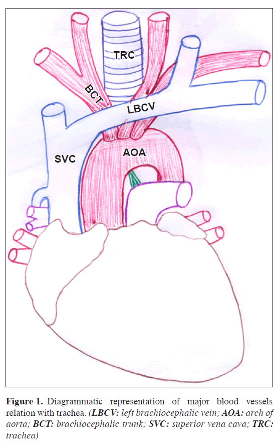
Brachiocephalic trunk is described as the largest branch of the arch of the aorta, 4 to 5 cm in length, and arises from the arch’s convexity, posterior to the center of the manubrium sterni. It ascends posterolaterally to the right, at first anterior to trachea, then on its right. At the level of right sternoclavicular joint’s upper border it divides into right common carotid and right subclavian arteries [1]. At the lower part of neck the two common carotid arteries are separated from each other by a very narrow interval, which contains the trachea (Figure 1).
Case Report
During routine educational dissection for undergraduate students in our department, this unusual origin of left brachiocephalic vein and brachiocephalic artery was observed in a male cadaver aged about 60 years.
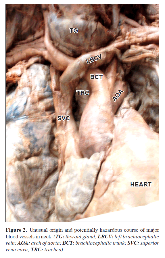
In the region of anterior triangle of the neck after removal of skin, superficial fascia, deep fascia and the infrahyoid muscles, it was found that the left brachiocephalic vein and brachiocephalic artery both were crossing obliquely casein front of trachea just below the thyroid gland (Figure 2).
Figure 2: Unusual origin and potentially hazardous course of major blood vessels in neck. (TG: thyroid gland; LBCV: left brachiocephalic vein; AOA: arch of aorta; BCT: brachiocephalic trunk; SVC: superior vena cava; TRC: trachea)
Discussion
There are few reports of anomalies in left brachiocephalic vein like anomalous sub-aortic brachiocephalic vein [2], rare retro-aortic brachiocephalic vein [3]. Muhammad et al. observed that high brachiocephalic vein was source of bleeding during percutaneous dilatational tracheostomy (PDT), in one case out of 6 bleeding patients in a series of 497 PDT procedures [4]. We also found high left brachiocephalic vein, crossing from left to right in front of trachea and just below thyroid gland.
There are also reports of variations of brachiocephalic artery [5] and subclavian artery [6], which may lead to problems during tracheostomy. Comert et al. [7] found an innominate artery crossing 4th and 5th tracheal ring while Iterezote et al. [8] observed brachiocephalic trunk originating from the aortic arch in front of the trachea. Gupta and Mehta also reported a case of brachiocephalic artery crossing in front of trachea [9]. In present case, the brachiocephalic artery and left brachiocephalic vein following an unusual course and lying in front of trachea such as occluding space for tracheostomy. The anatomy of aberrant great vessels is relevant in surgeries of the anterior neck, especially tracheostomy and thyroidectomy [10]. This case is a rare variant where the left brachiocephalic vein and brachiocephalic artery lie directly in the line of incision for tracheostomy or in the path of guide wire that is inserted in PDT.
Such a variant is of vital significance during surgeries and even more important in PDT, which has gained wide acceptance due to its relative speed, simplicity and the ability to perform it at the bedside but the major disadvantage is the increased risk of peri-operative complication of severe bleeding.
Acknowledgement
The Authors are thankful and grateful to Dr. Satyam Khare Professor and head, Department of Anatomy and Dr. A. K. Asthana, Dean of the institute for their advice and support.
References
- Giorgio Gabella. Angiology, The Arterial System. In: Williams PL, ed. Gray’s Anatomy. 38th Ed., Edinburgh, Churchill Livingstone. 2000; 1513.
- Fujimoto K, Abe T, Kumabe T, Hayabuchi N, Nozaki Y. Anomalous left brachiocephalic (innominate) vein: MR demonstration. AJR Am J Roentgenol. 1992; 159: 479–480.
- Yilmaz M, Sargon MF, Dogan OF, Pasaoglu I. A very rare anatomic variation of the left brachiocephalic vein: left retro-aortic brachiocephalic vein with tetralogy of Fallot. Surg Radiol Anat. 2003; 25: 158–160.
- Muhammad JK, Major E, Wood A, Patton DW. Percutaneous dilatational tracheostomy: haemorrhagic complications and the vascular anatomy of the anterior neck. A review based on 497 cases. Int J Oral Maxillofac Surg. 2000; 29: 217–222.
- Maldjian PD, Saric M, Tsai SC. High brachiocephalic artery: CT Appearance and clinical implications. J Thorac Imaging. 2007; 22: 192–194.
- Chadha NK, Chiti-Batelli S. Tracheostomy reveals a rare aberrant right subclavian artery; a case report. BMC Ear Nose Throat Disord. 2004; 4: 1.
- Comert A, Comert E, Ozlugedik S, Kendir S, Tekdemir I. High-located aberrant innominate artery: an unusual cause of serious hemorrhage of percutaneous tracheostomy. Am J Otolaryngol. 2004; 25: 368–369.
- Iterezote AM, de Medeiros AD, Filho RCCB, Petrella S, de Andrade Junior LC, Marques SR, Prates JC. Anatomical variation of the brachiocephalic trunk and common carotid artery in neck dissection. Int J Morphol. 2009; 27: 601–603.
- Kumar GR, Mehta CD. Anomalous origin and potentially hazardous course of the brachiocephalic artery. J Anat Soc India. 2007; 56: 38–41.
- Upadhyaya PK, Bertellotti R, Laeeq A, Sugimoto J. Beware of the aberrant innominate artery. Ann Thorac Surg. 2008; 85: 653–654.


