Unusual variation in the anatomy of the left lung: A Case Report
2 Assistant Professor, Department of Anatomy, Hamdard Institute of Medical Sciences and Research, New Delhi, India
3 Professor, Department of Anatomy, All India Institute of Medical Sciences, New Delhi, India
4 Assistant Professor, Indian Institute of Technology, Jammu, India
Received: 01-Nov-2024, Manuscript No. ijav-24-7309; Editor assigned: 04-Nov-2024, Pre QC No. ijav-24-7309 (PQ); Reviewed: 20-Nov-2024 QC No. ijav-24-7309; Revised: 26-Nov-2024, Manuscript No. ijav-24-7309 (R); Published: 30-Nov-2024, DOI: 10.37532/1308-4038.17(11).452
Citation: Arora P, et al. Unusual Variation in the Anatomy of the Left Lung: A Case Report. Int J Anat Var. 2024;17(11): 671-672.
This open-access article is distributed under the terms of the Creative Commons Attribution Non-Commercial License (CC BY-NC) (http://creativecommons.org/licenses/by-nc/4.0/), which permits reuse, distribution and reproduction of the article, provided that the original work is properly cited and the reuse is restricted to noncommercial purposes. For commercial reuse, contact reprints@pulsus.com
Abstract
Lungs are the functional units of respiration and are crucial to survival. They lie in the thoracic cavity, separated by the heart and mediastinum. While the two lungs are similar, they are not completely symmetrical, having different numbers of lobes and bronchial and vascular anatomy. In most individuals, the right lung comprises three lobes subdivided into ten segments, and the left lung consists of two lobes and 8-10 segments. The lobes of the lungs are also incompletely separated by fissures. Each lung has an oblique fissure, with a horizontal fissure in the right lung. We reported an accessory fissure and an additional accessory lobe in the left lung during routine dissection. As the fissures form boundaries for the lobes of the lungs, knowledge of their position is necessary for appreciation of lobar anatomy and, thus, for locating the bronchopulmonary segments, which are anatomically and clinically significant.
INTRODUCTION
The lungs, the organ for respiration, are paired cone-shaped organs in the thoracic cavity separated by the heart and other structures in the mediastinum [1]. The left lung is smaller in volume than the right lung, with a smaller transverse dimension but a larger longitudinal dimension [2]. The left main bronchus also differs from the right, as it is shorter, has a smaller caliber, and is more horizontal [2,3]. Each lung has an oblique fissure separating the upper lobes from the lower lobes, and the right lung has a horizontal fissure that separates the right upper lobe from the middle lobe [4]. Lung fissures are double-folds of visceral pleura that either completely or incompletely invaginate lung parenchyma to form lung lobes [4,5]. The oblique fissure/major fissure is similar for both lungs. It extends from the level of T4/T5 vertebrae postersuperiorly to the hemidiaphragms anteroinferiorly. The left oblique fissure has a more vertical course than the right oblique fissure [6]. The minor fissure is found in the right lung, separating the upper and middle lobes. It runs horizontally at the right 4th costal cartilage level from the hilum to the anterior and lateral surfaces of the right lung. Horizontal fissure is complete in only one-third of people and is absent in 10% of people [6].
CASE REPORT
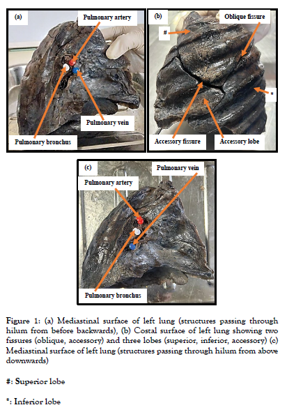
We reported an accessory horizontal fissure and an additional accessory lobe during routine dissection of a male cadaver in the gross anatomy laboratory in the Department of Anatomy, Hamdard Institute of Medical Sciences and Research, New Delhi. In the present case report, we observed two fissures (oblique, transverse/horizontal) and three lobes (superior, inferior, accessory) in the left lung (Figure 1). Imaging was done using a Canon EOS R50 with a 24.2-megapixel effective resolution. The camera supports high-speed electronic shutter capture, achieving up to 15 fps for JPEG (max 28 frames) and up to 7 fps for RAW. It also offers a burst rate of 12 fps for JPEG (max 42 frames) and seven fps for RAW. The focal length ranges from 18 to 45mm (29 to 72 mm in 35mm equivalent), with a maximum aperture of f/4.5 to 6.3.
Figure 1: (a) Mediastinal surface of left lung (structures passing through hilum from before backwards), (b) Costal surface of left lung showing two fissures (oblique, accessory) and three lobes (superior, inferior, accessory) (c) Mediastinal surface of left lung (structures passing through hilum from above downwards)
#: Superior lobe
*: Inferior lobe
DISCUSSION
In the fetal period, the bronchopulmonary segments are separated by spaces that later get obliterated except along the division of principal bronchi to give rise to major (oblique) and minor (horizontal) fissures in the fully developed lungs. Visceral pleura is reflected along these fissures and cover individual lobes on all sides [7]. In the field of anatomical variability, the lung is of particular interest for the presence of additional lobes and fissures [8]. Furthermore, in the study conducted by Manjunath et al., variations in fissures were predominant in males than females [9]. Defective pulmonary development will give rise to variations as encountered in fissures and lobes [10]. In the present case report, we observed an accessory fissure and an additional accessory lobe in the left lung. An accessory fissure is a cleft of varying depth lined by visceral pleura. Radiographically, it appears as a thin white line, resembling the major and minor fissures, except for location. This line can be mistaken for an interlobar fissure, scar, and wall of a bulla or for a pleural line made visible by pneumothorax [11]. The nature of fissure is of great importance in planning pulmonary surgeries. Accessory fissures in patients with endobronchial lesion, might alter the usual pattern of lung collapse and pose difficulty in diagnosing a lesion and its extent. Often, these accessory fissures act as a barrier to the spread of infection, creating sharply marginated pneumonia, which can wrongly be interpreted as atelectasis [11]. Sudikshya et al. observed different anatomical variations (accessory lobes and fissures) in both right and left lungs derived from cadavers of different ethnicities, which might be due to genetic and environmental factors during their development [12]. They also reported that an anomalous fissure can be mistaken for a lung lesion or an atypical appearance of pleural effusion [12]. Since lung anatomical variability is common in clinical practice and preclinical imaging studies can miss different morphologies, a deep and accurate knowledge of anatomical variations of the lung is of extreme importance to avoid difficulties or changes during the surgical procedure.
CONCLUSION
Deep knowledge of fissures and lobes helps appreciate lobar anatomy, thus helping clinicians and radiologists make correct diagnoses and better planning and execution of surgical procedures, decreasing morbidity and mortality induced by lung surgery.
ACKNOWLEDGEMENTS
Our sincere thanks to Hamdard Institute of Medical Sciences, New Delhi. The authors also sincerely thank those who donated their bodies to science so that anatomical research could be performed. Results from such research can potentially increase humankind’s overall knowledge, improving patient care. Therefore, these donors and their families deserve our highest gratitude.
ETHICAL STATEMENT
The authors state that every effort was made to follow all local and international ethical guidelines and laws that pertain to the use of human cadaveric donors in anatomical research.
CONFLICT OF INTEREST STATEMENT
The authors declare that they have no conflicts of interest with the contents of the case report.
FUNDING STATEMENT
This research received no external funding.
AUTHORS CONTRIBUTIONS
Conceptualization: PA; Methodology and Resources: PA, MAK, SK; Writing—original draft preparation: PA, RD; Writing—review and editing: PA, MAK, RD, SK. All authors have read and agreed to the published version of the manuscript.
REFERENCES
- McMinn. Last's Anatomy. Elsevier Australia. 2003 ISBN: 0729537528.
- Standring S. Gray's Anatomy (39th edition). Churchill Livingstone. 2005 ISBN: 0443066841.
- Moore KL, Agur AMR, Dalley AF. Clinically oriented anatomy. Lww. 2013 ISBN: 1451119453.
- Hayashi K, Aziz A, Ashizawa K. Radiographic and CT appearances of the major fissures. Radiographics 2001;21(4):861-874.
- Srichai MB. Computed Tomography and Magnetic Resonance of the Thorax. Lippincott Williams & Wilkins 2007 ISBN: 0781757657.
- Meenakshi S, Manjunath KY, Balasubramanyam V. Morphological variations of the lung fissures and lobes. Indian J Chest Dis Allied Sci 2004;46:179-82.
- Larsan WJ. Human embryology 3rd ed. Elsevier. New York Churchill Livingstone 1993.
- Paternostro F, Mangoni M, Allegra P, Capaccioli L, Gulisano M. In vivo evaluation of the lung scissures anatomy using 16 rows MDTC MPR (multiplanar reconstruction). In: Abstact of 61 Congresso Nazionale SIAI; Sassari, Italy. Italian Journal of Anatomy and Embryology 2007;112(1):204.
- Manjunath M, Sharma MV, John P, Anupama N, Harsha DS. Study on anatomical variations in fissures of lung by CT scan. Indian Journal of Radiology and Imaging 2021;31(4),797-804.
- Modgil V, Das S, Suri R. Anomalous lobar pattern of the right lung: a case report. Int J Mophol 2006;24:5-6.
- Godwin JD, Tarver RD. Accessory fissures of the lung. AJR Am J Roentgenol 1985;144:39-47.
- Kc S, Shrestha P, Shah AK, Jha AK. Variations in human pulmonary fissures and lobes: a study conducted in Nepalese cadavers. Anat Cell Biol 2018;51(2):85-92.
Indexed at, Google Scholar, Crossref
Indexed at, Google Scholar, Crossref
Indexed at, Google Scholar, Crossref


