Unusual variation of the extensor indicis muscle tendon*
Satya Prasad Venugopal*, Sree Bhanu Mallula
Department of Anatomy, M.N.R. Medical College, Sangareddy, Medak District, Andhra Pradesh, India.
- *Corresponding Author:
- Dr. Satya Prasad Venugopal
Department Of Anatomy, M.N.R. Medical College, Sangareddy, Medak District, Andhra Pradesh, India.
Tel: +91 8455 277055
Fax: +91 8455 277688
E-mail: satyaprasad33@yahoo.co.in
Date of Received: October 30th, 2008
Date of Accepted: January 19th, 2009
Published Online: January 31st, 2009
© IJAV. 2009; 2: 17–19.
[ft_below_content] =>Keywords
extensor indicis tendon, middle finger, capsule of metacarpophalangeal joint, extensor expansion
Introduction
The extensor indicis (EI) muscle is one of the known extensor for its variations. It normally arises from the posterior surface of the ulna and the adjoining interosseous membrane, and is inserted into the ulnar aspect of the extensor expansion of the index finger. The presence of two heads or complete duplication of EI, double tendon of the EI has been reported [1]. The presence of extensor indicis brevis in addition to normal EI and in the absence of the normal EI has also been reported [2,3].
Case Report
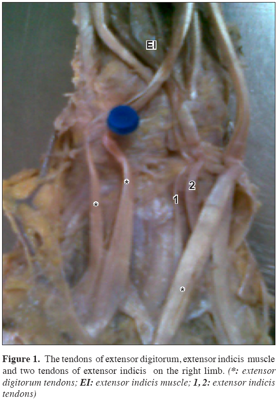
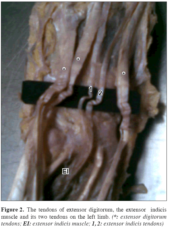
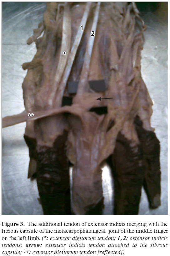
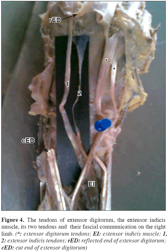
During routine dissection classes for undergraduate medical students, in an adult male cadaver fixed in 10% formalin, it was noted that the EI muscle showed the two tendons underneath the extensor retinaculum in both the upper limbs. The two tendons of EI travelled distally under the tendons of extensor digitorum. In both limbs one of the tendons got inserted into the ulnar aspect of the extensor expansion of the index finger (Figures 1 and 2). In the left upper limb the other tendon was inserted into the dorsal aspect of the capsule of the metacarpophalangeal joint of the middle finger and also few fibers passed on to the radial aspect of the extensor expansion of the same finger (Figure 3). Also, there is a thin, filamentous and fibrous communication between the two tendons over the proximal part of the metacarpal bones. While in the right upper limb the other tendon was attached to the extensor expansion of the middle finger. The uniqueness in this, was the formation of a very clear communicating sheath with the tendon that was having its usual normal attachment i.e., to the extensor expansion of the index finger (Figure 4).
Figure 3: The additional tendon of extensor indicis merging with the fibrous capsule of the metacarpophalangeal joint of the middle finger on the left limb. (*: extensor digitorum tendon; 1, 2: extensor indicis tendons; arrow: extensor indicis tendon attached to the fibrous capsule; **: extensor digitorum tendon [reflected])
Figure 4: The tendons of extensor digitorum, the extensor indicis muscle, its two tendons and their fascial communication on the right limb. (*: extensor digitorum tendons; EI: extensor indicis muscle; 1, 2: extensor indicis tendons; rED: reflected end of extensor digitorum; cED: cut end of extensor digitorum)
Discussion
The extensors of the forearm are known to exhibit wide range of variations. Among the extensors, the EI is well known to associate with the variation of its muscle belly as well as with tendon and its insertions [1-4]. In the present case, we are reporting a unique variation of the EI. The EI presenting two tendons is well documented but in the present case, among the two tendons one is inserted to the ulnar aspect of the extensor expansion of the index finger while the other in the left limb to the capsule of the metacarpophalangeal joint of the middle finger. It is documented that in some cases the tendons of long extensors are intercalating with the dorsal capsule of the metacarpophalangeal joint [5]. However, the additional tendon of EI having an insertion to the capsule of the middle finger is first of its kind to the best of our knowledge. This mode of attachment to the capsule may probably result in the pathology of the joint and/or capsule which may lead to the ganglion formation or may also restrict the movement of the joint. On the contrary with the tendon graft point of view this additional tendon may be helpful. In either case the knowledge of this variation is important not only for anatomists but also for the surgeons dealing with hand. The present case also highlights the attachment of the additional EI tendon in the right limb to the extensor expansion of the middle finger with the emphasis on the communication between the two tendons in the form of a sheath over the metacarpals. This is partially subdividing the subaponeurotic space on the dorsum of the hand.
To conclude this report with its rarity and uniqueness is adding on to the vast knowledge of variations with respect to the extensors of forearm and also very helpful to the surgeons dealing with the hand and its complications, repair of the tendons in surgical point of view and also diagnostic aspect.
References
- Bergman RA, Thompson SA, Afifi AK, Saadeh FA. Compendium of Human Anatomic Variation. Munich, Baltimore Urban and Schwarzenberg. 1988; 147.
- Rao MKG, Vollala VR, Bhat SM, Bolla S, Samuel VP, Pamidi N. Four cases of variations in the forearm extensor musculature in a study of hundred limbs and review of literature. Indian J Plast Surg. 2006; 39: 141–147.
- el- Badawi MG, Butt MM, al-Zuhair AG, Fadel RA. Extensor tendons of the fingers: Arrangement and variations-II. Clin Anat. 1995; 8: 391–398.
- Komiyama M, Nwe TM, Toyota N, Shimada Y. Variations of the extensor indicis muscle and tendon.
- J Hand Surg [Br]. 1999; 24: 575–578.
- Williams PL, Bannister LH, Berry MM, Collins P, Dyson M, Dussek JE, Ferguson MW, eds. Gray’s Anatomy. 38th Ed., London, Churchill Livingstone. 1995; 659.
Satya Prasad Venugopal*, Sree Bhanu Mallula
Department of Anatomy, M.N.R. Medical College, Sangareddy, Medak District, Andhra Pradesh, India.
- *Corresponding Author:
- Dr. Satya Prasad Venugopal
Department Of Anatomy, M.N.R. Medical College, Sangareddy, Medak District, Andhra Pradesh, India.
Tel: +91 8455 277055
Fax: +91 8455 277688
E-mail: satyaprasad33@yahoo.co.in
Date of Received: October 30th, 2008
Date of Accepted: January 19th, 2009
Published Online: January 31st, 2009
© IJAV. 2009; 2: 17–19.
Abstract
There are numerous reports regarding the variations of the extensor muscles. The present report is on the unusual variation of the extensor indicis muscle. The extensor indicis presented two tendons, one of which is inserted into the ulnar aspect of the dorsal digital expansion of the index finger. While the other tendon –especially in the left upper limb– was inserted into the dorsal part of the capsule of the metacarpophalangeal joint of the middle finger and also few tendinous fibers were merging with the dorsal expansion of the middle finger. The same variation was found in the right upper limb, but the insertion was to the dorsal digital expansion of the index and middle fingers and no attachment to the capsule was noticed.
-Keywords
extensor indicis tendon, middle finger, capsule of metacarpophalangeal joint, extensor expansion
Introduction
The extensor indicis (EI) muscle is one of the known extensor for its variations. It normally arises from the posterior surface of the ulna and the adjoining interosseous membrane, and is inserted into the ulnar aspect of the extensor expansion of the index finger. The presence of two heads or complete duplication of EI, double tendon of the EI has been reported [1]. The presence of extensor indicis brevis in addition to normal EI and in the absence of the normal EI has also been reported [2,3].
Case Report
During routine dissection classes for undergraduate medical students, in an adult male cadaver fixed in 10% formalin, it was noted that the EI muscle showed the two tendons underneath the extensor retinaculum in both the upper limbs. The two tendons of EI travelled distally under the tendons of extensor digitorum. In both limbs one of the tendons got inserted into the ulnar aspect of the extensor expansion of the index finger (Figures 1 and 2). In the left upper limb the other tendon was inserted into the dorsal aspect of the capsule of the metacarpophalangeal joint of the middle finger and also few fibers passed on to the radial aspect of the extensor expansion of the same finger (Figure 3). Also, there is a thin, filamentous and fibrous communication between the two tendons over the proximal part of the metacarpal bones. While in the right upper limb the other tendon was attached to the extensor expansion of the middle finger. The uniqueness in this, was the formation of a very clear communicating sheath with the tendon that was having its usual normal attachment i.e., to the extensor expansion of the index finger (Figure 4).
Figure 3: The additional tendon of extensor indicis merging with the fibrous capsule of the metacarpophalangeal joint of the middle finger on the left limb. (*: extensor digitorum tendon; 1, 2: extensor indicis tendons; arrow: extensor indicis tendon attached to the fibrous capsule; **: extensor digitorum tendon [reflected])
Figure 4: The tendons of extensor digitorum, the extensor indicis muscle, its two tendons and their fascial communication on the right limb. (*: extensor digitorum tendons; EI: extensor indicis muscle; 1, 2: extensor indicis tendons; rED: reflected end of extensor digitorum; cED: cut end of extensor digitorum)
Discussion
The extensors of the forearm are known to exhibit wide range of variations. Among the extensors, the EI is well known to associate with the variation of its muscle belly as well as with tendon and its insertions [1-4]. In the present case, we are reporting a unique variation of the EI. The EI presenting two tendons is well documented but in the present case, among the two tendons one is inserted to the ulnar aspect of the extensor expansion of the index finger while the other in the left limb to the capsule of the metacarpophalangeal joint of the middle finger. It is documented that in some cases the tendons of long extensors are intercalating with the dorsal capsule of the metacarpophalangeal joint [5]. However, the additional tendon of EI having an insertion to the capsule of the middle finger is first of its kind to the best of our knowledge. This mode of attachment to the capsule may probably result in the pathology of the joint and/or capsule which may lead to the ganglion formation or may also restrict the movement of the joint. On the contrary with the tendon graft point of view this additional tendon may be helpful. In either case the knowledge of this variation is important not only for anatomists but also for the surgeons dealing with hand. The present case also highlights the attachment of the additional EI tendon in the right limb to the extensor expansion of the middle finger with the emphasis on the communication between the two tendons in the form of a sheath over the metacarpals. This is partially subdividing the subaponeurotic space on the dorsum of the hand.
To conclude this report with its rarity and uniqueness is adding on to the vast knowledge of variations with respect to the extensors of forearm and also very helpful to the surgeons dealing with the hand and its complications, repair of the tendons in surgical point of view and also diagnostic aspect.
References
- Bergman RA, Thompson SA, Afifi AK, Saadeh FA. Compendium of Human Anatomic Variation. Munich, Baltimore Urban and Schwarzenberg. 1988; 147.
- Rao MKG, Vollala VR, Bhat SM, Bolla S, Samuel VP, Pamidi N. Four cases of variations in the forearm extensor musculature in a study of hundred limbs and review of literature. Indian J Plast Surg. 2006; 39: 141–147.
- el- Badawi MG, Butt MM, al-Zuhair AG, Fadel RA. Extensor tendons of the fingers: Arrangement and variations-II. Clin Anat. 1995; 8: 391–398.
- Komiyama M, Nwe TM, Toyota N, Shimada Y. Variations of the extensor indicis muscle and tendon.
- J Hand Surg [Br]. 1999; 24: 575–578.
- Williams PL, Bannister LH, Berry MM, Collins P, Dyson M, Dussek JE, Ferguson MW, eds. Gray’s Anatomy. 38th Ed., London, Churchill Livingstone. 1995; 659.


