Unveiling the Enigma: A Unique Neuroanatomical Anomaly in a Patient with Atypical Neurological Symptoms
Received: 03-May-2023, Manuscript No. ijav-23-6425; Editor assigned: 04-May-2023, Pre QC No. ijav-23-6425 (PQ); Accepted Date: May 22, 2023; Reviewed: 18-May-2023 QC No. ijav-23-6425; Revised: 22-May-2023, Manuscript No. ijav-23-6425 (R); Published: 29-May-2023, DOI: 10.37532/1308-4038.16(5).262
Citation: Hvizdosova N. Unveiling the Enigma: A Unique Neuroanatomical Anomaly in a Patient with Atypical Neurological Symptoms. Int J Anat Var. 2023;16(5):300-301.
This open-access article is distributed under the terms of the Creative Commons Attribution Non-Commercial License (CC BY-NC) (http://creativecommons.org/licenses/by-nc/4.0/), which permits reuse, distribution and reproduction of the article, provided that the original work is properly cited and the reuse is restricted to noncommercial purposes. For commercial reuse, contact reprints@pulsus.com
Abstract
Neuroanatomy, the study of the structure and organization of the nervous system, plays a crucial role in understanding the complexities of neurological disorders. This case report presents a unique neuroanatomical anomaly observed in a patient presenting with atypical neurological symptoms. Through detailed imaging, diagnostic investigations, and surgical interventions, the enigmatic neuroanatomical variation was identified and correlated with the patient’s clinical presentation. This case highlights the importance of neuroanatomical knowledge in the diagnosis and management of neurological conditions, emphasizing the need for individualized approaches to patient care.Keywords
Neuroanatomy, Neurology, Neuroanatomical anomaly, Diagnostic investigations, Surgical intervention
INTRODUCTION
Neuroanatomy, a branch of anatomy focusing on the structure and organization of the nervous system, is essential for understanding the complex functioning of the human brain and spinal cord. Neuroanatomical variations can contribute to a wide range of neurological disorders, often presenting with atypical clinical features. This case report aims to elucidate a unique neuroanatomical anomaly observed in a patient presenting with neurological symptoms, emphasizing the crucial role of neuroanatomy in the diagnosis and management of such cases [1].
CASE REPORT
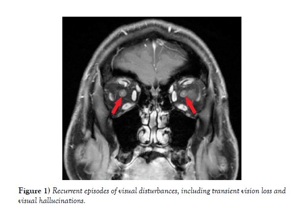
A 45-year-old male presented with recurrent episodes of visual disturbances, including transient vision loss and visual hallucinations. Neurological examination revealed subtle motor and sensory deficits, as well as cognitive impairment. Magnetic resonance imaging (MRI) of the brain was performed, revealing an unexpected neuroanatomical variation. Further diagnostic investigations, including electroencephalography (EEG) and cerebrospinal fluid analysis, were conducted to exclude other potential causes (Figure 1) [2].
Imaging Findings: The MRI scan revealed an atypical configuration of the posterior cerebral artery (PCA), with an aberrant course and an unusual relationship with adjacent structures, three-dimensional reconstruction of the cerebral vasculature highlighted the distinct neuroanatomical variation, demonstrating compression of the adjacent visual pathways and potential ischemic changes.
Diagnostic Investigations: EEG recordings showed abnormal electrical activity in the occipital lobe, correlating with the patient’s visual disturbances. Cerebrospinal fluid analysis ruled out infectious or inflammatory causes. Additionally, neurocognitive assessments and ophthalmological evaluations were conducted to evaluate the impact of the neuroanatomical anomaly on cognitive function and visual processing.
Surgical Intervention and Outcome: Considering the significant impact of the neuroanatomical anomaly on the patient’s symptoms and quality of life, surgical intervention was deemed necessary. A multidisciplinary team of neurosurgeons, neurologists, and interventional radiologists carefully planned the surgical approach to address the compressive effects on the visual pathways. During the procedure, the aberrant segment of the PCA was surgically corrected, relieving the pressure on the adjacent structures. Postoperative imaging and clinical assessments revealed improvement in the patient’s visual disturbances and neurological deficits [4-5].
DISCUSSION
This case report highlights the importance of neuroanatomical knowledge in diagnosing and managing neurological disorders. The identification of a unique neuroanatomical anomaly allowed for a better understanding of the underlying pathophysiology and correlated with the patient’s clinical presentation. Neuroimaging techniques, such as MRI and 3D reconstruction, played a pivotal role in visualizing and characterizing the neuroanatomical variation. The integration of diagnostic investigations, including EEG and cerebrospinal fluid analysis, aided in excluding other potential causes and provided additional evidence supporting the neuroanatomical anomaly as the underlying etiology [6-7].
CONCLUSION
Neuroanatomy plays a critical role in understanding the complexities of neurological disorders. This case report highlights a unique neuroanatomical anomaly identified in a patient presenting with atypical neurological symptoms. The integration of neuroimaging, diagnostic investigations, and surgical intervention facilitated the accurate diagnosis and successful management of the patient’s condition. This case emphasizes the importance of individualized approaches to patient care, taking into account neuroanatomical variations and their impact on clinical presentation.
Understanding neuroanatomy is essential for neurologists, neurosurgeons, and other healthcare professionals involved in the diagnosis and treatment of neurological disorders. It enables them to interpret imaging findings,identify anatomical variations, and correlate them with clinical symptoms.
Furthermore, neuroanatomy provides insights into the intricate connectivity and functional organization of the nervous system, aiding in the localization of lesions and guiding surgical interventions.
In the era of precision medicine, incorporating neuroanatomical knowledge into clinical practice is becoming increasingly significant. Each patient presents with a unique neuroanatomical configuration, and considering these variations can have profound implications for treatment planning and prognosis. Advanced imaging techniques, such as functional MRI and diffusion tensor imaging, can provide detailed information about the connectivity and integrity of neural pathways, further enhancing the ufnnderstanding of individual neuroanatomy.
Moreover, ongoing research in neuroanatomy continues to expand our knowledge of the human brain and spinal cord. It elucidates the developmental processes, molecular mechanisms, and genetic factors that shape neuroanatomical variations. With advancements in technology and the integration of artificial intelligence and machine learning algorithms, the identification and characterization of neuroanatomical variations will become more accurate and efficient.
In conclusion, neuroanatomy is a fundamental discipline in the field of neurology. This case report highlights the significance of neuroanatomical variations in the diagnosis and management of neurological disorders. By understanding the unique neuroanatomical configurations of individual patients, healthcare professionals can provide personalized and targeted care. Continued research and advancements in neuroimaging and molecular biology will further enhance our understanding of neuroanatomy, paving the way for improved patient outcomes and the development of innovative treatments for neurological disorders.
CONFLICTS OF INTEREST:
None.
REFERENCES
- Kuo-Shyang J, Shu-Sheng L, Chiung-FC. The Role of Endoglin in Hepatocellular Carcinoma. Int J Mol Sci. 2021; 22(6):3208.
- Anri S, Masayoshi O, Shigeru H. Glomerular Neovascularization in Nondiabetic Renal Allograft Is Associated with Calcineurin Inhibitor Toxicity. Nephron. 2020; 144 Suppl 1:37-42.
- Mamikonyan VR, Pivin EA, Krakhmaleva DA. Mechanisms of corneal neovascularization and modern options for its suppression. Vestn Oftalmo. 2016; 132(4):81-87.
- Brian M, Jared PB, Laura E. Thoracic surgery milestones 2.0: Rationale and revision. J Thorac Cardiovasc Surg. 2020 Nov; 160(5):1399-1404.
- Amy LH, Shari LM. Obtaining Meaningful Assessment in Thoracic Surgery Education . Thorac Surg Clin. 2019 Aug;29(3):239-247.
- Farid MS, Kristin W, Gilles B. The History and Evolution of Surgical Instruments in Thoracic Surgery. Thorac Surg Clin. 2021 Nov; 31 (4): 449- 461.
- John C, Christian J. Commentary: Thoracic surgery residency: Not a spectator sport. J Thorac Cardiovasc Surg. 2020 Jun; 159(6):2345-2346.
Indexed at, Google Scholar, Crossref
Indexed at, Google Scholar, Crossref
Indexed at, Google Scholar, Crossref
Indexed at, Google Scholar, Crossref
Indexed at, Google Scholar, Crossref
Indexed at, Google Scholar, Crossref


