Unveiling Uncommon Anatomy: A Case Report of Retrocecal Appendix in Appendectomy
Received: 03-May-2023, Manuscript No. ijav-23-6430; Editor assigned: 04-May-2023, Pre QC No. ijav-23-6430 (PQ); Accepted Date: May 22, 2023; Reviewed: 18-May-2023 QC No. ijav-23-6430; Revised: 22-May-2023, Manuscript No. ijav-23-6430 (R); Published: 29-May-2023, DOI: 10.37532/1308-4038.16(5).265
Citation: Karl M. Unveiling Uncommon Anatomy: A Case Report of Retrocecal Appendix in Appendectomy. Int J Anat Var. 2023;16(5):306-307.
This open-access article is distributed under the terms of the Creative Commons Attribution Non-Commercial License (CC BY-NC) (http://creativecommons.org/licenses/by-nc/4.0/), which permits reuse, distribution and reproduction of the article, provided that the original work is properly cited and the reuse is restricted to noncommercial purposes. For commercial reuse, contact reprints@pulsus.com
Abstract
Anatomical variations refer to deviations from the typical structure and arrangement of organs, tissues, and systems within the human body. These variations can have clinical implications and can pose challenges during surgical procedures, diagnosis, and treatment. This case report presents a unique anatomical variation encountered during a routine appendectomy procedure, highlighting the importance of recognizing and understanding such variations for successful surgical outcomes.
Keywords
Anatomical variations, Retrocecal appendix, Appendectomy, Surgical approach, Clinical implications, Surgical planning, Patient safety
INTRODUCTION
Anatomical variations are inherent differences in the structure, position, and arrangement of organs, tissues, and systems within the human body. These variations can occur at both macroscopic and microscopic levels and are a result of natural variations in human development. While there is a typical anatomical pattern considered as “normal,” it is essential to recognize that variations are common and can have clinical implications [1].
Understanding and being aware of anatomical variations are crucial for healthcare professionals, including surgeons, radiologists, anatomists, and clinicians. Anatomical variations can significantly impact medical practice, including surgical procedures, diagnostic imaging, and interpretation, as well as treatment planning and management [2].
This case report aims to present a unique anatomical variation encountered during a routine appendectomy procedure. The report underscores the significance of recognizing and comprehending such variations to ensure successful surgical outcomes. By highlighting the importance of awareness and adaptability, this case report contributes to the growing body of knowledge on anatomical variations and their impact on clinical practice [2].
It is worth noting that the incidence and prevalence of anatomical variations can vary widely across different populations and individuals. Factors such as genetic predisposition, ethnic background, and environmental influences can contribute to the occurrence of these variations. While some variations may be relatively common and well-documented, others may be rare and unique, requiring thorough investigation and understanding [2].
The discussion of this case report will focus on the specific anatomical variation encountered during the appendectomy procedure, providing insights into the challenges faced by the surgeon and the strategies employed to overcome them. Additionally, it will emphasize the importance of preoperative imaging studies, intraoperative vigilance, and adaptability in the context of anatomical variations [3].
By shedding light on this case, healthcare professionals can gain a better understanding of the potential anatomical variations they may encounter in their clinical practice. This knowledge enhances patient care by enabling precise diagnosis, tailored surgical approaches, and improved patient outcomes. Ultimately, a deeper understanding of anatomical variations contributes to the ongoing evolution of medical knowledge and practice [4].
CASE STUDY
A 32-year-old male patient presented to the emergency department with acute right lower quadrant abdominal pain, tenderness, and localized rebound tenderness. The patient’s medical history was unremarkable, with no previous abdominal surgeries or significant illnesses. Clinical examination findings were consistent with acute appendicitis, and the decision was made to proceed with an emergency appendectomy.
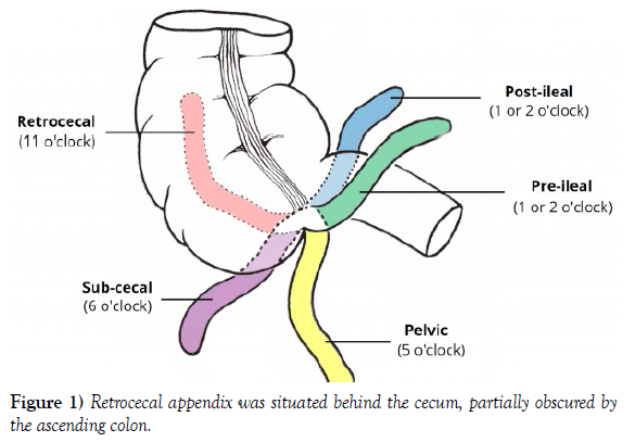
During the surgical procedure, an open appendectomy approach was chosen. A standard McBurney incision was made, and dissection through the layers was performed [5]. However, upon exposing the peritoneal cavity, an unexpected anatomical variation was observed. The appendix was found to be retrocecal, which is an atypical location compared to the typical intraperitoneal position. The retrocecal appendix was situated behind the cecum, partially obscured by the ascending colon (Figure 1) [6].
The surgeon adapted the surgical approach accordingly, carefully dissecting the retrocecal appendix free from its surrounding structures. Due to the altered anatomical location, the appendix was adherent to the posterior abdominal wall, making dissection technically challenging. However, with meticulous dissection, the retrocecal appendix was successfully removed without complications [7].
DISCUSSION
The retrocecal appendix is an anatomical variation encountered in approximately 20% of individuals. It occurs when the appendix is positioned behind the cecum, usually lying in close proximity to the right psoas muscle. This variation can be challenging during surgical interventions as it may require alterations in the surgical approach and careful identification to prevent inadvertent injury to surrounding structures.
An awareness of anatomical variations, such as the retrocecal appendix, is crucial for accurate diagnosis and surgical planning. Preoperative imaging studies, such as computed tomography (CT) scans, can provide valuable information about the appendix’s location and aid in surgical decisionmaking. However, in emergency cases, imaging studies may not always be feasible or readily available, making intraoperative identification of anatomical variations crucial.
In this case, the surgeon encountered a retrocecal appendix during a routine appendectomy procedure. Although it posed technical challenges, the surgeon successfully adapted the surgical approach and safely removed the appendix. Awareness of this anatomical variation, combined with intraoperative vigilance, ensured a favorable surgical outcome.
CONCLUSION
Anatomical variations are a common occurrence in clinical practice, and their recognition plays a vital role in surgical interventions. This case report highlights the importance of identifying and understanding anatomical variations, such as the retrocecal appendix, during surgical procedures. Surgeons and healthcare professionals should maintain a high level of vigilance to adapt surgical techniques when encountering such variations to ensure optimal patient outcomes. Increased awareness and knowledge of anatomical variations contribute to improved surgical planning, decreased complications, and enhanced patient safety.
CONFLICT OF INTEREST
None
ACKNOWLEDGEMENT
We would like to thank the patient and their family for their participation and consent in this case report. We also acknowledge the support and expertise of the laboratory personnel involved in the genetic testing and analysis.
REFERENCES
- Krause DA, Youdas JW. Bilateral presence of a variant subscapularis muscle. Int J Anat Var. 2017; 10(4):79-80.
- Mann MR, Plutecki D, Janda P, Pękala J, Malinowski K, et al. The subscapularis muscle‐a meta‐analysis of its variations, prevalence, and anatomy. Clin Anat. 2023; 36(3):527-541.
- Pillay M, Jacob SM. Bilateral presence of axillary arch muscle passing through the posterior cord of the brachial plexus. Int. J. Morphol., 27(4):1047-1050, 2009.
- Pires LAS, Souza CFC, Teixeira AR, Leite TFO, Babinski MA, et al. Accessory subscapularis muscle–A forgotten variation?. Morphologie. 2017; 101(333):101-104.
- John C, Christian J. Commentary: Thoracic surgery residency: Not a spectator sport. J Thorac Cardiovasc Surg. 2020 Jun; 159(6):2345-2346.
- Anri S, Masayoshi O, Shigeru H. Glomerular Neovascularization in Nondiabetic Renal Allograft Is Associated with Calcineurin Inhibitor Toxicity. Nephron. 2020; 144 Suppl 1:37-42.
- Mamikonyan VR, Pivin EA, Krakhmaleva DA. Mechanisms of corneal neovascularization and modern options for its suppression. Vestn Oftalmo. 2016; 132(4):81-87.
Indexed at, Google Scholar, Crossref
Indexed at, Google Scholar, Crossref
Indexed at, Google Scholar, Crossref
Indexed at, Google Scholar, Crossref
Indexed at, Google Scholar, Crossref


