Variant adductor muscle complex of thigh – a case report
Upasna1*, Ashwani Kumar2, Gurdeep Singh Kalyan1
1Departments of Anatomy, Government Medical College, Patiala, India.
2Departments of Anatomy and Surgery, Government Medical College, Patiala, India.
- *Corresponding Author:
- Dr. Upasna
C2, Medical College Campus, Government Medical College, Patiala, Punjab, India.
Tel: +91 987 2624818
E-mail: ashwaniupasna@yahoo.com
Date of Received: February 9th, 2012
Date of Accepted: October 17th, 2012
Published Online: February 14th, 2013
© Int J Anat Var (IJAV). 2013; 6: 36–40.
[ft_below_content] =>Keywords
pectineus, adductor longus, adductor brevis, variant muscular slip, obturator externus
Introduction
The medial thigh muscles –the adductor group– are in the medial compartment of the thigh. The adductor group of thigh muscles consist of adductor longus, adductor brevis, adductor magnus, gracilis and obturator externus muscles.
Collectively these muscles are the adductors of the thigh; however actions of some of these muscles are more complex [1]. Pectineus also belongs anatomically and physiologically and probably in part also morphologically with this group [2].
Present case report includes the variant origin and insertion of pectineus, adductor longus, adductor brevis and adductor magnus muscles. A variant muscular slip has also been observed beneath adductor brevis muscle.
Case Report
During routine dissection for undergraduate students at the Government Medical College, Patiala a variant adductor group of muscles of thigh was observed in a middle-aged female cadaver. The variations observed were bilateral. A variant muscular slip was also observed. The origin and insertion of the adductor group of muscles were studied and their nerve supply was traced. The variations were photographed.
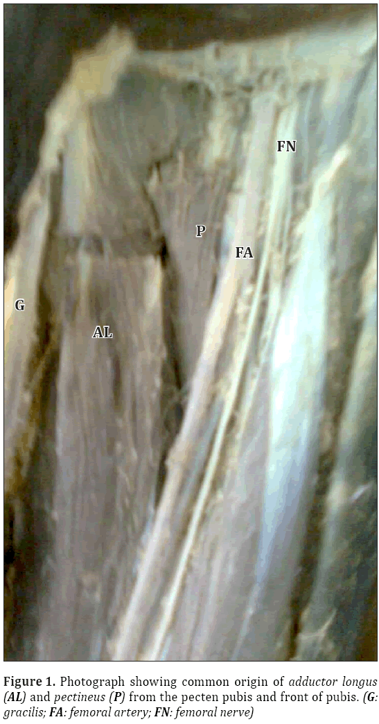
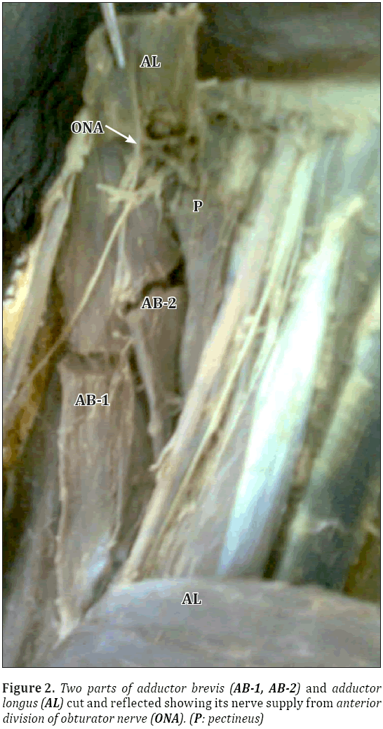
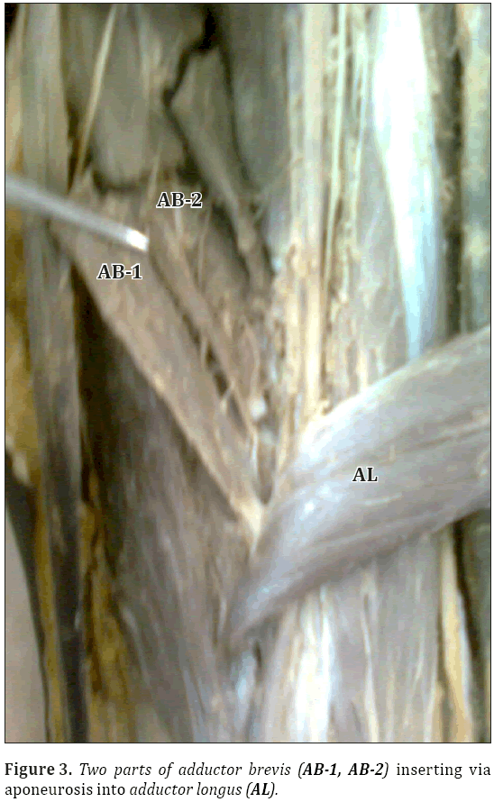
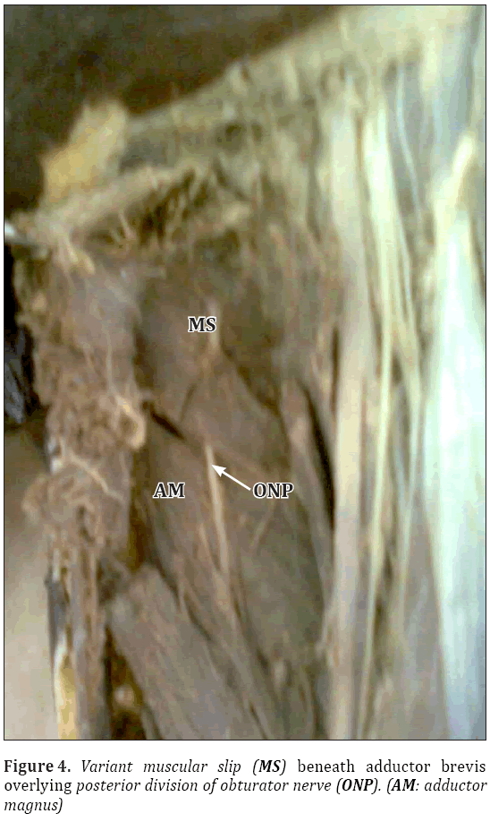
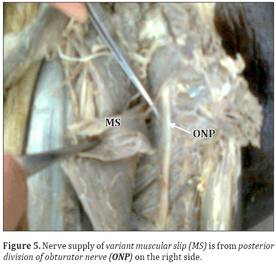
On left lower limb, the adductor longus and pectineus were having the combined origin from the front of the pecten pubis and front of pubis (Figure 1). The nerve supply was from femoral nerve and anterior division of obturator nerve. Insertion of adductor longus and pectineus was as usual. After sectioning and reflecting adductor longus and pectineus, adductor brevis was observed. It was having usual origin but was seen in two parts, and lower down its fibers merged with the fibers of adductor longus. The nerve supply was from anterior division of obturator nerve (Figures 2, 3). Then, the adductor brevis was reflected and a transverse muscular slip was observed beneath it. This muscular slip was overlying the posterior division of obturator nerve and the obturator externus muscle. The fibers were attached to the front of inferior pubic ramus and merged with aponeurosis of adductor magnus (Figure 4). The nerve supply was from the posterior division of obturator nerve (Figure 5). Adductor magnus was usual in origin and insertion.
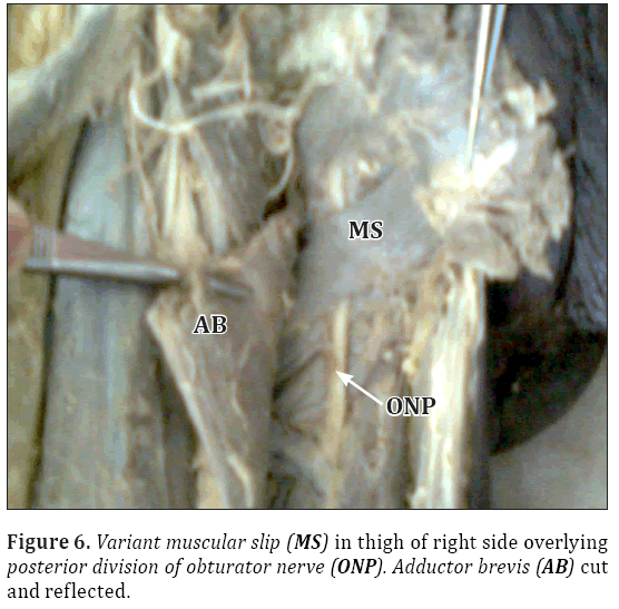
Similar variant muscular slip was also observed in thigh of right lower limb (Figures 5, 6).
Discussion
Pectineus arises from the pecten pubis, from the bone in front of it between the iliopubic ramus and the pubic tubercle and from the fascia on its own anterior surface. The fibers descend initially posteromedially and then posterolaterally to attach along a line from the lesser trochanter to the linea aspera. Pectineus may be bilaminar, in which case the two layers receive separate nerve supplies [3]. But in our case report, its fibers merged with the fibers of adductor longus at the site of origin. The insertion was usual (Figure 1). Nerve supply was from femoral nerve and anterior division of obturator nerve.
Adductor longus is attached to the front of the pubis in the angle between the crest and the symphysis. It inserts by an aponeurosis into the linea aspera in the middle third of femur, between vastus medialis and adductor magnus and brevis, usually blending with all of them. Adductor longus is occasionally double [3]. In our case, the nerve supply of adductor longus was from anterior division of obturator nerve.
Adductor brevis arises by a narrow attachment from the external aspect of the body and inferior ramus of pubis, between gracilis and obturator externus. It inserts via an aponeurosis into the femur, along a line from the lesser trochanter to the linea aspera and on the upper part of the line immediately behind pectineus and the upper part of adductor longus. Adductor brevis often has two or three separate parts or it may be integrated into adductor magnus [3]. In our case, it had two separate parts (Figure 2) and the fibers inserted via the aponeurosis into the adductor longus. The nerve supply was obturator nerve (Figure 3).
Adductor magnus, a massive triangular muscle arises from a small part of the inferior ramus of the pubis, from the conjoined ischial ramus, and from the inferolateral aspect of the ischial tuberosity. The short, horizontal fibers from the pubic ramus are inserted into the medial margin of the gluteal tuberosity of the femur, medial to the gluteus maximus; this part of the muscle, in the plane anterior to the rest, is sometimes called adductor minimus. The fibers from the ischial ramus fan out downwards and laterally to insert via a broad aponeurosis into the linea aspera and the proximal part of the medial supracondylar line [3]. The adductor minimus muscle has had scant and conflicting reports regarding its anatomy with some authors ignoring its existence altogether. Tubbs et al. found partial fusion between the adductor minimus and adductor magnus muscles in 24% of sides [4].
A distinct transverse muscular slip (Figure 4) described in our case report was not fused with quadratus femoris or obturator externus, instead it was lying in front of posterior division of obturator nerve and obturator externus. The nerve supply of this muscular slip was from posterior division of obturator nerve (Figure 5). Nakamura et al. have described a supernumerary muscle found between the adductor brevis and minimus muscles. They speculated that such a muscle most likely has an origin from either the obturator externus or the adductor minimus muscle. An incidence of 33% was recorded by the authors. It stemmed from the inferior pubis ramus and inserted into the aponeurosis of adductor minimus muscle, pectineal line or the posterior aspect of lesser trochanter base. It derived its innervation from its posterior aspect by a filament from the twig originating from the posterior branch of the obturator nerve. It was deduced further that the additional muscle might have formed as a result of detachment from the superficial layer of the obturator externus during the process of ontogeny [5]. It shows similarity in the origin of muscular slip from inferior pubic ramus but insertion was into aponeurosis of adductor magnus (Figure 5). The source of nerve supply is also similar in both cases.
Chopra et al. also reported a supernumerary muscle in the adductor compartment of thigh between adductor brevis and adductor magnus. The origin of this muscle was similar as in our case, from outer surface of the body and inferior ramus of pubis but insertion was not similar. The insertion was into the aponeurosis of adductor magnus not on the femur as reported by them. The authors described this supernumerary muscle as an additional adductor brevis muscle which might have been formed due to unusual splitting of original muscle anlagen [6].
A special pre-semimembranosus muscle which extends usually from the ischial tuberosity to the medial side of the distal end of the femur parallel with semimembranosus has also been described in the literature. In the gorilla, orang and gibbon the pre-semimembranosus is combined with the adductor magnus as it is in man. It may be looked upon as derived phylogenetically from the semimembranosus or medial flexor of the thigh [2].
In the different groups of mammals there is considerable variation in the number of individual muscles into which the adductor musculature is divisible, from one to six. In man the chief variations noted have to do with the greater or less fusion of different muscles into which the group is divided [2].
Myelodysplastic patients with subluxation or dislocation of the hip were treated surgically by transferring the origins of adductor brevis along with the other adductors to the ischial tuberosity. The presence of an additional belly of adductor brevis could possibly be utilized for the same purpose, thereby sparing the other adductors for the adduction function which otherwise weakens as a result of the transfer [7].
Adductor muscle strain may prove to be incapacitating for the athletes especially if improperly treated. Sport surgeons resort to adductor release and tenotomy only if other rehabilitative procedures fail to cure the patient of pain and debility [8]. Surgically, DiGeronimo has utilized the adductor minimus in a myocutaneous flap for scrotal reconstruction [9]. The surgeon might use the adductor minimus muscle as a landmark for identifying the posterior extent of medial femoral circumflex artery and first perforating artery for vascular anastomosis or avoidance [4].
Ultrasound imaging has been successful in localizing the medial femoral region with 100% clarity. Additionally this region is frequently used as a landmark for localization of the common obturator nerve with a success rate of 80%. There is considerable inconsistency in the obturator nerve’s divisions and subdivisions and this is reflected in the form of tribulations frequently encountered in the application of regional anesthetic techniques [10].
Conclusion
Authors arrived at a conclusion that a variant adductor complex of thigh occurred due to greater or less fusion of different muscles into which the adductor group is divided. The supernumerary muscle or muscular slip present formed as a result of detachment from the superficial layer of obturator externus during the process of ontogeny. Adequate anatomical knowledge of medial side of thigh with the possible muscular variations is needed for the proper performance of surgeries and reconstructions.
References
- Moore KL, Dalley AF. Clinically Oriented Anatomy. 4th Ed., Philadelphia, Lippincott Williams and Wilkins. 1999; 537.
- Bardeen CR. Development and variation of the nerves and the musculature of the inferior extremity and of the neighbouring regions of the trunk in man. Am J Anat. 1906; 6: 259–390.
- Strandring S, ed. Gray’s Anatomy. The Anatomical Basis of Clinical Practice. 39th Ed., Philadelphia, Elsevier Churchill Livingstone. 2008; 1375–1376.
- Shane Tubbs R, Griessenauer CJ, Marshall T, Dennison CP, Shoja MM, Loukas M, Apaydin N, Cohen-Gadol AA. The adductor minimus muscle revisited. Surg Radiol Anat. 2011; 33: 429–432.
- Nakamura E, Masumi S, Muara M, Kato S, Miyauchi R. A supernumerary muscle between the adductors brevis and minimus in humans. Okajimas Folia Anat Jpn. 1992; 69: 89–98.
- Jyoti C, Anita R, Archana R, Punita M. Supernumerary muscle in the adductor compartment of thigh – a case report. J Anat Soc India. 2009; 58: 195–198.
- London JT, Nichols O. Paralytic dislocation of the hip in myelodysplasia. The role of the adductor transfer. J Bone Joint Surg Am. 1975; 57: 501–506.
- Nicholas SJ, Tyler TF. Adductor muscle strains in sport. Sports Med. 2002; 32: 339–344.
- DiGeronimo EM. Scrotal reconstruction utilising a unilateral adductor minimus myocutaneous flap. Plast Reconstr Surg. 1982; 70: 749–751.
- Anagnostopoulou S, Kostopanagiotou G, Paraskeuopoulos T, Chantzi C, Lolis E, Saranteas T. Anatomic variations of the obturator nerve in the inguinal region: implications in conventional and ultrasound regional anaesthesia techniques. Reg Anesth Pain Med. 2009; 34: 33–39.
Upasna1*, Ashwani Kumar2, Gurdeep Singh Kalyan1
1Departments of Anatomy, Government Medical College, Patiala, India.
2Departments of Anatomy and Surgery, Government Medical College, Patiala, India.
- *Corresponding Author:
- Dr. Upasna
C2, Medical College Campus, Government Medical College, Patiala, Punjab, India.
Tel: +91 987 2624818
E-mail: ashwaniupasna@yahoo.com
Date of Received: February 9th, 2012
Date of Accepted: October 17th, 2012
Published Online: February 14th, 2013
© Int J Anat Var (IJAV). 2013; 6: 36–40.
Abstract
The medial thigh muscles –the adductor group– are in the medial compartment of thigh. This group consists of adductor longus, adductor brevis, adductor magnus, gracilis, obturator externus and pectineus muscles. Present case report includes the variant origin and insertion of these muscles. A variant muscular slip has also been observed beneath the adductor brevis muscle. This variation has been compared with variations reported by other authors. Embryological and clinical aspect has also been discussed.
-Keywords
pectineus, adductor longus, adductor brevis, variant muscular slip, obturator externus
Introduction
The medial thigh muscles –the adductor group– are in the medial compartment of the thigh. The adductor group of thigh muscles consist of adductor longus, adductor brevis, adductor magnus, gracilis and obturator externus muscles.
Collectively these muscles are the adductors of the thigh; however actions of some of these muscles are more complex [1]. Pectineus also belongs anatomically and physiologically and probably in part also morphologically with this group [2].
Present case report includes the variant origin and insertion of pectineus, adductor longus, adductor brevis and adductor magnus muscles. A variant muscular slip has also been observed beneath adductor brevis muscle.
Case Report
During routine dissection for undergraduate students at the Government Medical College, Patiala a variant adductor group of muscles of thigh was observed in a middle-aged female cadaver. The variations observed were bilateral. A variant muscular slip was also observed. The origin and insertion of the adductor group of muscles were studied and their nerve supply was traced. The variations were photographed.
On left lower limb, the adductor longus and pectineus were having the combined origin from the front of the pecten pubis and front of pubis (Figure 1). The nerve supply was from femoral nerve and anterior division of obturator nerve. Insertion of adductor longus and pectineus was as usual. After sectioning and reflecting adductor longus and pectineus, adductor brevis was observed. It was having usual origin but was seen in two parts, and lower down its fibers merged with the fibers of adductor longus. The nerve supply was from anterior division of obturator nerve (Figures 2, 3). Then, the adductor brevis was reflected and a transverse muscular slip was observed beneath it. This muscular slip was overlying the posterior division of obturator nerve and the obturator externus muscle. The fibers were attached to the front of inferior pubic ramus and merged with aponeurosis of adductor magnus (Figure 4). The nerve supply was from the posterior division of obturator nerve (Figure 5). Adductor magnus was usual in origin and insertion.
Similar variant muscular slip was also observed in thigh of right lower limb (Figures 5, 6).
Discussion
Pectineus arises from the pecten pubis, from the bone in front of it between the iliopubic ramus and the pubic tubercle and from the fascia on its own anterior surface. The fibers descend initially posteromedially and then posterolaterally to attach along a line from the lesser trochanter to the linea aspera. Pectineus may be bilaminar, in which case the two layers receive separate nerve supplies [3]. But in our case report, its fibers merged with the fibers of adductor longus at the site of origin. The insertion was usual (Figure 1). Nerve supply was from femoral nerve and anterior division of obturator nerve.
Adductor longus is attached to the front of the pubis in the angle between the crest and the symphysis. It inserts by an aponeurosis into the linea aspera in the middle third of femur, between vastus medialis and adductor magnus and brevis, usually blending with all of them. Adductor longus is occasionally double [3]. In our case, the nerve supply of adductor longus was from anterior division of obturator nerve.
Adductor brevis arises by a narrow attachment from the external aspect of the body and inferior ramus of pubis, between gracilis and obturator externus. It inserts via an aponeurosis into the femur, along a line from the lesser trochanter to the linea aspera and on the upper part of the line immediately behind pectineus and the upper part of adductor longus. Adductor brevis often has two or three separate parts or it may be integrated into adductor magnus [3]. In our case, it had two separate parts (Figure 2) and the fibers inserted via the aponeurosis into the adductor longus. The nerve supply was obturator nerve (Figure 3).
Adductor magnus, a massive triangular muscle arises from a small part of the inferior ramus of the pubis, from the conjoined ischial ramus, and from the inferolateral aspect of the ischial tuberosity. The short, horizontal fibers from the pubic ramus are inserted into the medial margin of the gluteal tuberosity of the femur, medial to the gluteus maximus; this part of the muscle, in the plane anterior to the rest, is sometimes called adductor minimus. The fibers from the ischial ramus fan out downwards and laterally to insert via a broad aponeurosis into the linea aspera and the proximal part of the medial supracondylar line [3]. The adductor minimus muscle has had scant and conflicting reports regarding its anatomy with some authors ignoring its existence altogether. Tubbs et al. found partial fusion between the adductor minimus and adductor magnus muscles in 24% of sides [4].
A distinct transverse muscular slip (Figure 4) described in our case report was not fused with quadratus femoris or obturator externus, instead it was lying in front of posterior division of obturator nerve and obturator externus. The nerve supply of this muscular slip was from posterior division of obturator nerve (Figure 5). Nakamura et al. have described a supernumerary muscle found between the adductor brevis and minimus muscles. They speculated that such a muscle most likely has an origin from either the obturator externus or the adductor minimus muscle. An incidence of 33% was recorded by the authors. It stemmed from the inferior pubis ramus and inserted into the aponeurosis of adductor minimus muscle, pectineal line or the posterior aspect of lesser trochanter base. It derived its innervation from its posterior aspect by a filament from the twig originating from the posterior branch of the obturator nerve. It was deduced further that the additional muscle might have formed as a result of detachment from the superficial layer of the obturator externus during the process of ontogeny [5]. It shows similarity in the origin of muscular slip from inferior pubic ramus but insertion was into aponeurosis of adductor magnus (Figure 5). The source of nerve supply is also similar in both cases.
Chopra et al. also reported a supernumerary muscle in the adductor compartment of thigh between adductor brevis and adductor magnus. The origin of this muscle was similar as in our case, from outer surface of the body and inferior ramus of pubis but insertion was not similar. The insertion was into the aponeurosis of adductor magnus not on the femur as reported by them. The authors described this supernumerary muscle as an additional adductor brevis muscle which might have been formed due to unusual splitting of original muscle anlagen [6].
A special pre-semimembranosus muscle which extends usually from the ischial tuberosity to the medial side of the distal end of the femur parallel with semimembranosus has also been described in the literature. In the gorilla, orang and gibbon the pre-semimembranosus is combined with the adductor magnus as it is in man. It may be looked upon as derived phylogenetically from the semimembranosus or medial flexor of the thigh [2].
In the different groups of mammals there is considerable variation in the number of individual muscles into which the adductor musculature is divisible, from one to six. In man the chief variations noted have to do with the greater or less fusion of different muscles into which the group is divided [2].
Myelodysplastic patients with subluxation or dislocation of the hip were treated surgically by transferring the origins of adductor brevis along with the other adductors to the ischial tuberosity. The presence of an additional belly of adductor brevis could possibly be utilized for the same purpose, thereby sparing the other adductors for the adduction function which otherwise weakens as a result of the transfer [7].
Adductor muscle strain may prove to be incapacitating for the athletes especially if improperly treated. Sport surgeons resort to adductor release and tenotomy only if other rehabilitative procedures fail to cure the patient of pain and debility [8]. Surgically, DiGeronimo has utilized the adductor minimus in a myocutaneous flap for scrotal reconstruction [9]. The surgeon might use the adductor minimus muscle as a landmark for identifying the posterior extent of medial femoral circumflex artery and first perforating artery for vascular anastomosis or avoidance [4].
Ultrasound imaging has been successful in localizing the medial femoral region with 100% clarity. Additionally this region is frequently used as a landmark for localization of the common obturator nerve with a success rate of 80%. There is considerable inconsistency in the obturator nerve’s divisions and subdivisions and this is reflected in the form of tribulations frequently encountered in the application of regional anesthetic techniques [10].
Conclusion
Authors arrived at a conclusion that a variant adductor complex of thigh occurred due to greater or less fusion of different muscles into which the adductor group is divided. The supernumerary muscle or muscular slip present formed as a result of detachment from the superficial layer of obturator externus during the process of ontogeny. Adequate anatomical knowledge of medial side of thigh with the possible muscular variations is needed for the proper performance of surgeries and reconstructions.
References
- Moore KL, Dalley AF. Clinically Oriented Anatomy. 4th Ed., Philadelphia, Lippincott Williams and Wilkins. 1999; 537.
- Bardeen CR. Development and variation of the nerves and the musculature of the inferior extremity and of the neighbouring regions of the trunk in man. Am J Anat. 1906; 6: 259–390.
- Strandring S, ed. Gray’s Anatomy. The Anatomical Basis of Clinical Practice. 39th Ed., Philadelphia, Elsevier Churchill Livingstone. 2008; 1375–1376.
- Shane Tubbs R, Griessenauer CJ, Marshall T, Dennison CP, Shoja MM, Loukas M, Apaydin N, Cohen-Gadol AA. The adductor minimus muscle revisited. Surg Radiol Anat. 2011; 33: 429–432.
- Nakamura E, Masumi S, Muara M, Kato S, Miyauchi R. A supernumerary muscle between the adductors brevis and minimus in humans. Okajimas Folia Anat Jpn. 1992; 69: 89–98.
- Jyoti C, Anita R, Archana R, Punita M. Supernumerary muscle in the adductor compartment of thigh – a case report. J Anat Soc India. 2009; 58: 195–198.
- London JT, Nichols O. Paralytic dislocation of the hip in myelodysplasia. The role of the adductor transfer. J Bone Joint Surg Am. 1975; 57: 501–506.
- Nicholas SJ, Tyler TF. Adductor muscle strains in sport. Sports Med. 2002; 32: 339–344.
- DiGeronimo EM. Scrotal reconstruction utilising a unilateral adductor minimus myocutaneous flap. Plast Reconstr Surg. 1982; 70: 749–751.
- Anagnostopoulou S, Kostopanagiotou G, Paraskeuopoulos T, Chantzi C, Lolis E, Saranteas T. Anatomic variations of the obturator nerve in the inguinal region: implications in conventional and ultrasound regional anaesthesia techniques. Reg Anesth Pain Med. 2009; 34: 33–39.


