Variant extensor muscles on the dorsum of the hand
Keerti Singh1*, Donnahue Dennie1, Suresh R Rao2
1Anatomy Section, Department of Basic Medical Science, University of West Indies Mona campus, Jamaica.
2Anatomy Unit, University of West Indies, St. Augustine, Trinidad.
- *Corresponding Author:
- Dr. Keerti Singh
Lecturer, Anatomy Section, Department of Basic Medical Science, University of West Indies, Mona Campus, Kingston-6, Jamaica.
Tel: +1 876 9270586
E-mail: keertisingh@hotmail.com
Date of Received: March 19th, 2009
Date of Accepted: July 10th, 2009
Published Online: August 5th, 2009
© IJAV. 2009; 2: 80–82.
[ft_below_content] =>Keywords
hand, dorsum, accessory muscles, variation
Introduction
Usually the dorsal surface of the hand is devoid of muscle bellies, it only contains the tendons of the extensor muscles of the hand. Between the dorsal carpal ligament and the carpal bones six compartments are formed for the passage of tendons. Found on the dorsum of the hand are the tendons of the extensor pollicis longus, extensor digitorum communis, extensor indicis (extensor indicis proprius), and the extensor digiti minimi (extensor digiti quinti proprius). However, the existence of variant muscles on the dorsum of the hand has been documented in numerous publications. Some of these variant muscles include; the accessory abductor digiti minimi, found in 24 % of all wrists; the extensor digitorum brevis manus found in 1-3% of the population [1], and the extensor medii proprius which has an incidence between 0.8% and 10.4% [2].
Case Report
Case I
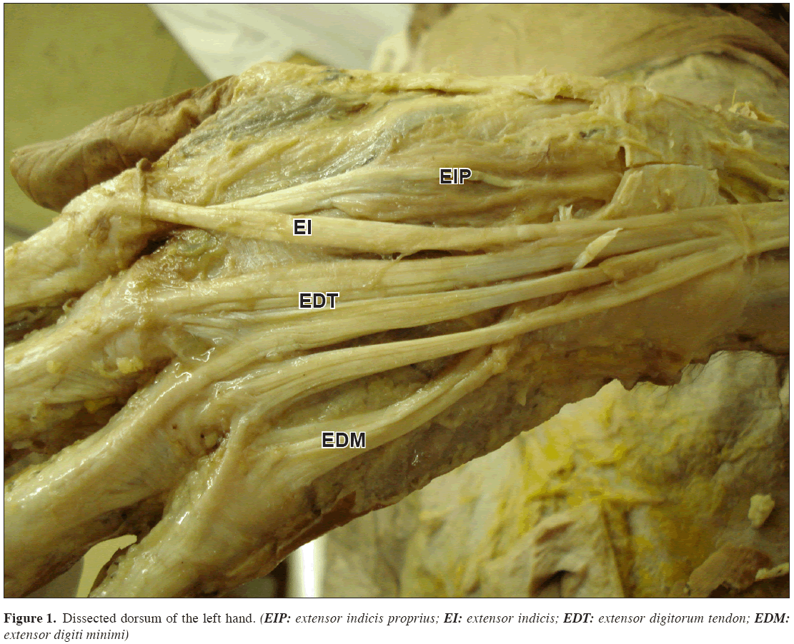
While performing routine dissections for undergraduate medical students in the Department of Anatomy of UWI, a variant muscle was found in the dorsum of the hand of a 78-year-old African female cadaver bilaterally. The muscle originated from the distal end of radius, wrist joint capsule, lying deep to the extensor retinaculum, and inserted on the base of the proximal phalanx of the index finger. It received its innervations and blood supply from the posterior interosseous nerve and artery (Figure 1).
Case II
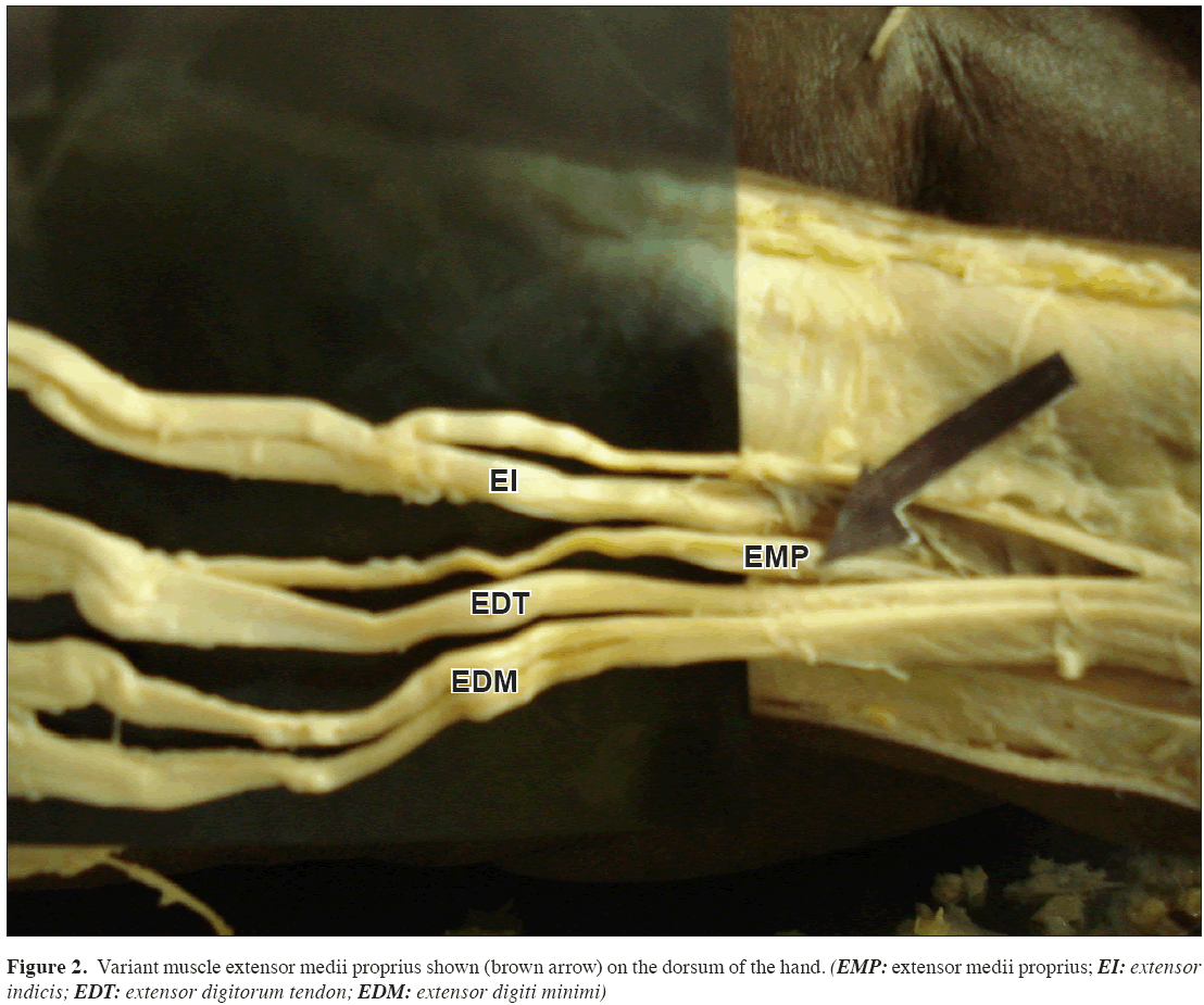
This case of additional muscle was observed in the dorsum of both hands of a 67-year-old African male cadaver. On the right side the muscle belly originated from interosseus membrane and shaft of ulna; on the left side from the dorsal surface of the carpal bones. On both sides the tendons inserted deep to the extensor digitorum tendons slips of the middle finger (Figure 2).
Discussion
The existence of this variant muscle has been reported by many authors. It originates at the joint capsule overlying the carpus and beneath the cover of the extensor retinaculum, and inserts into the extensor mechanism of the middle finger [3]. Another author describes it as having a single muscle belly, originating from the distal radius, dorsal radiocarpal ligament and the wrist joint capsule, and inserting into either the extensor hood of the second, third or both the second and the third digit [4]. In human, it represents a failure of the proximal migration of ulnacarpal elements of the antebrachial mass [5]. In amphibian, the digits are controlled only by the extensor digitorum brevis manus muscle of the forearm [2].
Often the presence of this muscle may be asymptomatic and one can live without knowing of its existence. It may be of some clinical significance, as it can cause compressive symptoms or appear like a mass [1]. Symptoms of its existence include pain and swelling of the dorsum of the hand due to heavy manual work, and at times the symptoms are only noticeable after minor injury [6]. These symptoms are often confused by clinicians as ganglion cysts or soft tissue tumors [4].
Analogous to the extensor indicis proprius, the extensor medii proprius (EMP) is a variant muscle and tendon inserts to the middle finger [7]. The EMP tendon inserts into the dorsal expansion of the middle finger, usually on the palmar and ulnar aspects of the extensor digitorum communis, and occasionally into the deep fibrous tissue proximal to the metacarpophalangeal joint [2]. The action of the muscle is extension of the proximal phalanx of the middle finger [8]. The incidence of extensor medii proprius is reported to be 0.8% to 10.4% in cadaver studies. In a study carried out by Von Schroder, an incidence of 12% of cases was reported [7]. In most cases, the muscle belly of the extensor medii proprius is separated from the extensor indicis proprius. However, in about 3.4% of cases, the extensor indicis et medii communis exist where there is one muscle belly with two split tendons inserted on the dorsal aponeurosis of the second and third fingers [5]. The common origin of the EMP and extensor indicis proprius along with occurrence of the extensor indicis medius communis suggests a common embryologic origin of these muscles. It was also suggested that the function of the two muscles become redundant as a result of the proximal migration of the distal group and the distal migration of the proximal group; thus this muscle was lost [8]. The EMP rarely gives rise to symptoms. Even during dissection, because of its small width which is very often covered by the extensor digitorum communis tendons, it is often not seen [2].
Conclusion
Even though the extensor digitorum manus brevis and extensor digitorum medii proprius are asymptomatic in most cases, authors feel it is still essential for surgeons to be aware of possible variations of the extensor tendons and to ensure these variations are not overlooked in the differential diagnosis. These variations may be asymptomatic but when requiring surgical interventions an extra care must be taken especially during routine hand surgeries. Presence of these types of variant tendons on the dorsum of the hand in persons who are involved in sports such as, golf, cricket, tennis, weight lifting were the excessive wrist movement is involved, may show some symptoms. If there is detailed knowledge of the anatomy and prevalence of this muscle, it can help to prevent diagnostic errors, influence surgical and interventional procedures and avoid surgical complications during hand surgery.
References
- Teh J, Whiteley G. MRI of soft tissue masses of the hand and wrist. Br J Radiol. 2007; 80: 47–63.
- Tan ST, Smith PJ. Anomalous extensor muscles of the hand: a review. J Hand Surg Am. 1999; 24: 449–455.
- Reef TC, Brestin SG. The extensor digitorum brevis manus and its clinical significance. J Bone Joint Surg Am. 1975; 57: 704–706.
- Ouellette H, Thomas BJ, Torriani M. Using dynamic sonography to diagnose extensor digitorum brevis manus. AJR Am J Roentgenol. 2003; 181: 1224–1226.
- Cigali BS, Kutoglu T, Cikmaz S. Musculus extensor digiti medii proprius and musculus extensor digitorum brevis manus - a case report of a rare variation. Anat Histol Embryol. 2002; 31: 126–127.
- Ross JA, Troy CA. The clinical significance of the extensor digitorum brevis manus. J Bone Joint Surg Br. 1969; 51: 473–478.
- von Schroeder HP, Botte MJ. Anatomy of the extensor tendons of the fingers: variations and multiplicity. J Hand Surg Am. 1995; 20: 27–34.
- Yalcin B, Kutoglu T, Ozan H, Gurbuz H. The extensor indicis et medii communis. Clin Anat. 2006; 19: 112–114.
Keerti Singh1*, Donnahue Dennie1, Suresh R Rao2
1Anatomy Section, Department of Basic Medical Science, University of West Indies Mona campus, Jamaica.
2Anatomy Unit, University of West Indies, St. Augustine, Trinidad.
- *Corresponding Author:
- Dr. Keerti Singh
Lecturer, Anatomy Section, Department of Basic Medical Science, University of West Indies, Mona Campus, Kingston-6, Jamaica.
Tel: +1 876 9270586
E-mail: keertisingh@hotmail.com
Date of Received: March 19th, 2009
Date of Accepted: July 10th, 2009
Published Online: August 5th, 2009
© IJAV. 2009; 2: 80–82.
Abstract
The presence of accessory muscle bellies and tendons in the hand is of great interest to the hand surgeons. Awareness of the variations of the muscles will serve as useful guide for both in studies of human anatomy and in clinical practice today; this knowledge can be borne in mind while performing hand surgeries, tendon rerouting or transplants. These extra muscles often present as a ganglion, soft tissue tumor or mass in the hand, which could be quite misleading to the surgeon. With the purpose of preparation of the teaching and museum specimens, in two of the cadavers of elderly Jamaican African male and female, we observed accessory muscles on the dorsum of the hand. One was inserted to the index finger (extensor digitorum brevis manus) and the other was arising deep to the extensor digitorum and inserted to the middle finger (extensor medii proprius).
-Keywords
hand, dorsum, accessory muscles, variation
Introduction
Usually the dorsal surface of the hand is devoid of muscle bellies, it only contains the tendons of the extensor muscles of the hand. Between the dorsal carpal ligament and the carpal bones six compartments are formed for the passage of tendons. Found on the dorsum of the hand are the tendons of the extensor pollicis longus, extensor digitorum communis, extensor indicis (extensor indicis proprius), and the extensor digiti minimi (extensor digiti quinti proprius). However, the existence of variant muscles on the dorsum of the hand has been documented in numerous publications. Some of these variant muscles include; the accessory abductor digiti minimi, found in 24 % of all wrists; the extensor digitorum brevis manus found in 1-3% of the population [1], and the extensor medii proprius which has an incidence between 0.8% and 10.4% [2].
Case Report
Case I
While performing routine dissections for undergraduate medical students in the Department of Anatomy of UWI, a variant muscle was found in the dorsum of the hand of a 78-year-old African female cadaver bilaterally. The muscle originated from the distal end of radius, wrist joint capsule, lying deep to the extensor retinaculum, and inserted on the base of the proximal phalanx of the index finger. It received its innervations and blood supply from the posterior interosseous nerve and artery (Figure 1).
Case II
This case of additional muscle was observed in the dorsum of both hands of a 67-year-old African male cadaver. On the right side the muscle belly originated from interosseus membrane and shaft of ulna; on the left side from the dorsal surface of the carpal bones. On both sides the tendons inserted deep to the extensor digitorum tendons slips of the middle finger (Figure 2).
Discussion
The existence of this variant muscle has been reported by many authors. It originates at the joint capsule overlying the carpus and beneath the cover of the extensor retinaculum, and inserts into the extensor mechanism of the middle finger [3]. Another author describes it as having a single muscle belly, originating from the distal radius, dorsal radiocarpal ligament and the wrist joint capsule, and inserting into either the extensor hood of the second, third or both the second and the third digit [4]. In human, it represents a failure of the proximal migration of ulnacarpal elements of the antebrachial mass [5]. In amphibian, the digits are controlled only by the extensor digitorum brevis manus muscle of the forearm [2].
Often the presence of this muscle may be asymptomatic and one can live without knowing of its existence. It may be of some clinical significance, as it can cause compressive symptoms or appear like a mass [1]. Symptoms of its existence include pain and swelling of the dorsum of the hand due to heavy manual work, and at times the symptoms are only noticeable after minor injury [6]. These symptoms are often confused by clinicians as ganglion cysts or soft tissue tumors [4].
Analogous to the extensor indicis proprius, the extensor medii proprius (EMP) is a variant muscle and tendon inserts to the middle finger [7]. The EMP tendon inserts into the dorsal expansion of the middle finger, usually on the palmar and ulnar aspects of the extensor digitorum communis, and occasionally into the deep fibrous tissue proximal to the metacarpophalangeal joint [2]. The action of the muscle is extension of the proximal phalanx of the middle finger [8]. The incidence of extensor medii proprius is reported to be 0.8% to 10.4% in cadaver studies. In a study carried out by Von Schroder, an incidence of 12% of cases was reported [7]. In most cases, the muscle belly of the extensor medii proprius is separated from the extensor indicis proprius. However, in about 3.4% of cases, the extensor indicis et medii communis exist where there is one muscle belly with two split tendons inserted on the dorsal aponeurosis of the second and third fingers [5]. The common origin of the EMP and extensor indicis proprius along with occurrence of the extensor indicis medius communis suggests a common embryologic origin of these muscles. It was also suggested that the function of the two muscles become redundant as a result of the proximal migration of the distal group and the distal migration of the proximal group; thus this muscle was lost [8]. The EMP rarely gives rise to symptoms. Even during dissection, because of its small width which is very often covered by the extensor digitorum communis tendons, it is often not seen [2].
Conclusion
Even though the extensor digitorum manus brevis and extensor digitorum medii proprius are asymptomatic in most cases, authors feel it is still essential for surgeons to be aware of possible variations of the extensor tendons and to ensure these variations are not overlooked in the differential diagnosis. These variations may be asymptomatic but when requiring surgical interventions an extra care must be taken especially during routine hand surgeries. Presence of these types of variant tendons on the dorsum of the hand in persons who are involved in sports such as, golf, cricket, tennis, weight lifting were the excessive wrist movement is involved, may show some symptoms. If there is detailed knowledge of the anatomy and prevalence of this muscle, it can help to prevent diagnostic errors, influence surgical and interventional procedures and avoid surgical complications during hand surgery.
References
- Teh J, Whiteley G. MRI of soft tissue masses of the hand and wrist. Br J Radiol. 2007; 80: 47–63.
- Tan ST, Smith PJ. Anomalous extensor muscles of the hand: a review. J Hand Surg Am. 1999; 24: 449–455.
- Reef TC, Brestin SG. The extensor digitorum brevis manus and its clinical significance. J Bone Joint Surg Am. 1975; 57: 704–706.
- Ouellette H, Thomas BJ, Torriani M. Using dynamic sonography to diagnose extensor digitorum brevis manus. AJR Am J Roentgenol. 2003; 181: 1224–1226.
- Cigali BS, Kutoglu T, Cikmaz S. Musculus extensor digiti medii proprius and musculus extensor digitorum brevis manus - a case report of a rare variation. Anat Histol Embryol. 2002; 31: 126–127.
- Ross JA, Troy CA. The clinical significance of the extensor digitorum brevis manus. J Bone Joint Surg Br. 1969; 51: 473–478.
- von Schroeder HP, Botte MJ. Anatomy of the extensor tendons of the fingers: variations and multiplicity. J Hand Surg Am. 1995; 20: 27–34.
- Yalcin B, Kutoglu T, Ozan H, Gurbuz H. The extensor indicis et medii communis. Clin Anat. 2006; 19: 112–114.


