Variant insertion of extensor carpi radialis longus in a South Indian cadaverwas
Raghu Jetti1*, Velayudhan Nair2, Rema Velyudhan Nair2, RV Mookambica2 and Krishnaraj Somayaji3
1Department of Anatomy, Melaka Manipal Medical College, Manipal University, Manipal, Karnataka, India
2Sri Mookambica Institute of Medical Sciences, Kulasekaram, India
3Manipal College of Dental Sciences, Manipal University, Manipal, Karnataka, India
- *Corresponding Author:
- Raghu Jetti, BPT, MSc
Department of Anatomy, Melaka Manipal Medical College (Manipal Campus) Madhava Nagar, Manipal, 576104, India
Tel: +91 820 2922642
E-mail: raghujetti@yahoo.co.in
Date of Received: November 4th, 2009
Date of Accepted: June 16th, 2010
Published Online: June 23rd, 2010
© Int J Anat Var (IJAV). 2010; 3: 86–87.
[ft_below_content] =>Keywords
antebrachial region, extensor carpi radilis longus, insertion, variation, clinical implications
Introduction
The extensor carpi radialis longus (ECRL) muscle originates from the distal third of the lateral supracondylar ridge of the humerus, front of lateral intermuscular septum and from the lateral epicondyle of humerus. The tendon of ECRL along with extensor carpi radialis brevis (ECRB) passes through the second compartment of extensor retinaculum and inserts into the base of second metacarpal bone [1]. Variation in muscles of antebrachial region has been widely reported by many authors. Two classical variations reported so far are the extensor carpi radialis accessorius (ECRA) and extensor carpi radialis intermedius (ECRI). ECRA was the name given by Wood to an accessory head of carpal extensor inserting into the base of the first metacarpal bone [2,3]. ECRI also named by Wood, refers to a tendon that arises between the radial extensors of wrist inserting into the second (or) third metacarpal bone [2].
Case Report
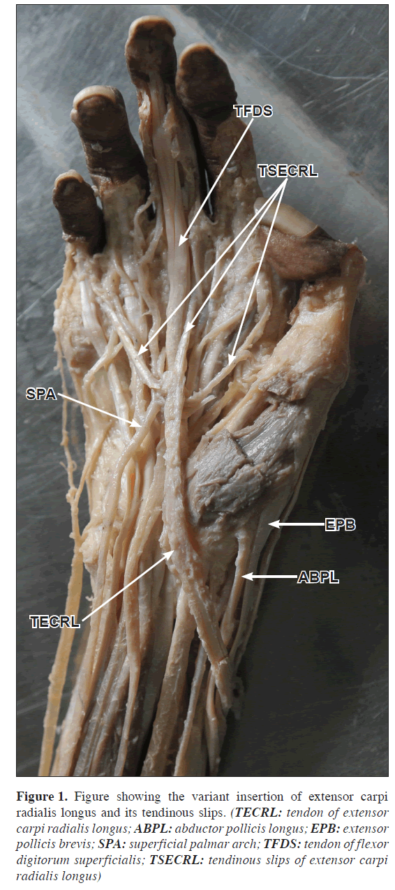
Regular dissection of right forearm of a 60-year-old male embalmed cadaver revealed a rare variation in insertion pattern of ECRL. The tendon of ECRL rounded the tendons of abductor pollicis longus and extensor pollicis brevis and coursed into the lower part of anterior forearm. Then descended anterior to the flexor retinaculum and appeared in the palm (Figure 1). It passed superficial to the superficial palmar arch, nerves and superficial flexor tendons in the palm to blend with proximal parts of digital fibrous flexor sheaths of middle, ring and little fingers. It cadaverwas supplied by radial nerve. The associated variation was the absence of palmaris longus muscle. The muscles of the opposite limb of same cadaver were as usual.
Figure 1: Figure showing the variant insertion of extensor carpi radialis longus and its tendinous slips. (TECRL: tendon of extensor carpi radialis longus; ABPL: abductor pollicis longus; EPB: extensor pollicis brevis; SPA: superficial palmar arch; TFDS: tendon of flexor digitorum superficialis; TSECRL: tendinous slips of extensor carpi radialis longus)
Discussion
Wood was the first author described the variation of radial extensor muscles [2]. MacAlister reviewed the work of past authors and described various forms of ECRI. He described the origin of ECRA as generally below the extensor carpi radialis longus, but its tendon run in the second dorsal compartment of extensor retinaculum. It usually inserts into the base of first metacarpal bone, but may insert into the outer part of abductor pollicis brevis giving a digastric appearance to it. It may also send a few fibers to first dorsal or palmar interosseus muscle or it may continue as the origin of flexor pollicis brevis [3]. Frohse and Frankel reported the ECRA tendon gave origin to the abductor pollicis brevis and passed in its own dorsal tunnel under extensor retinaculum inserted into the first metacarpophalangeal joint [4]. Classen and Wree reported the similar kind of variation of ECRA inserting into the first metacarpal bone [5]. Khaledpour and Schindelmeiser reported bilateral additional wrist extensors that were located between the extensor carpi radialis longus and brevis [6]. Hong and Hong observed two additional muscles in the lateral part of forearm [7]. Melling et al. reported an additional muscle in the forearm; which was branched from extensor carpi radialis brevis, its tendon crossed over the extensor retinaculum and inserted into the dorsal digital expansion of index finger [8]. Bergmann et al. described that rarely the extensor muscle tendons can be divided into two or three slips before their insertion [9]. Nyssen- Behets et al. reported that the tendon of extensor carpi radialis brevis could be split into two parts [10]. But in our present case there were no additional wrist extensors; neither extensor carpi radialis intermedius nor extensor carpi radialis accessorius was present. The origin, course of the ECRL was normal but there was a variation in the mode of insertion. ECRL crossed the tendons of abductor pollicis longus and extensor pollicis brevis to enter the flexor compartment of forearm blending with the digital fibrous flexor sheaths of middle, ring and little fingers.
This variant insertion may alter action of the ECRL muscle. According to classical description ECRL and ECRB are the chief extensors of wrist. It is also responsible for the abduction of wrist along with flexor carpi radialis. Out of the two radial extensors the ECRL is more powerful. Since its usual insertion is altered it may limit the abduction and extension of wrist. In addition to being an extensor muscle, based on its insertion it may assist in flexion of wrist and the medial three fingers. This abnormal tendon in palm may confuse radiologists in assessing MRI, CT scan. This may confuse anatomists during surface marking. The surgeons should be aware of this kind of variation during planning for flexor retinaculum release, tendon transfer procedures.
References
- Williams PL, ed. Gray’s Anatomy. 38th Ed., Edinburgh, Churchill Livingstone. 1995; 849.
- Wood VE. The extensor carpi radialis intermedius tendon. J Hand Surg Am. 1988; 13: 242–245.
- Macalister A. Additional observations on muscular anomalies in human anatomy with a catalogue of the principal muscular variations hitherto published (third series). Trans Roy Ir Acad. 1871; 25: 101–102.
- Frohse F, Frankel M. Die muskelen des menschlichen Armes. In: Bardeleban, Kv. (Ed.), Handbuchs der Anatomie des Menschen. Jeena, Fischer. 1908; 160–161.
- Claassen H, Wree A. Multiple variations in the region of Mm. extensores carpi radialis longus and brevis. Ann Anat. 2002; 184: 489–491.
- Khaledpour C, Schindelmeister J. Atypical course of the rare accessory extensor carpi radialis muscle. J Anat. 1994; 184: 161–163.
- Hong MK, Hong MK. An uncommon form of the rare extensor carpi radialis accessorius. Ann Anat. 2005; 187: 89–92.
- Melling M, Steindl M, Wilde J, Karimian-Teherani D. An anatomical variant of the extensor carpi radialis brevis muscle. Wien Klin Wochenschr. 2001; 113: 960–963.
- Bergman R, Thompson SA, Afifi AK, Saadeh FA. Compendium of human anatomic variation. Baltimore - Munich, Urban & Schwarzenberg. 1988; 15.
- Nyssen-Behets C, Lengele B, Dhem A. [Anatomical variations at the level of the muscles of the arm]. Arch Anat Histol Embryol. 1986; 69: 111–117. (French)
Raghu Jetti1*, Velayudhan Nair2, Rema Velyudhan Nair2, RV Mookambica2 and Krishnaraj Somayaji3
1Department of Anatomy, Melaka Manipal Medical College, Manipal University, Manipal, Karnataka, India
2Sri Mookambica Institute of Medical Sciences, Kulasekaram, India
3Manipal College of Dental Sciences, Manipal University, Manipal, Karnataka, India
- *Corresponding Author:
- Raghu Jetti, BPT, MSc
Department of Anatomy, Melaka Manipal Medical College (Manipal Campus) Madhava Nagar, Manipal, 576104, India
Tel: +91 820 2922642
E-mail: raghujetti@yahoo.co.in
Date of Received: November 4th, 2009
Date of Accepted: June 16th, 2010
Published Online: June 23rd, 2010
© Int J Anat Var (IJAV). 2010; 3: 86–87.
Abstract
Knowledge of muscular variations in the antebrachial region is clinically significant in certain operative procedures like tendon transfer, correction of hand deformities. Extensor carpi radialis accessorius and extensor carpi radialis intermedius were reported variations of extensor region. We present a rare variation of extensor carpi radialis longus, which was inserted into the fibrous flexor sheath of middle, ring, and little fingers on the palmar aspect. The present variation will influence the biomechanics of wrist joint.
-Keywords
antebrachial region, extensor carpi radilis longus, insertion, variation, clinical implications
Introduction
The extensor carpi radialis longus (ECRL) muscle originates from the distal third of the lateral supracondylar ridge of the humerus, front of lateral intermuscular septum and from the lateral epicondyle of humerus. The tendon of ECRL along with extensor carpi radialis brevis (ECRB) passes through the second compartment of extensor retinaculum and inserts into the base of second metacarpal bone [1]. Variation in muscles of antebrachial region has been widely reported by many authors. Two classical variations reported so far are the extensor carpi radialis accessorius (ECRA) and extensor carpi radialis intermedius (ECRI). ECRA was the name given by Wood to an accessory head of carpal extensor inserting into the base of the first metacarpal bone [2,3]. ECRI also named by Wood, refers to a tendon that arises between the radial extensors of wrist inserting into the second (or) third metacarpal bone [2].
Case Report
Regular dissection of right forearm of a 60-year-old male embalmed cadaver revealed a rare variation in insertion pattern of ECRL. The tendon of ECRL rounded the tendons of abductor pollicis longus and extensor pollicis brevis and coursed into the lower part of anterior forearm. Then descended anterior to the flexor retinaculum and appeared in the palm (Figure 1). It passed superficial to the superficial palmar arch, nerves and superficial flexor tendons in the palm to blend with proximal parts of digital fibrous flexor sheaths of middle, ring and little fingers. It cadaverwas supplied by radial nerve. The associated variation was the absence of palmaris longus muscle. The muscles of the opposite limb of same cadaver were as usual.
Figure 1: Figure showing the variant insertion of extensor carpi radialis longus and its tendinous slips. (TECRL: tendon of extensor carpi radialis longus; ABPL: abductor pollicis longus; EPB: extensor pollicis brevis; SPA: superficial palmar arch; TFDS: tendon of flexor digitorum superficialis; TSECRL: tendinous slips of extensor carpi radialis longus)
Discussion
Wood was the first author described the variation of radial extensor muscles [2]. MacAlister reviewed the work of past authors and described various forms of ECRI. He described the origin of ECRA as generally below the extensor carpi radialis longus, but its tendon run in the second dorsal compartment of extensor retinaculum. It usually inserts into the base of first metacarpal bone, but may insert into the outer part of abductor pollicis brevis giving a digastric appearance to it. It may also send a few fibers to first dorsal or palmar interosseus muscle or it may continue as the origin of flexor pollicis brevis [3]. Frohse and Frankel reported the ECRA tendon gave origin to the abductor pollicis brevis and passed in its own dorsal tunnel under extensor retinaculum inserted into the first metacarpophalangeal joint [4]. Classen and Wree reported the similar kind of variation of ECRA inserting into the first metacarpal bone [5]. Khaledpour and Schindelmeiser reported bilateral additional wrist extensors that were located between the extensor carpi radialis longus and brevis [6]. Hong and Hong observed two additional muscles in the lateral part of forearm [7]. Melling et al. reported an additional muscle in the forearm; which was branched from extensor carpi radialis brevis, its tendon crossed over the extensor retinaculum and inserted into the dorsal digital expansion of index finger [8]. Bergmann et al. described that rarely the extensor muscle tendons can be divided into two or three slips before their insertion [9]. Nyssen- Behets et al. reported that the tendon of extensor carpi radialis brevis could be split into two parts [10]. But in our present case there were no additional wrist extensors; neither extensor carpi radialis intermedius nor extensor carpi radialis accessorius was present. The origin, course of the ECRL was normal but there was a variation in the mode of insertion. ECRL crossed the tendons of abductor pollicis longus and extensor pollicis brevis to enter the flexor compartment of forearm blending with the digital fibrous flexor sheaths of middle, ring and little fingers.
This variant insertion may alter action of the ECRL muscle. According to classical description ECRL and ECRB are the chief extensors of wrist. It is also responsible for the abduction of wrist along with flexor carpi radialis. Out of the two radial extensors the ECRL is more powerful. Since its usual insertion is altered it may limit the abduction and extension of wrist. In addition to being an extensor muscle, based on its insertion it may assist in flexion of wrist and the medial three fingers. This abnormal tendon in palm may confuse radiologists in assessing MRI, CT scan. This may confuse anatomists during surface marking. The surgeons should be aware of this kind of variation during planning for flexor retinaculum release, tendon transfer procedures.
References
- Williams PL, ed. Gray’s Anatomy. 38th Ed., Edinburgh, Churchill Livingstone. 1995; 849.
- Wood VE. The extensor carpi radialis intermedius tendon. J Hand Surg Am. 1988; 13: 242–245.
- Macalister A. Additional observations on muscular anomalies in human anatomy with a catalogue of the principal muscular variations hitherto published (third series). Trans Roy Ir Acad. 1871; 25: 101–102.
- Frohse F, Frankel M. Die muskelen des menschlichen Armes. In: Bardeleban, Kv. (Ed.), Handbuchs der Anatomie des Menschen. Jeena, Fischer. 1908; 160–161.
- Claassen H, Wree A. Multiple variations in the region of Mm. extensores carpi radialis longus and brevis. Ann Anat. 2002; 184: 489–491.
- Khaledpour C, Schindelmeister J. Atypical course of the rare accessory extensor carpi radialis muscle. J Anat. 1994; 184: 161–163.
- Hong MK, Hong MK. An uncommon form of the rare extensor carpi radialis accessorius. Ann Anat. 2005; 187: 89–92.
- Melling M, Steindl M, Wilde J, Karimian-Teherani D. An anatomical variant of the extensor carpi radialis brevis muscle. Wien Klin Wochenschr. 2001; 113: 960–963.
- Bergman R, Thompson SA, Afifi AK, Saadeh FA. Compendium of human anatomic variation. Baltimore - Munich, Urban & Schwarzenberg. 1988; 15.
- Nyssen-Behets C, Lengele B, Dhem A. [Anatomical variations at the level of the muscles of the arm]. Arch Anat Histol Embryol. 1986; 69: 111–117. (French)


