Variant muscle to the little finger originating from the flexor digitorum superficialis Introduction Flexor
Nurcan Imre*,Selda Yildiz and Necdet Kocabiyik
Department of Anatomy, Faculty of Medicine, Gulhane Military Medical Academy, Ankara, TURKEY.
- *Corresponding Author:
- Nurcan Imre, MD
Department of Anatomy Faculty of Medicine Gulhane Military Medical Academy 06010 Etlik Ankara, Turkey
Tel: +90 (312) 304 3503
E-mail: nercikti@gata.edu.tr
Date of Received: March 12th, 2014
Date of Accepted: February 20th, 2015
Published Online: December 20th, 2015
© Int J Anat Var (IJAV). 2015; 8: 34–36.
[ft_below_content] =>Keywords
unusual muscle,compression,flexor digitorum superficialis,forearm,variation
Introduction
Flexor digitorum superficialis (FDS) is the muscle which responsible from the skill actions of the fingers. It has three heads including humeral, ulnar and radial; generally the humeral and ulnar heads are fused together and called as humeroulnar head. The muscle fibers reach the other fingers except the thumb. Each of the muscle beams divides into two parts at the level of the 1st phalanx, and they end at the lateral sides of the middle parts of the middle phalanx. It is innervated by the median nerve. In case of the function loss of this muscle, 2nd phalanx can not make flexion against force. Variations of the muscles of the anterior forearm are common [1,2,3,4,5,6]. The clinicians and the anatomist should know that the variations of FDS are more common than the other antebrachial muscles [7]. Clinical [1] and anatomical [8] studies have revealed variations of the portion of the FDS to the little finger (FDS -V) where the muscle belly is fused with that of the belly to the ring (fourth) finger or lacks a tendon. Even if most variations of FDS have been reported, some details might have been missed. Because of the importance of these anatomic details, we reported this case. Knowledge of such variations may be important during the hand surgery and in the preoperative diagnosis related to the muscles in the front side of the forearm and the tendons passing through the carpal tunnel.
Case Report
During an educational dissection of the front side of the right arm of a 78-year-old male cadaver, we observed an unusual muscle which originated from the muscle belly of FDS to the fingers 2 and 3, and inserted to the middle phalanx of the little finger.
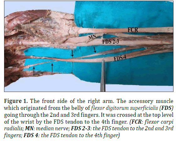
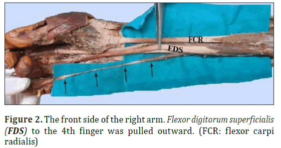
The upper limbs were carefully examined to ensure that they showed no signs of trauma, deformities, tumors or significant weight loss. The main tendon of FDS divided into three tendons. The FDS tendon to the little finger was absent. Instead, there was an unusual muscle to the little finger. The belly of the variant muscle was 4.9 mm in width and 48.8 mm in length. The total length of the tendon was 130 mm. While the first part of 83.5 mm of the tendon was in the shape of the thin slip (its width 1.3 mm); it continued as a thicker structure whose width was 4.4 mm at the metacarpophalangeal joint. The tendon was bifurcated in its synovial sheath, substituted the tendon of FDS to the little finger. It was crossed at the top of the level of the wrist by the FDS tendon to the 4th finger (Figure 1). The origin of the variant muscle (from the FDS belly to the 2nd and 3rd fingers) to the radiocarpal joint was 82.7 mm (Figure 2). In the left forearm, the FDS sent a thin tendon to the little finger with no unusual muscle belly. This variant muscle was innervated by the median nerve.
Figure 1: The front side of the right arm. The accessory muscle which originated from the belly of flexor digitorum superficialis (FDS) going through the 2nd and 3rd fingers. It was crossed at the top level of the wrist by the FDS tendon to the 4th finger. (FCR: flexor carpi radialis; MN: median nerve; FDS 2-3: the FDS tendon to the 2nd and 3rd fingers; FDS 4: the FDS tendon to the 4th finger)
Discussion
FDS has generally two heads as humeroulnar and radial. Median nerve and ulnar artery pass between these heads. The main tendon of the muscle divides into four tendons. Each of the tendons divides into two, over the proximal phalanx (hiatus tendineus) and it insert to both sides of the middle phalanx of 2nd-5th fingers. The tendon of flexor digitorum profundus passes through hiatus tendineus and inserts to the base of the distal phalanx. Standring et al. reported that the radial head or the tendon to little finger might be absent [9]. And they stated that some fibers originating from ulnar tuberosity would lie on the middle and ring fingers by participating in superficial part [9]. However, as seen in our case, it was not mentioned about the unusual muscle which inserted to the middle phalanx of the little finger, and which started from the FDS portion going to the 2nd and 3rd fingers. Gray’s Anatomy has the following description for FDS “the part of the muscle for the fifth digit may be absent and replaced by a separate slip arising from the ulna, flexor retinaculum, or palmar fascia” [10]. Yamada proposed that FDS would have a single origin or a pair of origins during the development of the muscle belly [11]. The variation of FDS related to the little finger is referred to as “brevis type”. Wesser et al. also reported the similar variation [12]. The observations account for the portions of FDS that go to the little and index fingers. Because the portions of FDS remained in the original localizations defined as “brevis type” without immigrating to the forearm. For this reason, these reports support the pair origin hypothesis in FDS development. Tan et al. studied on 500 cases in order to explain the different variations of FDS [4]. They noted that FDS function is independently at most in the ring and middle fingers, less in the index finger and at least in the little finger. Therefore, it is difficult to understand the functional loss of FDS in a single finger. Gonzales et al. studied the variations of FDS related to the little finger in 70 cadavers [8]. They reported the prevalence of variations as 13% of the hands, and the variations interfering with the function of little finger were seen in 10% of the hands. Gonzales et al. measured the distance from the side where tendon crossed the metacarpophalangeal joint to the insertion and argued that this distance was independent from the length of the phalanx [8]. Austin et al. suggested the clinic manifestation of the variations of FDS in 50 cases by using the standard and modified superficial flexion test [1]. They dissected 40 hands having the consistent anatomical variations with the clinical findings. Austin et al. found out that FDS did not have independent pattern in 58%, combined pattern in 21% and any pattern in 21% [1]. FDS were asymmetrically arranged on the right and left side in the 26% of the cases. FDS were present on the palmar side and the fingers in all cadaver hands. The variation in FDS function may be explained by both the interconnections between flexor digitorum profundus of the little finger and FDS of the ring finger. Absence of FDS function in the little finger is a relatively common congenital anomaly that can complicate assessment of little finger injuries [5]. Absence of the FDS-V tendon was reported in around 2% of Japanese cadavers [7]. Thompson et al. examined the incidence of the absent palmaris longus and FDS function related to the little finger in 300 cases [13]. They evaluated FDS with the standard and modified tests, and palmaris longus absence with the clinical observations. Modified tests demonstrated that FDS of the little finger was missing in 10/300 cases. They reported unilateral absence of palmaris longus in 49/300 and bilateral absence in 26/300 cases. They reported that there was no correlation between the absence of little finger FDS and the absence of palmaris longus [13]. Townley et al. studied on the prevalence of unilateral and bilateral absence of FDS [5]. They reported these variants as unilateral in 92 and bilateral in 81 of 1352 cases. If there is no FDS function in the little finger of a hand, probably the proportion of this absence on the contralateral hand has been detected as 0.64. If there is FDS function, probably the proportion of this absence of FDS on contralateral hand little finger has been detected as 0.02. Therefore, they emphasized that the little finger FDS absence was independent from that of the contralateral hand [5].
D’Costa et al. stated that FDS had normal origin and insertion except index finger in their cases [14]. The unusual muscle that they observed, started with the tendon of FDS coursing in the carpal tunnel and attached to the middle phalanx. This unusual muscle was innervated by median nerve. They emphasized that such unusual muscles should be considered in the etiology of carpal tunnel syndrome. Ciftcioglu et al. pointed out that the variant muscles had embryological tumoral source [15]. Therefore, they emphasized that such muscles should be analyzed clinically and anatomically in a good manner. Koizumi et al. observed unusual lumbrical muscle originated from the forearm in both arms of one cadaver. The muscle started with the FDS tendon to index finger. By passing from the carpal tunnel, it ended in 1st lumbrical muscle [16]. Kostakoglu et al. reported an unusual muscle starting with FDS tendon and combining with index finger by the accessory tendon [2]. Shoja et al. reported that there was a split which had belly portion and two different fusiform shape in the deep part of FDS [6]. They stated that medial portion started with a common flexor tendon from the medial epicondyle of humerus, and that held to the little finger by taking place of a thin tendon in the middle of the forearm. They noted that lateral portion combined with the deep side of the superficial portion of FDS and that it ended in the second finger with a thicker tendon. Kobayashi et al. mentioned unusual FDS on the right hand of a woman [17]. They reported that this muscle started with the front side of flexor retinaculum and ended on the palmar side of the hand by attaching to the middle phalanx of the little finger. And they noted that usual FDS did not have a tendon that goes to the little finger.
It is not surprising that FDS shows more variations than the other muscles on the front side of the forearm. Previously, it was mentioned that FDS would not have the radial head or the portion inserting to the little finger. And it was reported that some fibers starting with ulnar tuberosity joined in the superficial portion and would lie on the middle and ring fingers. There are some studies proposing that there are accessory muscles which go to the index finger by taking apart from FDS tendon and even there is a correlation between these accessory muscles and lumbrical muscles. However, no similar case has been reported as a muscle starting with the FDS portion which goes to the 2nd and 3rd fingers and inserting to the middle phalanx of little finger, as in our case.
Conclusion
The knowledge of these muscle variants is important in identifying the site of compression in neuropathies and in planning the appropriate treatment. Additionally, being aware of such variations and having adequate anatomical knowledge on the muscles of carpal region and flexor portion of forearm may be important in preoperative diagnosis, as well as during hand surgery. Therefore, for further studies on the the prevalence of unusual muscles of forearm and wrist in pediatric and adult series is essential to better understand the developmental patterns of this variant muscles.
Conflict of Interest
The authors declare that they have no conflict of interest.
References
- Austin GJ, Leslie BM, Ruby LK. Variations of the flexor digitorum superficialis of the small finger. J Hand Surg Am. 1989; 14: 262–267.
- Kostakoglu N, Borman H, Kecik A. Anomalous flexor digitorum superficialis muscle belly: an unusual case of mass in the palm. Br J Plast Surg. 1997; 50: 654-656.
- Lillmars SA, Bush DC. Flexor tendon rupture associated with an anomalous muscle. J Hand Surg Am. 1988; 13: 115–119.
- Tan JS, Oh L, Louis DS. Variations of the flexor digitorum superficialis as determined by an expanded clinical examination. J Hand Surg Am. 2009; 34: 900–906.
- Townley WA, Swan MC, Dunn RL. Congenital absence of flexor digitorum superficialis:implications for assessment of little finger lacerations. J Hand Surg Eur. 2010; 35: 417–418.
- Shoja MM, Tubbs RS, Loukas M, Shokouhi G. The split flexor digitorum superficialis. Ital J Anat Embryol. 2008; 113: 103–107.
- Ohtani O. Structure of the flexor digitorum superficialis. Okajimas Folia Anat Jpn. 1979; 56:277–288.
- Gonzalez MH, Whittum J, Kogan M, Weinzweig N. Variations of the flexor digitorum superficialis tendon of the little finger. J Hand Surg Br. 1997; 22: 277–280.
- Standring S, ed. Gray’s Anatomy. 40th Ed. Edinburg, Churchill Livingstone. 2008; 846.
- Williams PL, Bannister LH, Berry MM, Dyson M, Dussek JE, Ferguson MWJ, eds. Gray’s Anatomy. 38th Ed. Edinburgh, Churchill Livingstone. 1995; 847.
- Yamada TK. Re-evaluation of the flexor digitorum superficialis. Kaibogaku Zasshi. 1986; 61:283–298. (Japanese)
- Wesser DR, Calostypis F, Hoffman S. The evolutionary significance of an aberrant flexor superficialis muscle in the human palm. J Bone Joint Surg Am. 1969; 51: 396–398.
- Thompson NW, Mockford BJ, Rasheed T, Herbert KJ. Functional absence of flexor digitorum superficialis to the little finger and absence of palmaris longus-is there a link? J Hand Surg Br. 2002; 27: 433–434.
- D’Costa S, Jiji, Nayak SR, Sivanadan R, Abhishek. Anomalous muscle belly to the index finger.Ann Anat. 2006; 188: 473–475.
- Ciftcioglu E, Kopuz C, Corumlu U, Demir MT. Accessory muscle in the forearm: a clinical and embryological approach. Anat Cell Biol. 2011; 44: 160–163.
- Koizumi M, Kawai K, Honma S, Kodama K. Anomalous lumbrical muscles arising from the deep surface of flexor digitorum superficialis muscles in man. Ann Anat. 2002; 184:387–392.
- Kobayashi N, Saito S, Wakisaka H, Matsuda S. Anomalous flexor of the little finger. Clin Anat. 2003; 16: 40–43.
Nurcan Imre*,Selda Yildiz and Necdet Kocabiyik
Department of Anatomy, Faculty of Medicine, Gulhane Military Medical Academy, Ankara, TURKEY.
- *Corresponding Author:
- Nurcan Imre, MD
Department of Anatomy Faculty of Medicine Gulhane Military Medical Academy 06010 Etlik Ankara, Turkey
Tel: +90 (312) 304 3503
E-mail: nercikti@gata.edu.tr
Date of Received: March 12th, 2014
Date of Accepted: February 20th, 2015
Published Online: December 20th, 2015
© Int J Anat Var (IJAV). 2015; 8: 34–36.
Abstract
Flexor digitorum superficialis (FDS) is a muscle which exists on the front side of the forearm. The main tendon of the muscle divides into four tendons. It inserts to both sides of the middle phalanges of fingers 2 to 5. It provides rapid and strong flexion to fingers 2-5. Here we report findings of an unusual variant of FDS in a 78-year-old male cadaver, observed during educational dissection for the medical students. The main tendon of FDS divided into three tendons. The FDS tendon to the little finger was absent. The unusual muscle belly was 4.9 mm in width and 48.8 mm in length. The total length of the tendon was 130 mm. While the first part of 83.5 mm of the tendon from the (width 1.3 mm) was like a slip, its distal part from the metacarpophalangeal joint continued as a thick structure of 4.4 mm. It was crossed at the top of the level of the wrist by the FDS tendon to the 4th finger. The distance between the origin of this variant muscle originating from the belly of FDS for the 2nd and 3rd fingers to the radiocarpal joint was 82.7 mm. The knowledge of such muscle/tendon variantions is highly imperative in the management of compressive neuropathies, because of the close neighborhood of the FDS tendons with the median nerve in the wrist.
-Keywords
unusual muscle,compression,flexor digitorum superficialis,forearm,variation
Introduction
Flexor digitorum superficialis (FDS) is the muscle which responsible from the skill actions of the fingers. It has three heads including humeral, ulnar and radial; generally the humeral and ulnar heads are fused together and called as humeroulnar head. The muscle fibers reach the other fingers except the thumb. Each of the muscle beams divides into two parts at the level of the 1st phalanx, and they end at the lateral sides of the middle parts of the middle phalanx. It is innervated by the median nerve. In case of the function loss of this muscle, 2nd phalanx can not make flexion against force. Variations of the muscles of the anterior forearm are common [1,2,3,4,5,6]. The clinicians and the anatomist should know that the variations of FDS are more common than the other antebrachial muscles [7]. Clinical [1] and anatomical [8] studies have revealed variations of the portion of the FDS to the little finger (FDS -V) where the muscle belly is fused with that of the belly to the ring (fourth) finger or lacks a tendon. Even if most variations of FDS have been reported, some details might have been missed. Because of the importance of these anatomic details, we reported this case. Knowledge of such variations may be important during the hand surgery and in the preoperative diagnosis related to the muscles in the front side of the forearm and the tendons passing through the carpal tunnel.
Case Report
During an educational dissection of the front side of the right arm of a 78-year-old male cadaver, we observed an unusual muscle which originated from the muscle belly of FDS to the fingers 2 and 3, and inserted to the middle phalanx of the little finger.
The upper limbs were carefully examined to ensure that they showed no signs of trauma, deformities, tumors or significant weight loss. The main tendon of FDS divided into three tendons. The FDS tendon to the little finger was absent. Instead, there was an unusual muscle to the little finger. The belly of the variant muscle was 4.9 mm in width and 48.8 mm in length. The total length of the tendon was 130 mm. While the first part of 83.5 mm of the tendon was in the shape of the thin slip (its width 1.3 mm); it continued as a thicker structure whose width was 4.4 mm at the metacarpophalangeal joint. The tendon was bifurcated in its synovial sheath, substituted the tendon of FDS to the little finger. It was crossed at the top of the level of the wrist by the FDS tendon to the 4th finger (Figure 1). The origin of the variant muscle (from the FDS belly to the 2nd and 3rd fingers) to the radiocarpal joint was 82.7 mm (Figure 2). In the left forearm, the FDS sent a thin tendon to the little finger with no unusual muscle belly. This variant muscle was innervated by the median nerve.
Figure 1: The front side of the right arm. The accessory muscle which originated from the belly of flexor digitorum superficialis (FDS) going through the 2nd and 3rd fingers. It was crossed at the top level of the wrist by the FDS tendon to the 4th finger. (FCR: flexor carpi radialis; MN: median nerve; FDS 2-3: the FDS tendon to the 2nd and 3rd fingers; FDS 4: the FDS tendon to the 4th finger)
Discussion
FDS has generally two heads as humeroulnar and radial. Median nerve and ulnar artery pass between these heads. The main tendon of the muscle divides into four tendons. Each of the tendons divides into two, over the proximal phalanx (hiatus tendineus) and it insert to both sides of the middle phalanx of 2nd-5th fingers. The tendon of flexor digitorum profundus passes through hiatus tendineus and inserts to the base of the distal phalanx. Standring et al. reported that the radial head or the tendon to little finger might be absent [9]. And they stated that some fibers originating from ulnar tuberosity would lie on the middle and ring fingers by participating in superficial part [9]. However, as seen in our case, it was not mentioned about the unusual muscle which inserted to the middle phalanx of the little finger, and which started from the FDS portion going to the 2nd and 3rd fingers. Gray’s Anatomy has the following description for FDS “the part of the muscle for the fifth digit may be absent and replaced by a separate slip arising from the ulna, flexor retinaculum, or palmar fascia” [10]. Yamada proposed that FDS would have a single origin or a pair of origins during the development of the muscle belly [11]. The variation of FDS related to the little finger is referred to as “brevis type”. Wesser et al. also reported the similar variation [12]. The observations account for the portions of FDS that go to the little and index fingers. Because the portions of FDS remained in the original localizations defined as “brevis type” without immigrating to the forearm. For this reason, these reports support the pair origin hypothesis in FDS development. Tan et al. studied on 500 cases in order to explain the different variations of FDS [4]. They noted that FDS function is independently at most in the ring and middle fingers, less in the index finger and at least in the little finger. Therefore, it is difficult to understand the functional loss of FDS in a single finger. Gonzales et al. studied the variations of FDS related to the little finger in 70 cadavers [8]. They reported the prevalence of variations as 13% of the hands, and the variations interfering with the function of little finger were seen in 10% of the hands. Gonzales et al. measured the distance from the side where tendon crossed the metacarpophalangeal joint to the insertion and argued that this distance was independent from the length of the phalanx [8]. Austin et al. suggested the clinic manifestation of the variations of FDS in 50 cases by using the standard and modified superficial flexion test [1]. They dissected 40 hands having the consistent anatomical variations with the clinical findings. Austin et al. found out that FDS did not have independent pattern in 58%, combined pattern in 21% and any pattern in 21% [1]. FDS were asymmetrically arranged on the right and left side in the 26% of the cases. FDS were present on the palmar side and the fingers in all cadaver hands. The variation in FDS function may be explained by both the interconnections between flexor digitorum profundus of the little finger and FDS of the ring finger. Absence of FDS function in the little finger is a relatively common congenital anomaly that can complicate assessment of little finger injuries [5]. Absence of the FDS-V tendon was reported in around 2% of Japanese cadavers [7]. Thompson et al. examined the incidence of the absent palmaris longus and FDS function related to the little finger in 300 cases [13]. They evaluated FDS with the standard and modified tests, and palmaris longus absence with the clinical observations. Modified tests demonstrated that FDS of the little finger was missing in 10/300 cases. They reported unilateral absence of palmaris longus in 49/300 and bilateral absence in 26/300 cases. They reported that there was no correlation between the absence of little finger FDS and the absence of palmaris longus [13]. Townley et al. studied on the prevalence of unilateral and bilateral absence of FDS [5]. They reported these variants as unilateral in 92 and bilateral in 81 of 1352 cases. If there is no FDS function in the little finger of a hand, probably the proportion of this absence on the contralateral hand has been detected as 0.64. If there is FDS function, probably the proportion of this absence of FDS on contralateral hand little finger has been detected as 0.02. Therefore, they emphasized that the little finger FDS absence was independent from that of the contralateral hand [5].
D’Costa et al. stated that FDS had normal origin and insertion except index finger in their cases [14]. The unusual muscle that they observed, started with the tendon of FDS coursing in the carpal tunnel and attached to the middle phalanx. This unusual muscle was innervated by median nerve. They emphasized that such unusual muscles should be considered in the etiology of carpal tunnel syndrome. Ciftcioglu et al. pointed out that the variant muscles had embryological tumoral source [15]. Therefore, they emphasized that such muscles should be analyzed clinically and anatomically in a good manner. Koizumi et al. observed unusual lumbrical muscle originated from the forearm in both arms of one cadaver. The muscle started with the FDS tendon to index finger. By passing from the carpal tunnel, it ended in 1st lumbrical muscle [16]. Kostakoglu et al. reported an unusual muscle starting with FDS tendon and combining with index finger by the accessory tendon [2]. Shoja et al. reported that there was a split which had belly portion and two different fusiform shape in the deep part of FDS [6]. They stated that medial portion started with a common flexor tendon from the medial epicondyle of humerus, and that held to the little finger by taking place of a thin tendon in the middle of the forearm. They noted that lateral portion combined with the deep side of the superficial portion of FDS and that it ended in the second finger with a thicker tendon. Kobayashi et al. mentioned unusual FDS on the right hand of a woman [17]. They reported that this muscle started with the front side of flexor retinaculum and ended on the palmar side of the hand by attaching to the middle phalanx of the little finger. And they noted that usual FDS did not have a tendon that goes to the little finger.
It is not surprising that FDS shows more variations than the other muscles on the front side of the forearm. Previously, it was mentioned that FDS would not have the radial head or the portion inserting to the little finger. And it was reported that some fibers starting with ulnar tuberosity joined in the superficial portion and would lie on the middle and ring fingers. There are some studies proposing that there are accessory muscles which go to the index finger by taking apart from FDS tendon and even there is a correlation between these accessory muscles and lumbrical muscles. However, no similar case has been reported as a muscle starting with the FDS portion which goes to the 2nd and 3rd fingers and inserting to the middle phalanx of little finger, as in our case.
Conclusion
The knowledge of these muscle variants is important in identifying the site of compression in neuropathies and in planning the appropriate treatment. Additionally, being aware of such variations and having adequate anatomical knowledge on the muscles of carpal region and flexor portion of forearm may be important in preoperative diagnosis, as well as during hand surgery. Therefore, for further studies on the the prevalence of unusual muscles of forearm and wrist in pediatric and adult series is essential to better understand the developmental patterns of this variant muscles.
Conflict of Interest
The authors declare that they have no conflict of interest.
References
- Austin GJ, Leslie BM, Ruby LK. Variations of the flexor digitorum superficialis of the small finger. J Hand Surg Am. 1989; 14: 262–267.
- Kostakoglu N, Borman H, Kecik A. Anomalous flexor digitorum superficialis muscle belly: an unusual case of mass in the palm. Br J Plast Surg. 1997; 50: 654-656.
- Lillmars SA, Bush DC. Flexor tendon rupture associated with an anomalous muscle. J Hand Surg Am. 1988; 13: 115–119.
- Tan JS, Oh L, Louis DS. Variations of the flexor digitorum superficialis as determined by an expanded clinical examination. J Hand Surg Am. 2009; 34: 900–906.
- Townley WA, Swan MC, Dunn RL. Congenital absence of flexor digitorum superficialis:implications for assessment of little finger lacerations. J Hand Surg Eur. 2010; 35: 417–418.
- Shoja MM, Tubbs RS, Loukas M, Shokouhi G. The split flexor digitorum superficialis. Ital J Anat Embryol. 2008; 113: 103–107.
- Ohtani O. Structure of the flexor digitorum superficialis. Okajimas Folia Anat Jpn. 1979; 56:277–288.
- Gonzalez MH, Whittum J, Kogan M, Weinzweig N. Variations of the flexor digitorum superficialis tendon of the little finger. J Hand Surg Br. 1997; 22: 277–280.
- Standring S, ed. Gray’s Anatomy. 40th Ed. Edinburg, Churchill Livingstone. 2008; 846.
- Williams PL, Bannister LH, Berry MM, Dyson M, Dussek JE, Ferguson MWJ, eds. Gray’s Anatomy. 38th Ed. Edinburgh, Churchill Livingstone. 1995; 847.
- Yamada TK. Re-evaluation of the flexor digitorum superficialis. Kaibogaku Zasshi. 1986; 61:283–298. (Japanese)
- Wesser DR, Calostypis F, Hoffman S. The evolutionary significance of an aberrant flexor superficialis muscle in the human palm. J Bone Joint Surg Am. 1969; 51: 396–398.
- Thompson NW, Mockford BJ, Rasheed T, Herbert KJ. Functional absence of flexor digitorum superficialis to the little finger and absence of palmaris longus-is there a link? J Hand Surg Br. 2002; 27: 433–434.
- D’Costa S, Jiji, Nayak SR, Sivanadan R, Abhishek. Anomalous muscle belly to the index finger.Ann Anat. 2006; 188: 473–475.
- Ciftcioglu E, Kopuz C, Corumlu U, Demir MT. Accessory muscle in the forearm: a clinical and embryological approach. Anat Cell Biol. 2011; 44: 160–163.
- Koizumi M, Kawai K, Honma S, Kodama K. Anomalous lumbrical muscles arising from the deep surface of flexor digitorum superficialis muscles in man. Ann Anat. 2002; 184:387–392.
- Kobayashi N, Saito S, Wakisaka H, Matsuda S. Anomalous flexor of the little finger. Clin Anat. 2003; 16: 40–43.


