Variant obturator vessels
Satheesha Nayak B.1* and Soumya KV2
1Department of Anatomy, Melaka Manipal Medical College (Manipal Campus), Madhav Nagar, Manipal, Karnataka State, India
2Department of Mathematics, KMC International Centre, Madhav Nagar, Manipal, Karnataka State, India
- *Corresponding Author:
- Dr. Satheesha Nayak B., MSc, PhD
Professor of Anatomy, Melaka Manipal Medical College (Manipal Campus), International Centre for Health Sciences, Madhav Nagar, Manipal, Udupi District, 576104, Karnataka State, India
Tel: +91 820 2922519
E-mail: nayaksathish@yahoo.com
Date of Received: May 26th, 2009
Date of Accepted: September 30th, 2009
Published Online: October 9th, 2009
© IJAV. 2009; 2: 111–112.
[ft_below_content] =>Keywords
obturator artery, obturator vein, variant, pelvic vessels
Introduction
Obturator artery is a branch of anterior division of internal iliac artery. It normally runs anteroinferiorly on the lateral wall of pelvis to the upper part of the obturator foramen and leaves the pelvis by passing through the obturator canal. On its course, the artery is accompanied by the obturator nerve and vein. It supplies the muscles of the medial compartment of the thigh. In about 20% of cases it arises from the inferior epigastric artery. In such cases it is called “abnormal obturator artery”.
Case Report
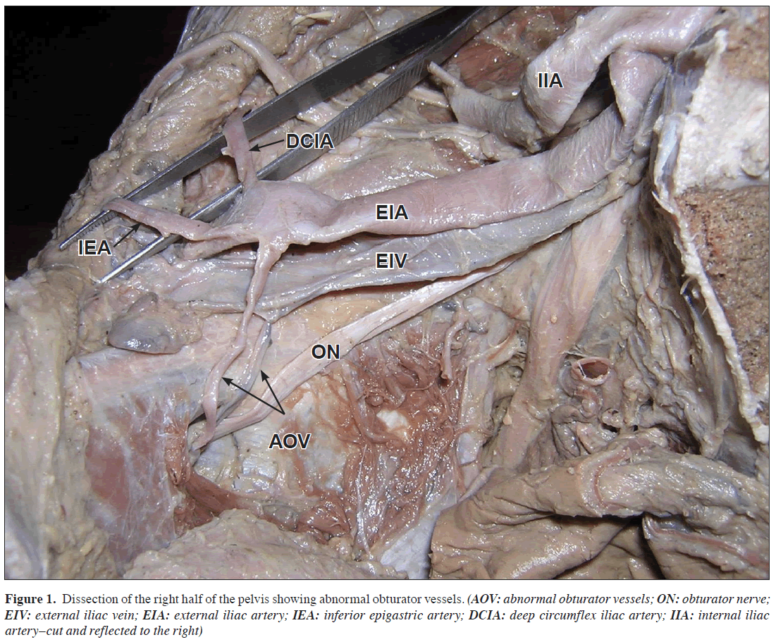
During routine dissections for the medical undergraduates at Melaka Manipal Medical College, Manipal, INDIA, we noted variant obturator vessels. The obturator artery took its origin from the external iliac artery and passed medially superficial to the external iliac vein. Then it crossed the pelvic brim and descended anteriorly into the obturator canal. The obturator vein and the nerve entered the canal below the artery (Figure 1). The external iliac artery also had the other two normal branches namely the inferior epigastric artery and deep circumflex iliac artery. The obturator vein terminated in the external iliac vein. The variations were found on the right side of an approximately 60-year-old male cadaver and they were unilateral.
Figure 1: Dissection of the right half of the pelvis showing abnormal obturator vessels. (AOV: abnormal obturator vessels; ON: obturator nerve; EIV: external iliac vein; EIA: external iliac artery; IEA: inferior epigastric artery; DCIA: deep circumflex iliac artery; IIA: internal iliac artery–cut and reflected to the right)
Discussion
Variations in the origin of obturator artery are common. The study by Bergman et al. reports its origin from common iliac or anterior division of internal iliac artery in 41.4% of cases, from inferior epigastric artery in 25% of cases, from superior gluteal artery in 10% of cases, from inferior gluteal or internal pudendal arteries in 10% of cases and from external iliac artery in 1.1% of cases [1]. In very rare cases, it may also arise from posterior division of the internal iliac artery [2].
The obturator artery develops from a plexus that is joined to the axial artery of the lower limb that accompanies the sciatic nerve [3]. In the current case, it would have probably developed from the plexus in relation to the external iliac artery.
The area of the pelvic brim and lateral pelvic wall is very important and it is the anchoring site for the repair of inguinal and femoral hernias. During surgery, the abdominal muscles are retracted laterally by applying pressure on the lateral pelvic wall. Hence a complete understanding of the anatomy in this area is very important [4].
The variant obturator vein tributary to external iliac vein is dangerous in Burch procedure, as it might bleed significantly [5].
The variant obturator artery being reported here is very important surgically as it descends down in relation to the lateral pelvic wall after crossing the external iliac artery and pelvic brim. It may cause serious complications during femoral ring procedures or laparoscopic interventions as it is a very rare variation. It may compress the external iliac vein and can result in venous stagnation in the lower limb.
References
- Bergman RA, Thompson SA, Afifi AK. Compendium of human anatomic variations. Munich, Urban and Schwarzenberg. 1988; 84.
- Kumar D, Rath G. Anomalous origin of oburator artery from the internal iliac artery. Int J Morphol. 2007; 25: 639–641.
- Sanudo JR, Roig M, Rodriguez A, Ferreira B, Domenech JM. Rare origin of the obturator, inferior epigastric and medial circumflex femoral arteries from a common trunk. J Anat. 1993; 183: 161–163.
- Gilroy AM, Hermey DC, DiBenedetto LM, Marks SC Jr, Page DW, Lei QF. Variability of the obturator vessels. Clin Anat. 1997; 10: 328–332.
- Negura A, Andreescu G, Marderos GG, Marderos GH and Margarit L. Hemorrhagic risks in the Burch procedure. Int Urogynecol J. 1993; 4: 310–313.
Satheesha Nayak B.1* and Soumya KV2
1Department of Anatomy, Melaka Manipal Medical College (Manipal Campus), Madhav Nagar, Manipal, Karnataka State, India
2Department of Mathematics, KMC International Centre, Madhav Nagar, Manipal, Karnataka State, India
- *Corresponding Author:
- Dr. Satheesha Nayak B., MSc, PhD
Professor of Anatomy, Melaka Manipal Medical College (Manipal Campus), International Centre for Health Sciences, Madhav Nagar, Manipal, Udupi District, 576104, Karnataka State, India
Tel: +91 820 2922519
E-mail: nayaksathish@yahoo.com
Date of Received: May 26th, 2009
Date of Accepted: September 30th, 2009
Published Online: October 9th, 2009
© IJAV. 2009; 2: 111–112.
Abstract
The obturator artery is a branch of anterior division of the internal iliac artery. However, in about 20% of cases it is replaced by the pubic branch of inferior epigastric artery, which is then known as abnormal obturator artery. The knowledge of abnormal obturator artery is of importance to the surgeons reducing the femoral hernia as it runs across the lacunar ligament. We saw an anomalous obturator artery arising from the external iliac artery. The obturator vein is usually a tributary of internal iliac vein. In the current case, the vein terminated in the external iliac vein. The knowledge of this variation is important anatomically, radiologically and surgically.
-Keywords
obturator artery, obturator vein, variant, pelvic vessels
Introduction
Obturator artery is a branch of anterior division of internal iliac artery. It normally runs anteroinferiorly on the lateral wall of pelvis to the upper part of the obturator foramen and leaves the pelvis by passing through the obturator canal. On its course, the artery is accompanied by the obturator nerve and vein. It supplies the muscles of the medial compartment of the thigh. In about 20% of cases it arises from the inferior epigastric artery. In such cases it is called “abnormal obturator artery”.
Case Report
During routine dissections for the medical undergraduates at Melaka Manipal Medical College, Manipal, INDIA, we noted variant obturator vessels. The obturator artery took its origin from the external iliac artery and passed medially superficial to the external iliac vein. Then it crossed the pelvic brim and descended anteriorly into the obturator canal. The obturator vein and the nerve entered the canal below the artery (Figure 1). The external iliac artery also had the other two normal branches namely the inferior epigastric artery and deep circumflex iliac artery. The obturator vein terminated in the external iliac vein. The variations were found on the right side of an approximately 60-year-old male cadaver and they were unilateral.
Figure 1: Dissection of the right half of the pelvis showing abnormal obturator vessels. (AOV: abnormal obturator vessels; ON: obturator nerve; EIV: external iliac vein; EIA: external iliac artery; IEA: inferior epigastric artery; DCIA: deep circumflex iliac artery; IIA: internal iliac artery–cut and reflected to the right)
Discussion
Variations in the origin of obturator artery are common. The study by Bergman et al. reports its origin from common iliac or anterior division of internal iliac artery in 41.4% of cases, from inferior epigastric artery in 25% of cases, from superior gluteal artery in 10% of cases, from inferior gluteal or internal pudendal arteries in 10% of cases and from external iliac artery in 1.1% of cases [1]. In very rare cases, it may also arise from posterior division of the internal iliac artery [2].
The obturator artery develops from a plexus that is joined to the axial artery of the lower limb that accompanies the sciatic nerve [3]. In the current case, it would have probably developed from the plexus in relation to the external iliac artery.
The area of the pelvic brim and lateral pelvic wall is very important and it is the anchoring site for the repair of inguinal and femoral hernias. During surgery, the abdominal muscles are retracted laterally by applying pressure on the lateral pelvic wall. Hence a complete understanding of the anatomy in this area is very important [4].
The variant obturator vein tributary to external iliac vein is dangerous in Burch procedure, as it might bleed significantly [5].
The variant obturator artery being reported here is very important surgically as it descends down in relation to the lateral pelvic wall after crossing the external iliac artery and pelvic brim. It may cause serious complications during femoral ring procedures or laparoscopic interventions as it is a very rare variation. It may compress the external iliac vein and can result in venous stagnation in the lower limb.
References
- Bergman RA, Thompson SA, Afifi AK. Compendium of human anatomic variations. Munich, Urban and Schwarzenberg. 1988; 84.
- Kumar D, Rath G. Anomalous origin of oburator artery from the internal iliac artery. Int J Morphol. 2007; 25: 639–641.
- Sanudo JR, Roig M, Rodriguez A, Ferreira B, Domenech JM. Rare origin of the obturator, inferior epigastric and medial circumflex femoral arteries from a common trunk. J Anat. 1993; 183: 161–163.
- Gilroy AM, Hermey DC, DiBenedetto LM, Marks SC Jr, Page DW, Lei QF. Variability of the obturator vessels. Clin Anat. 1997; 10: 328–332.
- Negura A, Andreescu G, Marderos GG, Marderos GH and Margarit L. Hemorrhagic risks in the Burch procedure. Int Urogynecol J. 1993; 4: 310–313.


