Variants of the cephalic arch: report of 2 cases
Russo A*, Cubas S, Mansilla A, Mansilla S and Olivera E
Anatomy Department, University of the Republic, Montevideo, Uruguay
- *Corresponding Author:
- Dr. Alejandro Russo
Anatomy Department
University of the Republic
1432 Achiras St. ZC: 11300. Montevideo, Uruguay
Tel: +5982 99 029 229
E-mail: aleru86@gmail.com
Citation: Russo A, Cubas S, Mansilla A, et al. Variants of the cephalic arch: report of 2 cases. Int J Anat Var. 2017;10(3):64-5.
Copyright: This open-access article is distributed under the terms of the Creative Commons Attribution Non-Commercial License (CC BY-NC) (http://creativecommons.org/licenses/by-nc/4.0/), which permits reuse, distribution and reproduction of the article, provided that the original work is properly cited and the reuse is restricted to noncommercial purposes. For commercial reuse, contact reprints@pulsus.com
[ft_below_content] =>Keywords
Anatomy; Cephalic vein; Cephalic arch; Abnormality
Introduction
The Cephalic Vein (CV) is part of the superficial venous system of the upper limb. It courses proximally following the radial aspect of the forearm reaching the anterior surface of the elbow where it communicates with the basilic vein by means of the antecubital vein. Later it ascends along the lateral surface of the arm to reach the deltopectoral groove. It passes beneath the clavicle and turns sharply to pierce the clavipectoral fascia and joins the axillary vein [1]. The Cephalic Arch (CA) is described as the final arch of the CV before it joins the axillary vein [2]. In addition, the CA has been defined in radiologic terms as the central perpendicular portion of the cephalic vein as it traverses the deltopectoral groove and joins the axillary vein [3].
Anatomic variants of the CA have been described. These include variations in morphology: Double (bifid) or triple arch, and variations in termination. The last include: CA that joins the subclavian, the external or even the internal jugular veins. Both morphologic and termination variants are associated with CA stenosis which may develop malfunction of vascular access in patients with arterio-venous fistulas [4]. The aim of this paper is to report 2 cases of variants of CA and briefly discuss its implications in CA stenosis.
Case Report
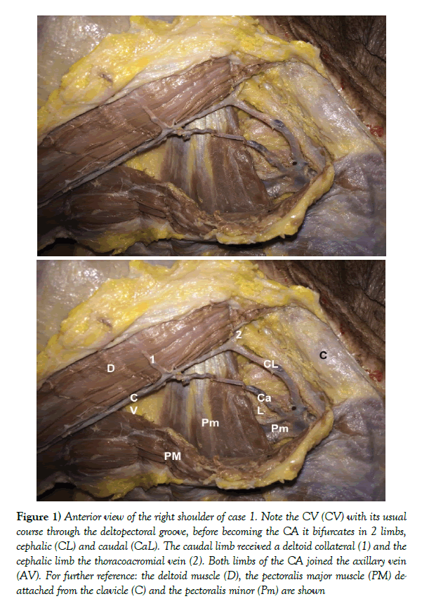
Case 1: During a routine dissection of an adult male formalin-fixed cadaver, a morphologic variant of the CA was found on the right side. The dissection was performed in the Anatomy Department, Facultad de Medicina, Universidad de la República, Montevideo, Uruguay by the authors. The right CV showed its typical pattern until it pierced the clavipectoral fascia by means of two venous channels that separately joined the axillary vein, separated by 10 mm from each other. As Figure 1 shows, it received a collateral vein from the deltoid muscle at its bifurcation, and the thoracoacromial vein joined the CA distally on the cephalic limb of the bifurcated CA. No other variants were found in the superficial venous system of the ipsilateral upper limb; the contralateral CV followed its normal course and joined the axillary vein.
Figure 1:Anterior view of the right shoulder of case 1. Note the CV (CV) with its usual course through the deltopectoral groove, before becoming the CA it bifurcates in 2 limbs, cephalic (CL) and caudal (CaL). The caudal limb received a deltoid collateral (1) and the cephalic limb the thoracoacromial vein (2). Both limbs of the CA joined the axillary vein (AV). For further reference: the deltoid muscle (D), the pectoralis major muscle (PM) deattached from the clavicle (C) and the pectoralis minor (Pm) are shown
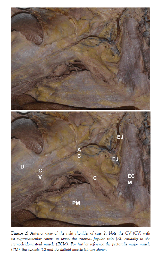
Case 2: During a routine dissection of an adult female formalin-fixed cadaver, a variant CV was found on the right side. The dissection was carried out in the Anatomy Department, Facultad de Medicina, Universidad de la República, Montevideo, Uruguay by the authors. The right CV had a common path through the forearm and elbow; however it did not course within the deltopectoral groove on the arm. As a matter of fact, the CA followed a supraclavicular course as a single conduit to reach the external jugular vein caudally to the sternocleidomastoid muscle (Figure 2). No other variants were found in the superficial venous system of the ipsilateral upper limb; the contralateral CV followed its normal course and joined the axillary vein.
Figure 2: Anterior view of the right shoulder of case 2. Note the CV (CV) with its supraclavicular course to reach the external jugular vein (EJ) caudally to the sternocleidomastoid muscle (ECM). For further reference the pectoralis major muscle (PM), the clavicle (C) and the deltoid muscle (D) are shown
Discussion and Conclusion
Typically, the CA courses underneath the clavicle, turning sharply to pierce the clavipectoral fascia and drain into the axillary vein [1]. Variants in this course as well as the surrounding structures, explain in part the development of CA stenosis, which may in turn cause malfunction of brachio-cephalic fistulas in dialysis patients [4].
Firstly, the deltopectoral fascia was noted to have a variable appearance, sometimes appearing thin or interrupted by segments of fat resembling subcutaneous tissue [5]. It has been reported that 80% of the CA were easily identified in the superficial deltopectoral triangle and the remaining 20% were located deep to or in the deltopectoral fascia [6]. Theoretically, in some patients, the fascia may prevent appropriate dilatation of CA by external compression [4]. Although there are no data to support that the angle of the CA causes CA stenosis, variations of the degree of the curvature of the CA have been reported [7].
Secondly, variants of the CA have been proposed to play a role in CA stenosis. A single channel that joins the subclavian vein is the most frequent reported variation. However, a double (bifid) arch occasionally is encountered. A bifid CA is one that bifurcates and both limbs may drain into the axillary vein, as reported in case 1, or one limb joins the axillary vein and the other joins the external or internal jugular veins. Triple circulation with complex collaterals have also been discovered [8]. Other variations reported include: a case of supraclavicular CA that drained into the subclavian vein found during permanent pacemaker implantation [9] and a cadaveric case of CA joining the external jugular vein but with an infraclavicular course [5] in contrast with case 2 of this communication. Noteworthy is a case of a patient under haemodialysis with brachio-cephalic fistulae that develop CA stenosis [10]. The angiogram showed a single conduit CA with a supraclavicular course to join the external jugular vein similar to case 2 above mentioned. The patient required several angioplasty of both CA and the external jugular vein to reach adequate fistulae flow [10].
In conclusion, we present 2 cadaveric cases of variations of CA. The proper knowledge of this entity is crucial from an anatomic stand point but also because of its relationship to angio access malfunction.
References
- Au F. The anatomy of the cephalic vein. Am Surg. 1989;55:638-9.
- Kian K, Asif A. Cephalic arch stenosis. Semin Dial. 2008;21:78-82.
- Rajan D, Clark T, Patel N, et al. Prevalence and treatment of cephalic arch stenosis in dysfunctional autogenous hemodialysis fistulas. J Vasc Interv Radiol. 2003;14:567-73.
- Sivananthan G, Mensahe L, Halin N. Cephalic arch stenosis in dialysis patients: review of clinical relevance, anatomy, current theories on etiology and management. J Vasc Access. 2014;15:157-62.
- Yeri L, Houghton E, Palmieri B, et al. Cephalic vein. Detail of its anatomy in the deltopectoral triangle. Int J Morphol. 2009;27:1037-42.
- Loukas M, Myers C, Wartmann C, et al. The clinical anatomy of the cephalic vein in the deltopectoral triangle. Folia Morphol. 2008;67:72-7.
- Mansilla S, Mansilla A, Pouy A, et al. Cephalic vein arch: Anatomy applied to vascular access. Unpublished Data. 2016.
- Bennett S, Hammens M, Blicharski T, et al. Characterization of the cephalic arch and location of stenosis. J Vasc Access. 2015;16:13-8.
- Lau E, Liew R, Harris S. An unusual case of the cephalic vein with a supraclavicular course. Pacing Clin Electrophysiol. 2007;30:719-20.
- Jun E, Lun A, Nikam M. A rare anatomic variant of a single-conduit supraclavicular cephalic arch draining into the external jugular vein presenting with recurrent arteriovenous fistula stenosis in a hemodialysis patient. J Vasc Surg Cases Innov Tech. 2017;3:20-2.
Russo A*, Cubas S, Mansilla A, Mansilla S and Olivera E
Anatomy Department, University of the Republic, Montevideo, Uruguay
- *Corresponding Author:
- Dr. Alejandro Russo
Anatomy Department
University of the Republic
1432 Achiras St. ZC: 11300. Montevideo, Uruguay
Tel: +5982 99 029 229
E-mail: aleru86@gmail.com
Citation: Russo A, Cubas S, Mansilla A, et al. Variants of the cephalic arch: report of 2 cases. Int J Anat Var. 2017;10(3):64-5.
Copyright: This open-access article is distributed under the terms of the Creative Commons Attribution Non-Commercial License (CC BY-NC) (http://creativecommons.org/licenses/by-nc/4.0/), which permits reuse, distribution and reproduction of the article, provided that the original work is properly cited and the reuse is restricted to noncommercial purposes. For commercial reuse, contact reprints@pulsus.com
Abstract
The cephalic arch is a unique anatomic structure. Multiple variants of its usual morphology and termination have been described in the literature. We present 2 cadaveric cases of variants of the cephalic arch; firstly, a morphologic variant, where the cephalic arch divided in two and joined the axillary vein, and secondly, a cephalic arch that drained directly into the external jugular vein. The variants of the cephalic arch are associated with arterio-venous fistulas malfunction, thus the proper anatomic knowledge is essential for both diagnosis and treatment of this condition.
-Keywords
Anatomy; Cephalic vein; Cephalic arch; Abnormality
Introduction
The Cephalic Vein (CV) is part of the superficial venous system of the upper limb. It courses proximally following the radial aspect of the forearm reaching the anterior surface of the elbow where it communicates with the basilic vein by means of the antecubital vein. Later it ascends along the lateral surface of the arm to reach the deltopectoral groove. It passes beneath the clavicle and turns sharply to pierce the clavipectoral fascia and joins the axillary vein [1]. The Cephalic Arch (CA) is described as the final arch of the CV before it joins the axillary vein [2]. In addition, the CA has been defined in radiologic terms as the central perpendicular portion of the cephalic vein as it traverses the deltopectoral groove and joins the axillary vein [3].
Anatomic variants of the CA have been described. These include variations in morphology: Double (bifid) or triple arch, and variations in termination. The last include: CA that joins the subclavian, the external or even the internal jugular veins. Both morphologic and termination variants are associated with CA stenosis which may develop malfunction of vascular access in patients with arterio-venous fistulas [4]. The aim of this paper is to report 2 cases of variants of CA and briefly discuss its implications in CA stenosis.
Case Report
Case 1: During a routine dissection of an adult male formalin-fixed cadaver, a morphologic variant of the CA was found on the right side. The dissection was performed in the Anatomy Department, Facultad de Medicina, Universidad de la República, Montevideo, Uruguay by the authors. The right CV showed its typical pattern until it pierced the clavipectoral fascia by means of two venous channels that separately joined the axillary vein, separated by 10 mm from each other. As Figure 1 shows, it received a collateral vein from the deltoid muscle at its bifurcation, and the thoracoacromial vein joined the CA distally on the cephalic limb of the bifurcated CA. No other variants were found in the superficial venous system of the ipsilateral upper limb; the contralateral CV followed its normal course and joined the axillary vein.
Figure 1:Anterior view of the right shoulder of case 1. Note the CV (CV) with its usual course through the deltopectoral groove, before becoming the CA it bifurcates in 2 limbs, cephalic (CL) and caudal (CaL). The caudal limb received a deltoid collateral (1) and the cephalic limb the thoracoacromial vein (2). Both limbs of the CA joined the axillary vein (AV). For further reference: the deltoid muscle (D), the pectoralis major muscle (PM) deattached from the clavicle (C) and the pectoralis minor (Pm) are shown
Case 2: During a routine dissection of an adult female formalin-fixed cadaver, a variant CV was found on the right side. The dissection was carried out in the Anatomy Department, Facultad de Medicina, Universidad de la República, Montevideo, Uruguay by the authors. The right CV had a common path through the forearm and elbow; however it did not course within the deltopectoral groove on the arm. As a matter of fact, the CA followed a supraclavicular course as a single conduit to reach the external jugular vein caudally to the sternocleidomastoid muscle (Figure 2). No other variants were found in the superficial venous system of the ipsilateral upper limb; the contralateral CV followed its normal course and joined the axillary vein.
Figure 2: Anterior view of the right shoulder of case 2. Note the CV (CV) with its supraclavicular course to reach the external jugular vein (EJ) caudally to the sternocleidomastoid muscle (ECM). For further reference the pectoralis major muscle (PM), the clavicle (C) and the deltoid muscle (D) are shown
Discussion and Conclusion
Typically, the CA courses underneath the clavicle, turning sharply to pierce the clavipectoral fascia and drain into the axillary vein [1]. Variants in this course as well as the surrounding structures, explain in part the development of CA stenosis, which may in turn cause malfunction of brachio-cephalic fistulas in dialysis patients [4].
Firstly, the deltopectoral fascia was noted to have a variable appearance, sometimes appearing thin or interrupted by segments of fat resembling subcutaneous tissue [5]. It has been reported that 80% of the CA were easily identified in the superficial deltopectoral triangle and the remaining 20% were located deep to or in the deltopectoral fascia [6]. Theoretically, in some patients, the fascia may prevent appropriate dilatation of CA by external compression [4]. Although there are no data to support that the angle of the CA causes CA stenosis, variations of the degree of the curvature of the CA have been reported [7].
Secondly, variants of the CA have been proposed to play a role in CA stenosis. A single channel that joins the subclavian vein is the most frequent reported variation. However, a double (bifid) arch occasionally is encountered. A bifid CA is one that bifurcates and both limbs may drain into the axillary vein, as reported in case 1, or one limb joins the axillary vein and the other joins the external or internal jugular veins. Triple circulation with complex collaterals have also been discovered [8]. Other variations reported include: a case of supraclavicular CA that drained into the subclavian vein found during permanent pacemaker implantation [9] and a cadaveric case of CA joining the external jugular vein but with an infraclavicular course [5] in contrast with case 2 of this communication. Noteworthy is a case of a patient under haemodialysis with brachio-cephalic fistulae that develop CA stenosis [10]. The angiogram showed a single conduit CA with a supraclavicular course to join the external jugular vein similar to case 2 above mentioned. The patient required several angioplasty of both CA and the external jugular vein to reach adequate fistulae flow [10].
In conclusion, we present 2 cadaveric cases of variations of CA. The proper knowledge of this entity is crucial from an anatomic stand point but also because of its relationship to angio access malfunction.
References
- Au F. The anatomy of the cephalic vein. Am Surg. 1989;55:638-9.
- Kian K, Asif A. Cephalic arch stenosis. Semin Dial. 2008;21:78-82.
- Rajan D, Clark T, Patel N, et al. Prevalence and treatment of cephalic arch stenosis in dysfunctional autogenous hemodialysis fistulas. J Vasc Interv Radiol. 2003;14:567-73.
- Sivananthan G, Mensahe L, Halin N. Cephalic arch stenosis in dialysis patients: review of clinical relevance, anatomy, current theories on etiology and management. J Vasc Access. 2014;15:157-62.
- Yeri L, Houghton E, Palmieri B, et al. Cephalic vein. Detail of its anatomy in the deltopectoral triangle. Int J Morphol. 2009;27:1037-42.
- Loukas M, Myers C, Wartmann C, et al. The clinical anatomy of the cephalic vein in the deltopectoral triangle. Folia Morphol. 2008;67:72-7.
- Mansilla S, Mansilla A, Pouy A, et al. Cephalic vein arch: Anatomy applied to vascular access. Unpublished Data. 2016.
- Bennett S, Hammens M, Blicharski T, et al. Characterization of the cephalic arch and location of stenosis. J Vasc Access. 2015;16:13-8.
- Lau E, Liew R, Harris S. An unusual case of the cephalic vein with a supraclavicular course. Pacing Clin Electrophysiol. 2007;30:719-20.
- Jun E, Lun A, Nikam M. A rare anatomic variant of a single-conduit supraclavicular cephalic arch draining into the external jugular vein presenting with recurrent arteriovenous fistula stenosis in a hemodialysis patient. J Vasc Surg Cases Innov Tech. 2017;3:20-2.


