Variation in branching pattern of the axillary artery – a case report
Mahendra Kumar Pant1*, Saim Hasan2, Sarangdhar3 and Syed Hasan Hyder Zaidi4
1Departments of Anatomy, Govt. Med. Coll., Haldwani, Nainital
2S.H.K.M. Government Med. Coll., Nuh, India
3B.S.D.B.A., Govt. Med. Coll., Kannauj UP, India
4Rohilkhand Med. Coll. & Hospital, Bareilly, UP, India
- *Corresponding Author:
- Dr. Mahendra Kumar Pant
Assistant Professor Department of Anatomy Government Medical College Haldwani, Nainital, (Uttarakhand) 263139, India
Tel: +91 989 7470722
E-mail: pant.mahendra@gmail.com
Date of Received: February 25th, 2012
Date of Accepted: October 20th, 2012
Published Online: February 14th, 2013
© Int J Anat Var (IJAV). 2013; 6: 47–48.
[ft_below_content] =>Keywords
axillary artery, variation, vascular variations
Introduction
Axillary artery (AA) is the continuation of subclavian artery from the outer border of the first rib and continues as the brachial artery at the inferior border of teres major. Pectoralis minor muscle divides the artery into three parts, as the first part (proximal), second part (posterior) and third part (distal) to the muscle. As classically described in anatomical textbooks, axillary artery gives six branches; first part giving rise to superior thoracic, second part to thoraco-acromial (TAA) and lateral thoracic arteries (LTA), and the third part to subscapular artery (SSA), anterior circumflex humeral artery (ACHA) and posterior circumflex humeral artery (PCHA) [1].
Case Report
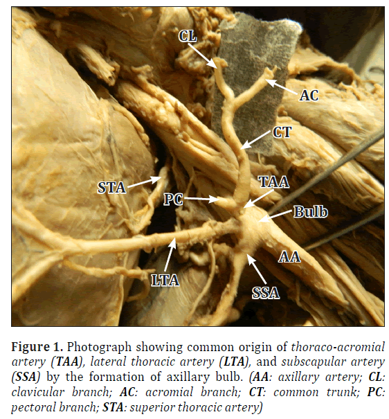
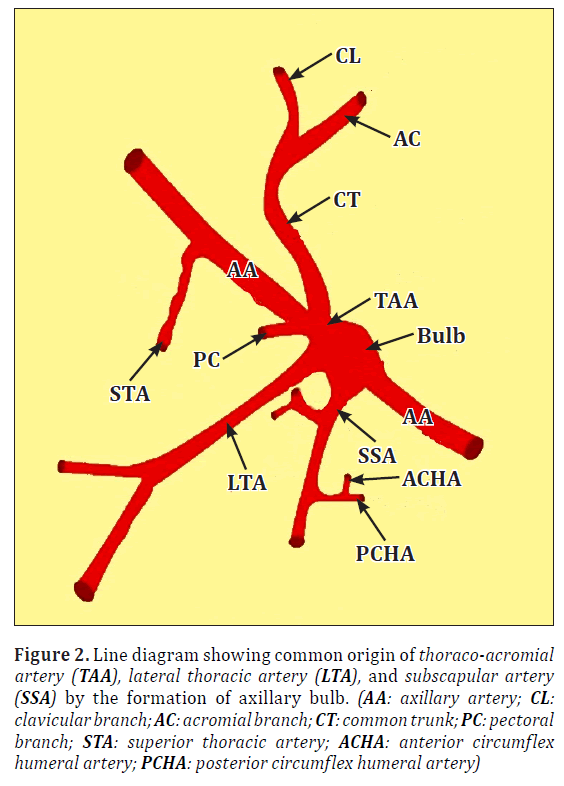
Anatomical variation in branching pattern of AA has been observed in the left upper limb of an old male cadaver, during routine dissection in dissection hall of RMCH, Bareilly, UP, India. It was observed that the branching pattern of the AA was not in usual pattern as described by Gray’s Anatomy 40th edition [1]. It has been observed, after removing the pectoralis minor muscle, that TAA and LTA were not taking origin from 2nd part of the AA. On keen observation it had been revealed that that TAA and LTA both originated along with SSA, by forming a common origin, below the inferior border of pectoralis minor muscle. This common origin of TAA, LTA and SSA was shown as more dilated part (axillary bulb) in AA. TAA after originated from 3rd part of AA, soon divided into two branches, one is pectoral branch of TAA, other was a common trunk, which was sigmoid in shape and gave two branches as clavicular and acromial branch of TAA. Diameter of TAA and its branches was much more than LTA. ACHA, and PCHA were originated from SSA in this cadaver (Figures 1, 2).
Figure 1: Photograph showing common origin of thoraco-acromial artery (TAA), lateral thoracic artery (LTA), and subscapular artery (SSA) by the formation of axillary bulb. (AA: axillary artery; CL: clavicular branch; AC: acromial branch; CT: common trunk; PC: pectoral branch; STA: superior thoracic artery)
Figure 2: Line diagram showing common origin of thoraco-acromial artery (TAA), lateral thoracic artery (LTA), and subscapular artery (SSA) by the formation of axillary bulb. (AA: axillary artery; CL: clavicular branch; AC: acromial branch; CT: common trunk; PC: pectoral branch; STA: superior thoracic artery; ACHA: anterior circumflex humeral artery; PCHA: posterior circumflex humeral artery)
Discussion
Anatomical variations are very common regarding branching pattern of AA as described by many previous researchers [2–4]. There is no fix pattern of origin and number of branches of AA. Any branch may originate from any part of AA [5–7]. Six major vessels usually arise from AA, as a separate manner [5]. ACHA, PCHA, SSA, radial collateral, middle collateral and superior ulnar collateral arteries arose by a common arterial trunk in the third part of the AA [8]. In the present study TAA, LTA and SSA were originated by a common point.
The TAA pierces the clavipectoral fascia and divides into four branches two major (pectoral and deltoid) and two minor (acromial and clavicular) [5]. In the present study branches of TAA showed the variation in origin. TAA just after originating divided into pectoral branch and a common stem, which further divided into acromial and clavicular branch.
In the present study there was formation of bulbous structure in AA from where TAA, LTA and SSA were originated, which is named as axillary bulb. In the present study we have also observed the diameter. Diameter of TAA and its branches was more than LTA. this is first reported case regarding diameter.
According to Patnaik et al. LTA arising from second part of AA in 92% of the upper limbs, and in 6% cases LTA arise from first part [4]. In the present case, it was arising from the third part of the AA. Pectoral branch is the largest branch of TAA as described by Gray’s anatomy 40th edition [1]. In the present study it was revealed that common trunk from which clavicular and acromial branches arose was quite large.
Such variations are considerable surgical, angiographic, clinical and statistical significant.
References
- Standring S, ed. Gray’s Anatomy. 40th Ed., London, Churchill Livingstone. 2008; 815–817.
- Saeed M, Rufai AA, Elsayed SE, Sadiq MS. Variations in subclavian-axillary arterial system. Saudi Med J. 2002; 23: 206–212.
- Yang HJ, Gil YC, Jung WS, Lee HY. Variations of the superficial brachial artery in Korean cadavers. J Korean Med Sci. 2008; 23: 884–887.
- Patnaik VVG, Kalsey G, Singla RK. Branching pattern of axillary artery – a morphological study. J Anat Soc India. 2000; 49: 127–132.
- Trotter M, Henderson JL, Gass H, Brua RS, eisman S, Agress H, Curtis GH, Westbrook ER. The origins of branches of the axillary artery in whites and in American negroes. Anat Rec. 1930; 46: 133–137.
- Hollinshead WH. Anatomy for Surgeons. New York, A Heber Harper Book. 1958; 290–300.
- Pandey SK, Shukla VK. Anatomical variation in origin and course of the thoracoacromial trunk and its branches. Nepal Med Coll J. 2004; 6: 88–91.
- Samuel VP, Vollala VR, Nayak S, Rao M, Bolla SR, Pammidi N. A rare variation in the branching pattern of the axillary artery. Indian J Plast Surg. 2006; 39: 222–223.
Mahendra Kumar Pant1*, Saim Hasan2, Sarangdhar3 and Syed Hasan Hyder Zaidi4
1Departments of Anatomy, Govt. Med. Coll., Haldwani, Nainital
2S.H.K.M. Government Med. Coll., Nuh, India
3B.S.D.B.A., Govt. Med. Coll., Kannauj UP, India
4Rohilkhand Med. Coll. & Hospital, Bareilly, UP, India
- *Corresponding Author:
- Dr. Mahendra Kumar Pant
Assistant Professor Department of Anatomy Government Medical College Haldwani, Nainital, (Uttarakhand) 263139, India
Tel: +91 989 7470722
E-mail: pant.mahendra@gmail.com
Date of Received: February 25th, 2012
Date of Accepted: October 20th, 2012
Published Online: February 14th, 2013
© Int J Anat Var (IJAV). 2013; 6: 47–48.
Abstract
Anatomical variation in branching pattern of the axillary artery has been observed in the left arm of an old male cadaver, in the department of Anatomy, Rohilkhand Medical College &Hospital (RMCH), Bareilly, UP, India, during routine undergraduate dissection. Pectoralis minor crossed in front of the axillary artery and divided it into three parts. It has been observed that the left side, the thoracoacromial artery and lateral thoracic artery were not originated from 2nd part, but arising from 3rd part along with subscapular artery by a common point of origin. Anterior circumflex humeral artery and posterior circumflex humeral artery were originated directly from subscapular artery by a common stem. Such variation might be due to the unusual developmental vascular pattern in the region. Such variation of the peripheral vascular system is of considerable surgical significance and might be used to explain unusual clinical signs and symptoms.
-Keywords
axillary artery, variation, vascular variations
Introduction
Axillary artery (AA) is the continuation of subclavian artery from the outer border of the first rib and continues as the brachial artery at the inferior border of teres major. Pectoralis minor muscle divides the artery into three parts, as the first part (proximal), second part (posterior) and third part (distal) to the muscle. As classically described in anatomical textbooks, axillary artery gives six branches; first part giving rise to superior thoracic, second part to thoraco-acromial (TAA) and lateral thoracic arteries (LTA), and the third part to subscapular artery (SSA), anterior circumflex humeral artery (ACHA) and posterior circumflex humeral artery (PCHA) [1].
Case Report
Anatomical variation in branching pattern of AA has been observed in the left upper limb of an old male cadaver, during routine dissection in dissection hall of RMCH, Bareilly, UP, India. It was observed that the branching pattern of the AA was not in usual pattern as described by Gray’s Anatomy 40th edition [1]. It has been observed, after removing the pectoralis minor muscle, that TAA and LTA were not taking origin from 2nd part of the AA. On keen observation it had been revealed that that TAA and LTA both originated along with SSA, by forming a common origin, below the inferior border of pectoralis minor muscle. This common origin of TAA, LTA and SSA was shown as more dilated part (axillary bulb) in AA. TAA after originated from 3rd part of AA, soon divided into two branches, one is pectoral branch of TAA, other was a common trunk, which was sigmoid in shape and gave two branches as clavicular and acromial branch of TAA. Diameter of TAA and its branches was much more than LTA. ACHA, and PCHA were originated from SSA in this cadaver (Figures 1, 2).
Figure 1: Photograph showing common origin of thoraco-acromial artery (TAA), lateral thoracic artery (LTA), and subscapular artery (SSA) by the formation of axillary bulb. (AA: axillary artery; CL: clavicular branch; AC: acromial branch; CT: common trunk; PC: pectoral branch; STA: superior thoracic artery)
Figure 2: Line diagram showing common origin of thoraco-acromial artery (TAA), lateral thoracic artery (LTA), and subscapular artery (SSA) by the formation of axillary bulb. (AA: axillary artery; CL: clavicular branch; AC: acromial branch; CT: common trunk; PC: pectoral branch; STA: superior thoracic artery; ACHA: anterior circumflex humeral artery; PCHA: posterior circumflex humeral artery)
Discussion
Anatomical variations are very common regarding branching pattern of AA as described by many previous researchers [2–4]. There is no fix pattern of origin and number of branches of AA. Any branch may originate from any part of AA [5–7]. Six major vessels usually arise from AA, as a separate manner [5]. ACHA, PCHA, SSA, radial collateral, middle collateral and superior ulnar collateral arteries arose by a common arterial trunk in the third part of the AA [8]. In the present study TAA, LTA and SSA were originated by a common point.
The TAA pierces the clavipectoral fascia and divides into four branches two major (pectoral and deltoid) and two minor (acromial and clavicular) [5]. In the present study branches of TAA showed the variation in origin. TAA just after originating divided into pectoral branch and a common stem, which further divided into acromial and clavicular branch.
In the present study there was formation of bulbous structure in AA from where TAA, LTA and SSA were originated, which is named as axillary bulb. In the present study we have also observed the diameter. Diameter of TAA and its branches was more than LTA. this is first reported case regarding diameter.
According to Patnaik et al. LTA arising from second part of AA in 92% of the upper limbs, and in 6% cases LTA arise from first part [4]. In the present case, it was arising from the third part of the AA. Pectoral branch is the largest branch of TAA as described by Gray’s anatomy 40th edition [1]. In the present study it was revealed that common trunk from which clavicular and acromial branches arose was quite large.
Such variations are considerable surgical, angiographic, clinical and statistical significant.
References
- Standring S, ed. Gray’s Anatomy. 40th Ed., London, Churchill Livingstone. 2008; 815–817.
- Saeed M, Rufai AA, Elsayed SE, Sadiq MS. Variations in subclavian-axillary arterial system. Saudi Med J. 2002; 23: 206–212.
- Yang HJ, Gil YC, Jung WS, Lee HY. Variations of the superficial brachial artery in Korean cadavers. J Korean Med Sci. 2008; 23: 884–887.
- Patnaik VVG, Kalsey G, Singla RK. Branching pattern of axillary artery – a morphological study. J Anat Soc India. 2000; 49: 127–132.
- Trotter M, Henderson JL, Gass H, Brua RS, eisman S, Agress H, Curtis GH, Westbrook ER. The origins of branches of the axillary artery in whites and in American negroes. Anat Rec. 1930; 46: 133–137.
- Hollinshead WH. Anatomy for Surgeons. New York, A Heber Harper Book. 1958; 290–300.
- Pandey SK, Shukla VK. Anatomical variation in origin and course of the thoracoacromial trunk and its branches. Nepal Med Coll J. 2004; 6: 88–91.
- Samuel VP, Vollala VR, Nayak S, Rao M, Bolla SR, Pammidi N. A rare variation in the branching pattern of the axillary artery. Indian J Plast Surg. 2006; 39: 222–223.


