Variation in the branching pattern of axillary artery – a case report
Divya Agrawal*, Narendra Singh, Biswa Bhusan Mohanty, Prafulla Kumar Chinara
Department of Anatomy, IMS and SUM Hospital, SOA University, B.O. Ghatikia, Bhubaneswar, India.
- *Corresponding Author:
- Dr. Divya Agrawal
Assistant Professor, Department of Anatomy, Institute of Medical Sciences and Sum Hospital, SOA University, K-8, Kalinganagar, B.O. Ghatikia, Bhubaneswar, 751003, India.
Tel: +91 993 7098825
E-mail: drdivjay@gmail.com
Date of Received: January 30th, 2012
Date of Accepted: July 12th, 2012
Published Online: February 14th, 2013
© Int J Anat Var (IJAV). 2013; 6: 31–33.
[ft_below_content] =>Keywords
axillary artery, variation, branches
Introduction
The axillary artery and its gross anatomy are well known to anatomists. The artery begins as a continuation of subclavian artery at the outer border of first rib and ends by becoming the brachial artery at the inferior border of teres major.
The course of the artery varies with the position of the arm in relation to the body. When the arm is held at right angles to the body, the artery is almost straight; when the arm is raised above the head, the artery is concave upwards; when the arm is held beside the body, the artery is concave downwards [1]. During its course, the artery passes deep to the pectoralis minor muscle, a fact that is used to divide the artery into three parts, proximal, posterior and distal to the muscle.
The axillary artery is conveniently described as giving six branches, but this is subject to considerable variations; two or more of its standard branches may arise from a common trunk or a new artery may arise separately.
The commonly named branches of axillary artery are superior thoracic, thoraco-acromial, lateral thoracic, subscapular, anterior circumflex humeral and posterior circumflex humeral artery [2].
Case Report
During routine dissection of a 45-year-old male cadaver in the Department of Anatomy IMS & SUM Hospital, Bhubaneswar, we came across unusual branching pattern of the axillary artery in the right upper limb. The case was photographed and findings were noted. The left upper limb showed no variation. At dissection it was seen that the 1st part of axillary artery gave one branch, i.e., superior thoracic artery at the normal level near the lower border of subclavius muscle and was seen to supply the lymph node and muscle of upper portion of thoracic wall.
The 2nd part of axillary artery gave the thoraco-acromial artery deep to the medial margin of pectoralis minor muscle, ran upward to pierce the clavipectoral fascia was divided into acromial, pectoral, clavicular and deltoid branches.
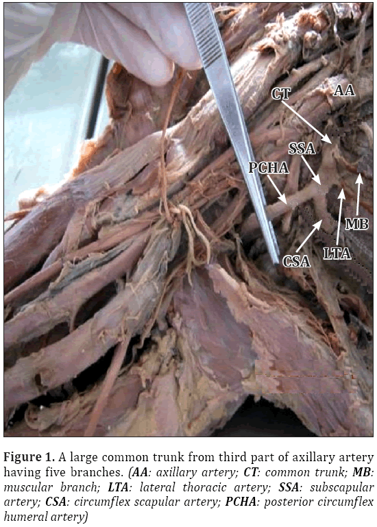
The 3rd part of axillary artery was seen to give two branches, a large common trunk which ran down medial to the axillary vein and another anterior circumflex humeral artery (Figure 1). The common trunk gave the following branches: a) a muscular branch, b) lateral thoracic artery, c) subscapular artery, d) circumflex scapular artery and e) posterior circumflex humeral artery.
The lateral thoracic artery arose from the common trunk deep to pectoralis minor muscle and descended on the chest wall to supply the lower part of chest wall.
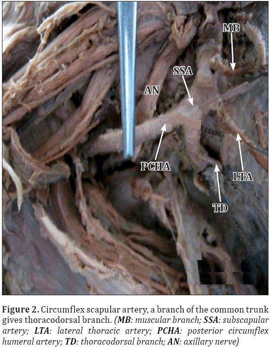
The circumflex scapular artery passed through the triangular space and emerged out on the posterior aspect near the lateral border of the scapula. Before piercing through the triangular space, it gave a thoracodorsal branch (Figure 2).
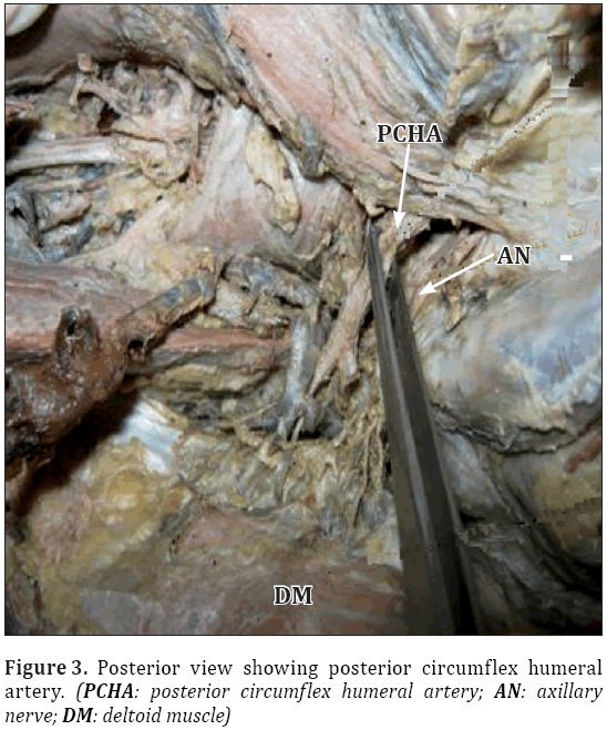
The posterior circumflex humeral artery was seen as a continuation of subscapular artery and passed through the quadrangular space along with the axillary nerve and emerged out on the posterior aspect and round the surgical neck of humerus below the deltoid muscle (Figure 3).
Anterior circumflex humeral artery was present as another branch from the third part, which ran in front of the surgical neck of humerus deep to the coracobrachialis and biceps brachii. It gave one ascending branch in the intertubercular groove to supply the shoulder joint.
Discussion
Anatomic variations of major arteries of the upper limb have been reported. It is not uncommon to find variation in the branching pattern of axillary artery. The present study revealed a variation in the branching pattern of axillary artery as also documented by many other workers.
The axillary artery is usually described as giving off six branches, although the number varies because two or more arteries often arise together instead of separately, or two branches of an artery arise separately instead of from the usual common trunk. Thus, instead of six, there may be anywhere from 5 to 11 branches. A rare but striking anomaly of the axillary artery is for it to divide into two branches as it proceeds down the arm. When this occurs, usually one of the arterial stems in the arm runs more superficially than does the other, and they are, therefore sometimes distinguished as brachial artery and superficial brachial artery. This apparent doubling of the brachial artery more commonly occurs in the arm than in the axilla [3]. High division of axillary artery into radial and ulnar arteries with the absence of brachial artery has also been reported [4,5].
The review of literature shows many variations, in which two or more branches arising from the common trunk are reported. However, all the branches of axillary artery (except superior thoracic) arising from a separate collateral branch is not reported adequately except for few cases. A case reported by Venieratos and Lolis [6] shows common subscapular trunk giving origin to circumflex scapular, thoracodorsal, anterior and posterior circumflex humeral, profunda brachii and ulnar collateral arteries. In another report by Samuel et al. [2] the third part of the axillary artery gave a common arterial trunk, which further gave anterior and posterior circumflex humeral, subscapular, radial collateral, middle collateral and superior ulnar collateral arteries with absence of profunda brachii artery.
In up to 30% of cases, the subscapular artery can arise from a common trunk with the posterior circumflex humeral artery. Occasionally the subscapular, circumflex humeral and profunda brachii arteries arise in common, in which case, branches of the brachial plexus surround this common vessel instead of axillary artery. The posterior circumflex humeral artery may arise from the profunda brachii artery and pass back below the teres major instead of passing through the quadrangular space [7].
The increasing use of invasive diagnostic and interventional procedures in cardiovascular diseases makes it important that the type and frequency of vascular variations are well documented and understood. Branches of the upper limb arteries have been used for coronary bypass and flaps in reconstructive surgery. Knowledge of branching pattern of axillary artery is useful during antegrade cerebral perfusion in aortic surgery [8], while treating axillary artery thrombosis, reconstructing axillary artery after trauma, using the artery for microvascular graft to replace damaged arteries, creating axillary coronary bypass shunt in high risk patients [9], and during surgical procedures of fractured upper end of humerus. Thus we see that accurate knowledge of the normal and variant arterial pattern of the human upper extremities is important both for reparative surgery and for angiography [2]. Cases of these kinds should be examined or evaluated carefully during surgical or electrophysiological procedures.
References
- Tan CB, Tan CK. An unusual course and relations of the human axillary artery. Singapore Med J. 1994; 35: 263–264.
- Samuel VP, Vollala VR, Nayak S, Rao M, Bolla SR, Pammidi N. A rare variation in the branching pattern of the axillary artery. Indian J Plast Surg. 2006; 39: 222–223.
- Hollingshead WH, Rossa C. Textbook of Anatomy. 4th Ed., Philadelphia, Harper and Row Publishers Inc. 1985; 187–189.
- Williams PL, Warwick R, Dyson M, Bannister. H. Gray’s Anatomy. 37th Ed., London, Churchill Livingstone. 1989; 756–758.
- Zuckerman S. A New System of Anatomy. 2nd Ed., USA, Oxford University Press. 1981; 15.
- Venieratos D, Lolis ED. Abnormal ramification of the axillary artery: sub-scapular common trunk. Morphologie. 2001; 85: 23–24. (French)
- Johnson D, Ellis H. Pectoral girdle and upper limb. In: Standrin S, ed., Gray’s Anatomy. 39th Ed., Edinburgh, Elsevier. 2005; 845.
- Sanioglu S, Sokullu O, Ozay B, Gullu AU, Sargin M, Albeyoglu S, Ozgen A, Bilgen F. Safety of unilateral antegrade cerebral perfusion at 22 degrees C systemic hypothermia. Heart Surg Forum. 2008; 11: E184–187.
- Charitou A, Athanasiou T, Morgan I S, DeL Stanbridge R. Use of Cough Lok can predispose to axillary artery thrombosis after a Robicsek procedure. Interact Cardiovasc Thorac Surg. 2003; 2: 68–69.
Divya Agrawal*, Narendra Singh, Biswa Bhusan Mohanty, Prafulla Kumar Chinara
Department of Anatomy, IMS and SUM Hospital, SOA University, B.O. Ghatikia, Bhubaneswar, India.
- *Corresponding Author:
- Dr. Divya Agrawal
Assistant Professor, Department of Anatomy, Institute of Medical Sciences and Sum Hospital, SOA University, K-8, Kalinganagar, B.O. Ghatikia, Bhubaneswar, 751003, India.
Tel: +91 993 7098825
E-mail: drdivjay@gmail.com
Date of Received: January 30th, 2012
Date of Accepted: July 12th, 2012
Published Online: February 14th, 2013
© Int J Anat Var (IJAV). 2013; 6: 31–33.
Abstract
Routine dissection of right upper limb in a 45-year-old male cadaver revealed a variation in the branching pattern of the axillary artery. Normally the 2nd part of axillary artery gives two branches: the lateral thoracic and the thoraco-acromial, whereas the third part gives the anterior circumflex humeral, posterior circumflex humeral and subscapular arteries. But in this case the second part gave only one branch, i.e., thoraco-acromial and the third part gave the anterior circumflex humeral and a common trunk which gave the lateral thoracic, subscapular, posterior circumflex humeral, circumflex scapular and a muscular branch. Awareness about details and topographic anatomy of variation of axillary artery may serve as a guide for both radiologists and vascular surgeons. During surgeries for lymph nodes in the axilla and surgeries for pectoral region, presence of such variations must be kept in mind.
-Keywords
axillary artery, variation, branches
Introduction
The axillary artery and its gross anatomy are well known to anatomists. The artery begins as a continuation of subclavian artery at the outer border of first rib and ends by becoming the brachial artery at the inferior border of teres major.
The course of the artery varies with the position of the arm in relation to the body. When the arm is held at right angles to the body, the artery is almost straight; when the arm is raised above the head, the artery is concave upwards; when the arm is held beside the body, the artery is concave downwards [1]. During its course, the artery passes deep to the pectoralis minor muscle, a fact that is used to divide the artery into three parts, proximal, posterior and distal to the muscle.
The axillary artery is conveniently described as giving six branches, but this is subject to considerable variations; two or more of its standard branches may arise from a common trunk or a new artery may arise separately.
The commonly named branches of axillary artery are superior thoracic, thoraco-acromial, lateral thoracic, subscapular, anterior circumflex humeral and posterior circumflex humeral artery [2].
Case Report
During routine dissection of a 45-year-old male cadaver in the Department of Anatomy IMS & SUM Hospital, Bhubaneswar, we came across unusual branching pattern of the axillary artery in the right upper limb. The case was photographed and findings were noted. The left upper limb showed no variation. At dissection it was seen that the 1st part of axillary artery gave one branch, i.e., superior thoracic artery at the normal level near the lower border of subclavius muscle and was seen to supply the lymph node and muscle of upper portion of thoracic wall.
The 2nd part of axillary artery gave the thoraco-acromial artery deep to the medial margin of pectoralis minor muscle, ran upward to pierce the clavipectoral fascia was divided into acromial, pectoral, clavicular and deltoid branches.
The 3rd part of axillary artery was seen to give two branches, a large common trunk which ran down medial to the axillary vein and another anterior circumflex humeral artery (Figure 1). The common trunk gave the following branches: a) a muscular branch, b) lateral thoracic artery, c) subscapular artery, d) circumflex scapular artery and e) posterior circumflex humeral artery.
The lateral thoracic artery arose from the common trunk deep to pectoralis minor muscle and descended on the chest wall to supply the lower part of chest wall.
The circumflex scapular artery passed through the triangular space and emerged out on the posterior aspect near the lateral border of the scapula. Before piercing through the triangular space, it gave a thoracodorsal branch (Figure 2).
The posterior circumflex humeral artery was seen as a continuation of subscapular artery and passed through the quadrangular space along with the axillary nerve and emerged out on the posterior aspect and round the surgical neck of humerus below the deltoid muscle (Figure 3).
Anterior circumflex humeral artery was present as another branch from the third part, which ran in front of the surgical neck of humerus deep to the coracobrachialis and biceps brachii. It gave one ascending branch in the intertubercular groove to supply the shoulder joint.
Discussion
Anatomic variations of major arteries of the upper limb have been reported. It is not uncommon to find variation in the branching pattern of axillary artery. The present study revealed a variation in the branching pattern of axillary artery as also documented by many other workers.
The axillary artery is usually described as giving off six branches, although the number varies because two or more arteries often arise together instead of separately, or two branches of an artery arise separately instead of from the usual common trunk. Thus, instead of six, there may be anywhere from 5 to 11 branches. A rare but striking anomaly of the axillary artery is for it to divide into two branches as it proceeds down the arm. When this occurs, usually one of the arterial stems in the arm runs more superficially than does the other, and they are, therefore sometimes distinguished as brachial artery and superficial brachial artery. This apparent doubling of the brachial artery more commonly occurs in the arm than in the axilla [3]. High division of axillary artery into radial and ulnar arteries with the absence of brachial artery has also been reported [4,5].
The review of literature shows many variations, in which two or more branches arising from the common trunk are reported. However, all the branches of axillary artery (except superior thoracic) arising from a separate collateral branch is not reported adequately except for few cases. A case reported by Venieratos and Lolis [6] shows common subscapular trunk giving origin to circumflex scapular, thoracodorsal, anterior and posterior circumflex humeral, profunda brachii and ulnar collateral arteries. In another report by Samuel et al. [2] the third part of the axillary artery gave a common arterial trunk, which further gave anterior and posterior circumflex humeral, subscapular, radial collateral, middle collateral and superior ulnar collateral arteries with absence of profunda brachii artery.
In up to 30% of cases, the subscapular artery can arise from a common trunk with the posterior circumflex humeral artery. Occasionally the subscapular, circumflex humeral and profunda brachii arteries arise in common, in which case, branches of the brachial plexus surround this common vessel instead of axillary artery. The posterior circumflex humeral artery may arise from the profunda brachii artery and pass back below the teres major instead of passing through the quadrangular space [7].
The increasing use of invasive diagnostic and interventional procedures in cardiovascular diseases makes it important that the type and frequency of vascular variations are well documented and understood. Branches of the upper limb arteries have been used for coronary bypass and flaps in reconstructive surgery. Knowledge of branching pattern of axillary artery is useful during antegrade cerebral perfusion in aortic surgery [8], while treating axillary artery thrombosis, reconstructing axillary artery after trauma, using the artery for microvascular graft to replace damaged arteries, creating axillary coronary bypass shunt in high risk patients [9], and during surgical procedures of fractured upper end of humerus. Thus we see that accurate knowledge of the normal and variant arterial pattern of the human upper extremities is important both for reparative surgery and for angiography [2]. Cases of these kinds should be examined or evaluated carefully during surgical or electrophysiological procedures.
References
- Tan CB, Tan CK. An unusual course and relations of the human axillary artery. Singapore Med J. 1994; 35: 263–264.
- Samuel VP, Vollala VR, Nayak S, Rao M, Bolla SR, Pammidi N. A rare variation in the branching pattern of the axillary artery. Indian J Plast Surg. 2006; 39: 222–223.
- Hollingshead WH, Rossa C. Textbook of Anatomy. 4th Ed., Philadelphia, Harper and Row Publishers Inc. 1985; 187–189.
- Williams PL, Warwick R, Dyson M, Bannister. H. Gray’s Anatomy. 37th Ed., London, Churchill Livingstone. 1989; 756–758.
- Zuckerman S. A New System of Anatomy. 2nd Ed., USA, Oxford University Press. 1981; 15.
- Venieratos D, Lolis ED. Abnormal ramification of the axillary artery: sub-scapular common trunk. Morphologie. 2001; 85: 23–24. (French)
- Johnson D, Ellis H. Pectoral girdle and upper limb. In: Standrin S, ed., Gray’s Anatomy. 39th Ed., Edinburgh, Elsevier. 2005; 845.
- Sanioglu S, Sokullu O, Ozay B, Gullu AU, Sargin M, Albeyoglu S, Ozgen A, Bilgen F. Safety of unilateral antegrade cerebral perfusion at 22 degrees C systemic hypothermia. Heart Surg Forum. 2008; 11: E184–187.
- Charitou A, Athanasiou T, Morgan I S, DeL Stanbridge R. Use of Cough Lok can predispose to axillary artery thrombosis after a Robicsek procedure. Interact Cardiovasc Thorac Surg. 2003; 2: 68–69.


