Variation in the origin and branching pattern of the musculocutaneous nerve and accessory head of the biceps brachii muscle
Ricardo E.Esparragoza*, Ana E.Nava, Marialvis Nava and Corina C. Nava
Cathedra of Anatomy, Department of Morphological Sciences, Medical College, Medical Faculty, University of Zulia, BOLIVARIAN REPUBLIC OF VENEZUELA.
- *Corresponding Author:
- Ricardo E. Esparragoza, MD, PhD
Titular Professor Cathedra of Anatomy Department of Morphological Sci. Medical College, Medical Faculty University of Zulia (LUZ),Bolivarianrep.of Venezuela.
Tel: +58 2642415808
E-mail: ricardoeem@gmail.com
Date of Received: October 24th, 2013
Date of Accepted: March 28th, 2015d
Published Online: December 30th, 2015
© Int J Anat Var (IJAV). 2015; 8: 40–42.
[ft_below_content] =>Keywords
musculocutaneous nerve,patron branching,biceps brachii,accessory head,anatomical variation
Introduction
Anatomical variants of the brachial plexus, than interesting lateral cord of plexus and the musculocutaneous and median nerves have been described [1]. The communications between median and musculocutanous nerves have been classified into different types [2,3], including the variant origin of the musculocutaneous nerve (MCN), which can be absent or originate from median nerve, instead lateral cord [4]. Also, variations in trajectory and branching pattern of MCN have been reported, including absence of piercing the coracobrachialis muscle [5]. The MCN is responsible for motor innervations of the muscles of anterior compartment of arm and provides sensitive innervation through of the lateral cutaneous nerve of forearm. In other hand, the biceps brachii muscle (BB) is one of the muscles most subject to variations. The most common variation is a third head; the accessory head may have distinct origins. It arises from humerus, between the insertion of the coracobrachialis and the upper part of the origin of the brachialis, intertubercular groove or less frequently from the tendon of the pectoralis major or deltoid, or from the articular capsule or even a dual origination. The accessory heads typically join the common belly, or the aponeurosis of the BB. Some heads unite with the belly of the long head or that of the short head. The presence of supernumerary heads of the BB has been associated with variations of the MCN [6,7]. Although, it is not usual find the association of such variations.
Case Report
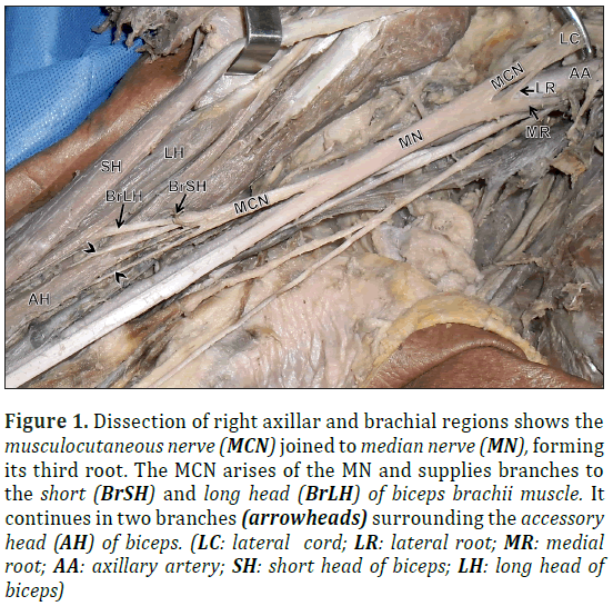
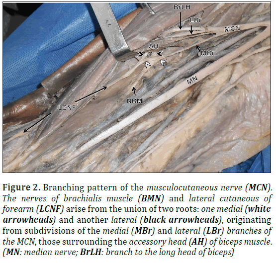
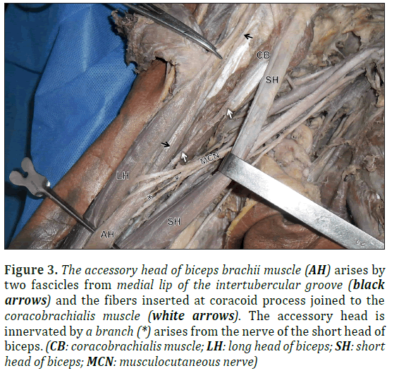
During routine dissection of the right upper limb of approximately 60-years-old formaline-fixed male cadaver, we found an unusual combination of anatomical variations of the MCN and BB. The nerve joined to median nerve, near its origin. Later, MCN divided from the median nerve at the proximal third of right arm, 6.8 cm distal to median nerve origin. After its origin, it passed from medial to lateral through the posterior aspect of the short head of the BB, medially and anteriorly to coracobrachialis, which did not pierce. At the middle third of the arm the MCN, after supplying branches for the long and short heads of BB, continued in two branches, one medial and other lateral, which formed a loop around the accessory head of BB (Figure 1).The medial and lateral branches were divided, each one, into two bundles, constituting the medial and lateral roots, respectively. Then, each medial root joined with the lateral root corresponding to form the nerve of brachialis muscle and the lateral cutaneous nerve of forearm (Figure 2). The accessory head of BB originated in the form of two fascicles from the medial lip of the intertubercular groove and tendon fibers inserted at coracoid process joined to the short head of biceps and coracobrachialis (Figure 3). Both fascicles united at middle third of arm, placed posterior to the short head of BB. The accessory head was innervated by a branch arose from the nerve of the short head of BB. It joined to deep aspect of the short head of BB. On the left side, the MCN originated from lateral cord, pierced the coracobrachialis. Also, BB showed usual anatomic configuration.
Figure 1: Dissection of right axillar and brachial regions shows the musculocutaneous nerve (MCN) joined to median nerve (MN), forming its third root. The MCN arises of the MN and supplies branches to the short (BrSH) and long head (BrLH) of biceps brachii muscle. It continues in two branches (arrowheads) surrounding the accessory head (AH) of biceps. (LC: lateral cord; LR: lateral root; MR: medial root; AA: axillary artery; SH: short head of biceps; LH: long head of biceps)
Figure 2: Branching pattern of the musculocutaneous nerve (MCN). The nerves of brachialis muscle (BMN) and lateral cutaneous of forearm (LCNF) arise from the union of two roots: one medial (white arrowheads) and another lateral (black arrowheads), originating from subdivisions of the medial (MBr) and lateral (LBr) branches of the MCN, those surrounding the accessory head (AH) of biceps muscle. (MN: median nerve; BrLH: branch to the long head of biceps)
Figure 3: The accessory head of biceps brachii muscle (AH) arises by two fascicles from medial lip of the intertubercular groove (black arrows) and the fibers inserted at coracoid process joined to the coracobrachialis muscle (white arrows). The accessory head is innervated by a branch (*) arises from the nerve of the short head of biceps. (CB: coracobrachialis muscle; LH: long head of biceps; SH: short head of biceps; MCN: musculocutaneous nerve)
Discussion
Brachial plexus variations are frequently referred in literature, including variant origin of MCN. It arises from the median nerve in 2% [1]. Variations of the MCN and median nerves have been described and classified by Le Minor into five types [2]. Our case corresponds to type 4, which the fibers of the MCN united to the median nerve at its origin. It can be described as the third root or the double lateral root of the median nerve [8]. Also, this type of nervous communication establishes than after some distance, the fibers of MCN arise from the median nerve.
Another finding was that MCN did not pierce the coracobrachialis muscle, being a different variation about the nervous trajectory. This variation is described in 11% on dissections of upper limbs [5]. The nerve can pass posterior or anterior to the coracobrachialis, the latter occurs in our case. Because MCN was originated in a different way, more distally, at the brachial region, from median nerve, it is expected to occur variations in nervous trajectory. In other similar case report of MCN originating from median nerve, also described not piercing of the coracobrachialis [4].
The most remarkable finding about MCN was its branching pattern, where the nerves of brachialis muscle and lateral cutaneous of forearm were constituted by converging subdivided branches of the MCN, forming a loop around the third head of BB. Kosugi [7] divided the branching patron of the MCN and described in some cases the loop formation from the communicating branch or from the trunk of the nerve. The accessory head of biceps was surrounded by the nerve loop. Nevertheless, the branching pattern of the MCN reported in our case, it was not found between many types and subtypes described by Kosugui. Neither, in the rest of literature reviewed, we found such branching pattern of the MCN.
The accessory head of the BB has been reported with varying frequency. The third head arises from the humerus, shoulder articulation, deltoid and pectoralis major muscles. In this case, the third head arose from medial lip of intertubecular groove and coracoids process. Dual origin of accessory head is less frequently reported. Asvat et al. reported as the least common two cases with a dual fascial origin from the short head of BB and the medial aspect of the deltoid and its insertion area [6].
The presence of accessory heads of the BB has been associated with variations of the MCN, affecting its course and branching [7]. Embryologically, these variations are explained, during the fifth week of development, mesoderm invades the upper limp bud to further condense into ventral and dorsal muscle masses. The biceps musculature is derived from the ventral muscle masses of the upper limb bud. It would be during this period of development that accessory muscles may have formed [9]. Likewise, at the fifth week the brachial plexus appears as a single radicular cone of the upper limb that is divided longitudinally into ventral and dorsal segments. Musculocutaneous and median nerves arise from ventral segment, because both nerves originate from the same segment, undergoing variations during their embryological division process [10].
Knowledge of the variations in the brachial plexus and its branches is useful for alert and guide, avoiding nerve injuries during diagnoses, surgical or therapeutic procedures accomplished in this region.
References
- Bergman RA, Thompson SA, Afifi AK, Saadeh FA. Compendium of human anatomic variation.Munich, Urban and Schwarzenberg. 1988; 140–142.
- Le Minor JM. Une variation rare des nerfs médian et musculo-cutané chez l’homme. Arch Anat Histol Embryol. 1990; 73: 33–42.
- Venieratos D, Anagnostopoulou S. Classification of communications between the median and musculocutaneous nerves. Clin Anat. 1998; 11: 327–331.
- Akhilandeswari B, Shubha R. Study of origin of musculocutaneous nerve. Anatomica Karnataka. 2009; 3: 30–34.
- Kazi AK. Muscular innervation of muscles of the upper extremities by the musculocutaneous nerve and the motor deficit arising from lesions of this nerve. Int J Biol Med Res. 2011; 2:1094–1097.
- Asvat R, Candler P, Sarmiento EE. High incidence of the third head of biceps brachii in South African populations. J Anat. 1993; 182(Pt 1): 101–104.
- Kosugi K, Shibata S, Yamashita H. Supernumerary head of biceps brachii and branching pattern of the musculocutaneus nerve in Japanese. Surg Radiol Anat.1992; 14: 175–185.
- Khullar M, Sharma S, Khullar S. Multiple bilateral neuroanatomical variations of the nerves of the arm-a case report. Int J Med Health Sci. 2012; 1: 75–84.
- Nayak SR, Krishnamurthy A, Kumar M, Prabhu LV, Saralaya V, Thomas MM. Four-headed biceps and triceps brachii muscles, with neurovascular variation. Anat Sci Int. 2008; 83:107–111.
- Iwamoto S, Kimura K, Takahashi Y, Konishi M. Some aspects of the communicating branch between the musculocutaneous and median nerves in man. Okajimas Folia Anat. Jpn. 1990;67: 47–52.
Ricardo E.Esparragoza*, Ana E.Nava, Marialvis Nava and Corina C. Nava
Cathedra of Anatomy, Department of Morphological Sciences, Medical College, Medical Faculty, University of Zulia, BOLIVARIAN REPUBLIC OF VENEZUELA.
- *Corresponding Author:
- Ricardo E. Esparragoza, MD, PhD
Titular Professor Cathedra of Anatomy Department of Morphological Sci. Medical College, Medical Faculty University of Zulia (LUZ),Bolivarianrep.of Venezuela.
Tel: +58 2642415808
E-mail: ricardoeem@gmail.com
Date of Received: October 24th, 2013
Date of Accepted: March 28th, 2015d
Published Online: December 30th, 2015
© Int J Anat Var (IJAV). 2015; 8: 40–42.
Abstract
Anatomical variants interesting the musculocutaneous nerve (MCN) have been described and classified. It is less common find association of variations about origin and branching pattern of the MCN and the presence of accessory head of biceps brachii muscle. During routine dissection of the right upper limb on an adult male cadaver, we observed the MCN constituting a third root of median nerve, which originated after. The MCN passed anterior to the coracobrachialis muscle, without piercing it, after supplying branches for the long and short heads of biceps and continued in two branches rounding the accessory head of biceps brachii muscle; these two branches subdivided into roots that originated the nerves of brachialis muscle and lateral cutaneous nerve of forearm. Accessory head of biceps had dual origin. The presence of accessory heads of biceps is associated with variations of the MCN. Knowledge of these variations is useful to clinical practice.
-Keywords
musculocutaneous nerve,patron branching,biceps brachii,accessory head,anatomical variation
Introduction
Anatomical variants of the brachial plexus, than interesting lateral cord of plexus and the musculocutaneous and median nerves have been described [1]. The communications between median and musculocutanous nerves have been classified into different types [2,3], including the variant origin of the musculocutaneous nerve (MCN), which can be absent or originate from median nerve, instead lateral cord [4]. Also, variations in trajectory and branching pattern of MCN have been reported, including absence of piercing the coracobrachialis muscle [5]. The MCN is responsible for motor innervations of the muscles of anterior compartment of arm and provides sensitive innervation through of the lateral cutaneous nerve of forearm. In other hand, the biceps brachii muscle (BB) is one of the muscles most subject to variations. The most common variation is a third head; the accessory head may have distinct origins. It arises from humerus, between the insertion of the coracobrachialis and the upper part of the origin of the brachialis, intertubercular groove or less frequently from the tendon of the pectoralis major or deltoid, or from the articular capsule or even a dual origination. The accessory heads typically join the common belly, or the aponeurosis of the BB. Some heads unite with the belly of the long head or that of the short head. The presence of supernumerary heads of the BB has been associated with variations of the MCN [6,7]. Although, it is not usual find the association of such variations.
Case Report
During routine dissection of the right upper limb of approximately 60-years-old formaline-fixed male cadaver, we found an unusual combination of anatomical variations of the MCN and BB. The nerve joined to median nerve, near its origin. Later, MCN divided from the median nerve at the proximal third of right arm, 6.8 cm distal to median nerve origin. After its origin, it passed from medial to lateral through the posterior aspect of the short head of the BB, medially and anteriorly to coracobrachialis, which did not pierce. At the middle third of the arm the MCN, after supplying branches for the long and short heads of BB, continued in two branches, one medial and other lateral, which formed a loop around the accessory head of BB (Figure 1).The medial and lateral branches were divided, each one, into two bundles, constituting the medial and lateral roots, respectively. Then, each medial root joined with the lateral root corresponding to form the nerve of brachialis muscle and the lateral cutaneous nerve of forearm (Figure 2). The accessory head of BB originated in the form of two fascicles from the medial lip of the intertubercular groove and tendon fibers inserted at coracoid process joined to the short head of biceps and coracobrachialis (Figure 3). Both fascicles united at middle third of arm, placed posterior to the short head of BB. The accessory head was innervated by a branch arose from the nerve of the short head of BB. It joined to deep aspect of the short head of BB. On the left side, the MCN originated from lateral cord, pierced the coracobrachialis. Also, BB showed usual anatomic configuration.
Figure 1: Dissection of right axillar and brachial regions shows the musculocutaneous nerve (MCN) joined to median nerve (MN), forming its third root. The MCN arises of the MN and supplies branches to the short (BrSH) and long head (BrLH) of biceps brachii muscle. It continues in two branches (arrowheads) surrounding the accessory head (AH) of biceps. (LC: lateral cord; LR: lateral root; MR: medial root; AA: axillary artery; SH: short head of biceps; LH: long head of biceps)
Figure 2: Branching pattern of the musculocutaneous nerve (MCN). The nerves of brachialis muscle (BMN) and lateral cutaneous of forearm (LCNF) arise from the union of two roots: one medial (white arrowheads) and another lateral (black arrowheads), originating from subdivisions of the medial (MBr) and lateral (LBr) branches of the MCN, those surrounding the accessory head (AH) of biceps muscle. (MN: median nerve; BrLH: branch to the long head of biceps)
Figure 3: The accessory head of biceps brachii muscle (AH) arises by two fascicles from medial lip of the intertubercular groove (black arrows) and the fibers inserted at coracoid process joined to the coracobrachialis muscle (white arrows). The accessory head is innervated by a branch (*) arises from the nerve of the short head of biceps. (CB: coracobrachialis muscle; LH: long head of biceps; SH: short head of biceps; MCN: musculocutaneous nerve)
Discussion
Brachial plexus variations are frequently referred in literature, including variant origin of MCN. It arises from the median nerve in 2% [1]. Variations of the MCN and median nerves have been described and classified by Le Minor into five types [2]. Our case corresponds to type 4, which the fibers of the MCN united to the median nerve at its origin. It can be described as the third root or the double lateral root of the median nerve [8]. Also, this type of nervous communication establishes than after some distance, the fibers of MCN arise from the median nerve.
Another finding was that MCN did not pierce the coracobrachialis muscle, being a different variation about the nervous trajectory. This variation is described in 11% on dissections of upper limbs [5]. The nerve can pass posterior or anterior to the coracobrachialis, the latter occurs in our case. Because MCN was originated in a different way, more distally, at the brachial region, from median nerve, it is expected to occur variations in nervous trajectory. In other similar case report of MCN originating from median nerve, also described not piercing of the coracobrachialis [4].
The most remarkable finding about MCN was its branching pattern, where the nerves of brachialis muscle and lateral cutaneous of forearm were constituted by converging subdivided branches of the MCN, forming a loop around the third head of BB. Kosugi [7] divided the branching patron of the MCN and described in some cases the loop formation from the communicating branch or from the trunk of the nerve. The accessory head of biceps was surrounded by the nerve loop. Nevertheless, the branching pattern of the MCN reported in our case, it was not found between many types and subtypes described by Kosugui. Neither, in the rest of literature reviewed, we found such branching pattern of the MCN.
The accessory head of the BB has been reported with varying frequency. The third head arises from the humerus, shoulder articulation, deltoid and pectoralis major muscles. In this case, the third head arose from medial lip of intertubecular groove and coracoids process. Dual origin of accessory head is less frequently reported. Asvat et al. reported as the least common two cases with a dual fascial origin from the short head of BB and the medial aspect of the deltoid and its insertion area [6].
The presence of accessory heads of the BB has been associated with variations of the MCN, affecting its course and branching [7]. Embryologically, these variations are explained, during the fifth week of development, mesoderm invades the upper limp bud to further condense into ventral and dorsal muscle masses. The biceps musculature is derived from the ventral muscle masses of the upper limb bud. It would be during this period of development that accessory muscles may have formed [9]. Likewise, at the fifth week the brachial plexus appears as a single radicular cone of the upper limb that is divided longitudinally into ventral and dorsal segments. Musculocutaneous and median nerves arise from ventral segment, because both nerves originate from the same segment, undergoing variations during their embryological division process [10].
Knowledge of the variations in the brachial plexus and its branches is useful for alert and guide, avoiding nerve injuries during diagnoses, surgical or therapeutic procedures accomplished in this region.
References
- Bergman RA, Thompson SA, Afifi AK, Saadeh FA. Compendium of human anatomic variation.Munich, Urban and Schwarzenberg. 1988; 140–142.
- Le Minor JM. Une variation rare des nerfs médian et musculo-cutané chez l’homme. Arch Anat Histol Embryol. 1990; 73: 33–42.
- Venieratos D, Anagnostopoulou S. Classification of communications between the median and musculocutaneous nerves. Clin Anat. 1998; 11: 327–331.
- Akhilandeswari B, Shubha R. Study of origin of musculocutaneous nerve. Anatomica Karnataka. 2009; 3: 30–34.
- Kazi AK. Muscular innervation of muscles of the upper extremities by the musculocutaneous nerve and the motor deficit arising from lesions of this nerve. Int J Biol Med Res. 2011; 2:1094–1097.
- Asvat R, Candler P, Sarmiento EE. High incidence of the third head of biceps brachii in South African populations. J Anat. 1993; 182(Pt 1): 101–104.
- Kosugi K, Shibata S, Yamashita H. Supernumerary head of biceps brachii and branching pattern of the musculocutaneus nerve in Japanese. Surg Radiol Anat.1992; 14: 175–185.
- Khullar M, Sharma S, Khullar S. Multiple bilateral neuroanatomical variations of the nerves of the arm-a case report. Int J Med Health Sci. 2012; 1: 75–84.
- Nayak SR, Krishnamurthy A, Kumar M, Prabhu LV, Saralaya V, Thomas MM. Four-headed biceps and triceps brachii muscles, with neurovascular variation. Anat Sci Int. 2008; 83:107–111.
- Iwamoto S, Kimura K, Takahashi Y, Konishi M. Some aspects of the communicating branch between the musculocutaneous and median nerves in man. Okajimas Folia Anat. Jpn. 1990;67: 47–52.


