Variation of branching pattern of axillary artery
Satabdi Sarkar*, Banani Kundu, Alpana de Bose and Pallab Kumar Saha
Department of Anatomy, R. G. Kar Medical College, West Bengal, India
- *Corresponding Author:
- Dr. Satabdi Sarkar
R. G. Kar Medical College 1, Khudiram Bose Sarani Kolkata, 700004 West Bengal, India
Tel: +91 9830503734
E-mail: dr.satabdi2010@rediffmail.com
Date of Received: February 21st, 2013
Date of Accepted: October 20th, 2013
Published Online: June 1st, 2014
© Int J Anat Var (IJAV). 2014; 7: 27–29.
[ft_below_content] =>Keywords
axillary artery, branch, thoracodorsal nerve
Introduction
Axillary artery begins as a continuation of 3rd part of subclavian artery at the outer border of the first rib. It extends at the lower border of teres major muscle where it continues as the brachial artery. The pectoralis minor muscle divides the axillary artery into three parts. It usually gives off 6 branches. The 1st part which is medial to pectoralis minor muscle gives superior thoracic artery; the 2nd part which is deep to the above-mentioned muscle gives thoracoacromial and lateral thoracic arteries. The 3rd part lays lateral to the pectoralis minor and gives subscapular, anterior circumflex humeral and posterior circumflex humeral arteries. An extensive anastomosis is present between the branches of subclavian and axillary arteries around the dorsal surface of scapula.
Case Report
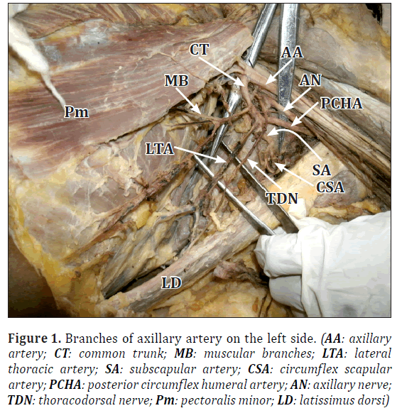
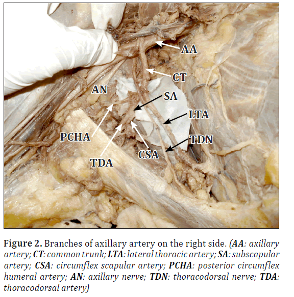
During routine dissection in a 64-year-old male cadaver we found bilateral variations in the branching pattern of the axillary artery. The course and branching of the first part of the artery was as usual. The second part gave only thoraco-acromial artery and provided its usual branches. Third part of the artery gave anterior circumflex humeral artery at a higher level than usual. It then gave a common trunk from where first muscular branches arose then it gave lateral thoracic artery, subscapular artery and posterior circumflex humeral artery. Lateral thoracic artery instead of subscapular artery was the largest branch of the axillary artery. The thoracodorsal nerve usually accompanied the subscapular artery but in this case it followed the lateral thoracic artery. The relationship of axillary vein and cords of brachial plexus were normal with respect to axillary artery. Similar variations were observed on opposite side though the posterior circumflex humeral artery was thin in comparison to other side.
Discussion
Earlier studies showed that variations in branching pattern of the axillary artery were quite common. George et al. observed axillary artery bifurcated into almost equal sized trunks [1]. The superficial one continued as the brachial artery. The deep trunk bifurcated into common circumflex humeral–subscapular trunk and profunda brachii artery. The above said common trunk divided into three branches: anterior circumflex humeral, posterior circumflex humeral and subscapular arteries.
Saeed et al. reported the origin of a common subscapular-circumflex humeral trunk from the third part of axillary artery, which divided into subscapular, anterior circumflex humeral and posterior circumflex humeral arteries in 3.8% of cases [2]. In this case, the common trunk gave lateral thoracic artery instead of anterior circumflex humeral artery.
Figure 1: Branches of axillary artery on the left side. (AA: axillary artery; CT: common trunk; MB: muscular branches; LTA: lateral thoracic artery; SA: subscapular artery; CSA: circumflex scapular artery; PCHA: posterior circumflex humeral artery; AN: axillary nerve; TDN: thoracodorsal nerve; Pm: pectoralis minor; LD: latissimus dorsi)
Magden et al. reported unusual branching pattern of the axillary artery [3]. The first branch went to serratus anterior muscle; the lateral thoracic and thoracodorsal arteries arose as a common trunk from third part of axillary artery. The circumflex scapular artery originated directly from the third part of axillay artery. The subscapular artery was absent.
Variations in branching pattern of axillary artery were due to the defects in the embryonic development of the vascular plexus of upper limb bud. The axial artery derived from lateral branch of seventh intersegmental artery and the proximal part of it formed the axillary and the brachial artery. This variation may be due to an arrest at any stage of development of vessels of upper limb. This may be due to regression, retention, or reappearance of new vessels and may lead to variations in the arterial origins and courses [4].
According to Arey, the unusual blood vessels may be due to [5]:
• The choice of unusual paths in the primitive vascular plexuses
• The persistence of vessels normally obliterated
• The disappearance of vessels normally retained
• Incomplete development and fusions and absorption of the parts usually distinct.
Variations in the course and branching pattern of the axillary artery had practical importance for the radiologists and vascular surgeons. Brachial plexus injury was a common condition which required exploration and repair. During surgery of the pectoral and axillary region the abnormal branch may be a definite cause of concern if its presence was not known. Branches of the upper limb arteries have been used while carrying out coronary bypass and other reconstructive surgery. Thus accurate knowledge of the normal and variant arterial anatomy of the axillary artery is important for clinical procedures in this region [6].
Knowledge of branching pattern of axillary artery is necessary while doing the antegrade cerebral perfusion in aortic surgery in case of axillary artery thrombosis [7,8].
When the reconstruction of the axillary artery is necessary after trauma, surgeons use microvascular graft taken from the various branches of axillary artery, and use the medial arm skin flap [9]. Therefore, both the usual as well as the variation of the axillary artery should be precisely known for accurate diagnostic interpretation and surgical intervention.
Acknowledgement
The authors express their heartfelt gratitude to all the members of the Department of Anatomy, R G Kar Medical College, Kolkata for their kind cooperation and generosity in the conduct of this study.
References
- George BM, Nayak S, Kumar P. Clinically significant neurovascular variations in the axilla and the arm – a case report. Neuroanatomy. 2007; 6: 36–38.
- Saeed M, Rufai AA, Elsayed SE, Sadiq MS. Variations in the subclavian-axillary arterial system. Saudi Med J. 2002; 23: 206–212.
- Magden O, Gocmen-Mas N, Caglar B. Multiple variations in the axillary arterial tree relevant to plastic surgery: A Case report. Int J Morphol. 2007; 25: 357–361.
- Decker GAG, du plessis DJ, Lee Mc Gregor. Shoulder joint. In: Synopsis of Surgical Anatomy. Mumbai, K. M. Varghese Company. 1986; 451.
- Arey LB. Developmental Anatomy. 6th Ed., Philadelphia, W.B. Saunders. 1957; 375–375.
- Cavdar S, Zeybek A, Bayramicli M. Rare variation of the axillary artery. Clin Anat. 2000; 13: 66–68.
- Sanioglu S, Sokullu O, Ozay B, Gullu AU, Sargin M, Albeyoglu S, Ozgen A, Bilgen F. Safety of unilateral antegrade cerebral perfusion at 22 degrees C systemic hypothermia. Heart Surg Forum. 2008; 11: 184–187.
- Charitou A, Athanasiou T, Morgan IS, Del SR. Use of Cough Lok can predispose to axillary artery thrombosis after a Robicsek procedure. Interact Cardiovasc Thorac Surg. 2003; 2: 68–69.
- Karamursel S, Bagdatli D, Demir Z, Tuccar E, Celebioglu S. Use of medial arm skin as a free flap. Plast Reconstr Surg. 2005; 115: 2025–2031.
Satabdi Sarkar*, Banani Kundu, Alpana de Bose and Pallab Kumar Saha
Department of Anatomy, R. G. Kar Medical College, West Bengal, India
- *Corresponding Author:
- Dr. Satabdi Sarkar
R. G. Kar Medical College 1, Khudiram Bose Sarani Kolkata, 700004 West Bengal, India
Tel: +91 9830503734
E-mail: dr.satabdi2010@rediffmail.com
Date of Received: February 21st, 2013
Date of Accepted: October 20th, 2013
Published Online: June 1st, 2014
© Int J Anat Var (IJAV). 2014; 7: 27–29.
Abstract
Bilateral variations of the branches of the axillary artery were found on routine anatomical dissection in a 64-year-old male cadaver in the Anatomy Department of R. G. Kar Medical College, Kolkata. From the first part superior thoracic artery, from the second part only thoracoacromial artery and from the third part anterior circumflex humeral artery and a common trunk arose, this common trunk gave four branches namely muscular branch, lateral thoracic artery, subscapular artery and posterior circumflex humeral artery. Thoracodorsal nerve accompanied the lateral thoracic artery instead of subscapular artery. The relationship of the axillary artery with axillary vein and cords of brachial plexus were as usual. Knowledge regarding this kind of variations is important for surgeries and imaging procedures.
-Keywords
axillary artery, branch, thoracodorsal nerve
Introduction
Axillary artery begins as a continuation of 3rd part of subclavian artery at the outer border of the first rib. It extends at the lower border of teres major muscle where it continues as the brachial artery. The pectoralis minor muscle divides the axillary artery into three parts. It usually gives off 6 branches. The 1st part which is medial to pectoralis minor muscle gives superior thoracic artery; the 2nd part which is deep to the above-mentioned muscle gives thoracoacromial and lateral thoracic arteries. The 3rd part lays lateral to the pectoralis minor and gives subscapular, anterior circumflex humeral and posterior circumflex humeral arteries. An extensive anastomosis is present between the branches of subclavian and axillary arteries around the dorsal surface of scapula.
Case Report
During routine dissection in a 64-year-old male cadaver we found bilateral variations in the branching pattern of the axillary artery. The course and branching of the first part of the artery was as usual. The second part gave only thoraco-acromial artery and provided its usual branches. Third part of the artery gave anterior circumflex humeral artery at a higher level than usual. It then gave a common trunk from where first muscular branches arose then it gave lateral thoracic artery, subscapular artery and posterior circumflex humeral artery. Lateral thoracic artery instead of subscapular artery was the largest branch of the axillary artery. The thoracodorsal nerve usually accompanied the subscapular artery but in this case it followed the lateral thoracic artery. The relationship of axillary vein and cords of brachial plexus were normal with respect to axillary artery. Similar variations were observed on opposite side though the posterior circumflex humeral artery was thin in comparison to other side.
Discussion
Earlier studies showed that variations in branching pattern of the axillary artery were quite common. George et al. observed axillary artery bifurcated into almost equal sized trunks [1]. The superficial one continued as the brachial artery. The deep trunk bifurcated into common circumflex humeral–subscapular trunk and profunda brachii artery. The above said common trunk divided into three branches: anterior circumflex humeral, posterior circumflex humeral and subscapular arteries.
Saeed et al. reported the origin of a common subscapular-circumflex humeral trunk from the third part of axillary artery, which divided into subscapular, anterior circumflex humeral and posterior circumflex humeral arteries in 3.8% of cases [2]. In this case, the common trunk gave lateral thoracic artery instead of anterior circumflex humeral artery.
Figure 1: Branches of axillary artery on the left side. (AA: axillary artery; CT: common trunk; MB: muscular branches; LTA: lateral thoracic artery; SA: subscapular artery; CSA: circumflex scapular artery; PCHA: posterior circumflex humeral artery; AN: axillary nerve; TDN: thoracodorsal nerve; Pm: pectoralis minor; LD: latissimus dorsi)
Magden et al. reported unusual branching pattern of the axillary artery [3]. The first branch went to serratus anterior muscle; the lateral thoracic and thoracodorsal arteries arose as a common trunk from third part of axillary artery. The circumflex scapular artery originated directly from the third part of axillay artery. The subscapular artery was absent.
Variations in branching pattern of axillary artery were due to the defects in the embryonic development of the vascular plexus of upper limb bud. The axial artery derived from lateral branch of seventh intersegmental artery and the proximal part of it formed the axillary and the brachial artery. This variation may be due to an arrest at any stage of development of vessels of upper limb. This may be due to regression, retention, or reappearance of new vessels and may lead to variations in the arterial origins and courses [4].
According to Arey, the unusual blood vessels may be due to [5]:
• The choice of unusual paths in the primitive vascular plexuses
• The persistence of vessels normally obliterated
• The disappearance of vessels normally retained
• Incomplete development and fusions and absorption of the parts usually distinct.
Variations in the course and branching pattern of the axillary artery had practical importance for the radiologists and vascular surgeons. Brachial plexus injury was a common condition which required exploration and repair. During surgery of the pectoral and axillary region the abnormal branch may be a definite cause of concern if its presence was not known. Branches of the upper limb arteries have been used while carrying out coronary bypass and other reconstructive surgery. Thus accurate knowledge of the normal and variant arterial anatomy of the axillary artery is important for clinical procedures in this region [6].
Knowledge of branching pattern of axillary artery is necessary while doing the antegrade cerebral perfusion in aortic surgery in case of axillary artery thrombosis [7,8].
When the reconstruction of the axillary artery is necessary after trauma, surgeons use microvascular graft taken from the various branches of axillary artery, and use the medial arm skin flap [9]. Therefore, both the usual as well as the variation of the axillary artery should be precisely known for accurate diagnostic interpretation and surgical intervention.
Acknowledgement
The authors express their heartfelt gratitude to all the members of the Department of Anatomy, R G Kar Medical College, Kolkata for their kind cooperation and generosity in the conduct of this study.
References
- George BM, Nayak S, Kumar P. Clinically significant neurovascular variations in the axilla and the arm – a case report. Neuroanatomy. 2007; 6: 36–38.
- Saeed M, Rufai AA, Elsayed SE, Sadiq MS. Variations in the subclavian-axillary arterial system. Saudi Med J. 2002; 23: 206–212.
- Magden O, Gocmen-Mas N, Caglar B. Multiple variations in the axillary arterial tree relevant to plastic surgery: A Case report. Int J Morphol. 2007; 25: 357–361.
- Decker GAG, du plessis DJ, Lee Mc Gregor. Shoulder joint. In: Synopsis of Surgical Anatomy. Mumbai, K. M. Varghese Company. 1986; 451.
- Arey LB. Developmental Anatomy. 6th Ed., Philadelphia, W.B. Saunders. 1957; 375–375.
- Cavdar S, Zeybek A, Bayramicli M. Rare variation of the axillary artery. Clin Anat. 2000; 13: 66–68.
- Sanioglu S, Sokullu O, Ozay B, Gullu AU, Sargin M, Albeyoglu S, Ozgen A, Bilgen F. Safety of unilateral antegrade cerebral perfusion at 22 degrees C systemic hypothermia. Heart Surg Forum. 2008; 11: 184–187.
- Charitou A, Athanasiou T, Morgan IS, Del SR. Use of Cough Lok can predispose to axillary artery thrombosis after a Robicsek procedure. Interact Cardiovasc Thorac Surg. 2003; 2: 68–69.
- Karamursel S, Bagdatli D, Demir Z, Tuccar E, Celebioglu S. Use of medial arm skin as a free flap. Plast Reconstr Surg. 2005; 115: 2025–2031.


