Variation of posterior deltoid muscle
Received: 22-Dec-2017 Accepted Date: Jan 15, 2018; Published: 22-Jan-2018, DOI: 10.37532/1308-4038.18.11.11
Citation: Baillio MR, Reeves RE, Fisher CL. Variation of posterior deltoid muscle. Int J Anat Var. 2018;11(1):011-012.
This open-access article is distributed under the terms of the Creative Commons Attribution Non-Commercial License (CC BY-NC) (http://creativecommons.org/licenses/by-nc/4.0/), which permits reuse, distribution and reproduction of the article, provided that the original work is properly cited and the reuse is restricted to noncommercial purposes. For commercial reuse, contact reprints@pulsus.com
Summary
During a routine dissection, bilateral posterior variants of the deltoid muscle, with an interesting orientation of muscle fibers, was discovered on a 78-year-old female cadaver. These variants show an almost perpendicular orientation to the expected direction of deltoid muscle fibers and are not consistent with any other expected muscle bellies in the region. Consideration should be given to variant muscles during clinical assessments and surgical procedures.
Keywords
Variant; Deltoid; Muscle; Posterior
Introduction
Aberrant deltoid muscles have been noted on cadaveric bodies, particularly as extensions from the posterior deltoid muscles to the dorsal aspect of the scapula. The deltoid muscle develops from the posterior condensation of myotomes migrating towards the limb bud during week 5 of gestation [1]. Normal shoulder anatomy demonstrates the deltoid muscle encompassing the glenohumeral joint with three heads.
These heads arise from the anteriolateral 1/3 of the clavicle, the lateral aspect of the acromion, and the spine of the scapula yielding an anterior, medial and posterior head, respectively. These heads meet and insert on the deltoid tuberosity on the lateral aspect of the humeral shaft. The position of the muscle fibers allows the humerus to be abducted, particularly after the initial first 15 degrees of abduction.
The direction of the muscle fibers allows for supplemental flexion and extension which yields the many planes of motion characteristic of the shoulder joint. In addition to the deltoid muscle, the posterior portion of the shoulder contains the majority of the rotator cuff muscles [2]. The muscles responsible for external rotation of the humerus is the infraspinatus and the teres minor, both of which lie on the dorsal aspect of the scapula below its spine. They reach across to the lateral aspect of the humerus to allow for lateral rotation when shortened [3]. This anatomic case demonstrates aberrant posterior deltoid muscle fibers that lay superficially to the normal anatomy of the posterior deltoid. This variant occurred bilaterally, with no other muscular variant noted in the body.
Case Report
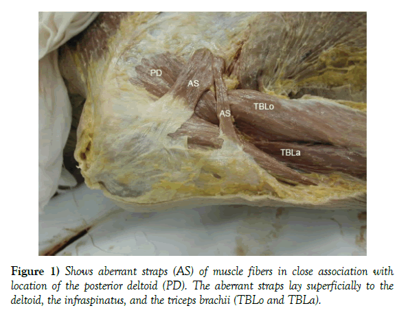
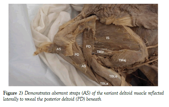
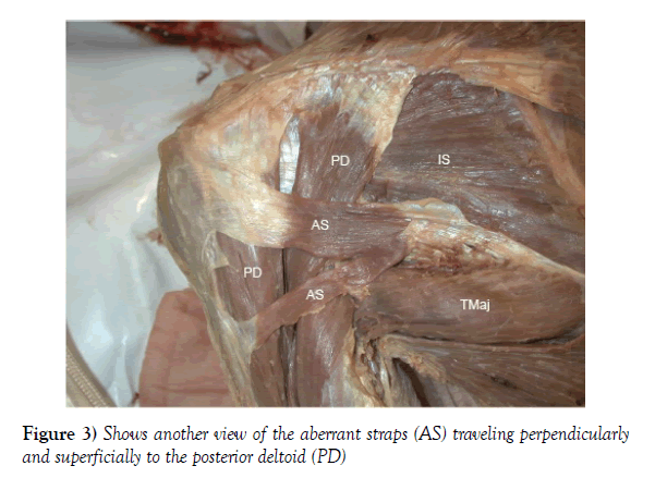
While performing a routine dissection in the gross anatomy laboratory, aberrant deltoid muscle straps were observed bilaterally on a 78-year-old female cadaver. These fibers, seen in the figures below, extend superficially from the lateral aspect of the overlying deltoid and lateral triceps brachii fascias (Figures 1-3). These fibers then lay on top of the infraspinatus muscle belly. Directionally they run at a perpendicular angle to the posterior deltoid fibers and are contained within a separate fascial sheath lying superficially over the existing muscle bellies. An additional, albeit rather small, set of fibers run from the same deep deltoid and triceps brachii fascias to then lay over the scapular spine origin of the posterior deltoid. Although these fibers are contained within a separate fascial sheath, there appears to be no change in the neurovascular anatomy associated with the posterior shoulder. A separate neurovascular bundle was not noted to accompany either of the variant muscle straps.
Discussion
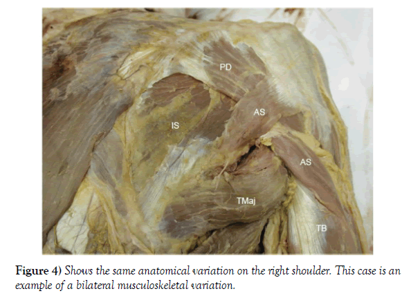
Aberrant straps of muscle in the deltoid region have been described on several occasions. One case study in 2016 demonstrated that an entire additional deltoid head accompanied the posterior deltoid muscle and was noted to be in the same orientation [4]. Another case study in 2008 presented a case of a unique unilateral aberrant muscle body on the right upper extremity of a 45 year old specimen [5]. While this case report demonstrates bilateral variants, it is clear that there are a wide range of variations possible, including the direction of the fibers as well as the symmetry (Figure 4).
Posterior abnormalities are not the sole consideration for the deltoid. Even anterior aberrant muscles have been noted. One study, in 1999, analyzed a sample of 13 cases of aberrant anterior shoulder muscles encountered during surgery and discussed their influence on the procedure [6]. The shoulder joint is an unstable joint with one of the widest ranges of motion of any joint in the body. Its mobility is derived from the construction of the capsule, which is composed of a network of tendon and connective tissue. What the shoulder joint offers in mobility, however, it suffers in stability.
Consequently, this joint is the source of a great number of acute and chronic pain cases [6]. Perhaps a portion of these cases may be attributed to the presence of variant muscles influencing the shoulder joint. In addition to pain, the shoulder is a frequent location for a variety of surgical procedures. The junction of the middle and posterior thirds of the muscle is a recommended location for the posterior splitting deltoid approach due to the ability to separate the muscle to a greater extent compared to the anterior approach [7]. It is clear that if the surgeon is not aware of the variance in musculature of the shoulder, then confusion may ensue and the surgeon may become disoriented. Considering this case study, an approach to the quadrangular space may be impeded by the muscle fibers which travel in an unusual fashion. Effective clinical management of patients with concerning shoulder issues should include consideration of atypical structures, whether intra operatively or not.
Acknowledgement
The faculty and students at the University of North Texas Health Science Center would like to take this opportunity to express a wealth of gratitude to the donors for our Willed Body Program. The gifts given here extend far beyond words and we are grateful for their generosity.
REFERENCES
- Dudek RW. Board Review Series: Embryology. Lippincott Williams & Wilkins. 2011;pp:230.
- Drake RL, Vogl W, Mitchell AWM. Gray’s Anatomy for Students. Churchill Livingstone Elsevier. 2010;pp:676.
- Ramakrishnan G. A Case Report on Variant Origin of Deltoid Muscle. Int J Adv Case Reports. 2016;3:269-72.
- Kamburoglu HO, Boran OF, Sargon MF, et al. An Unusual Variation of Deltoid Muscle. Int J Shoulder Surg. 2008;2:62-3.
- Ogawa MDK, Takahashi MDM, Yoshida MD A. Aberrant Muscle Anterior to the Shoulder Joint: Its Clinical Relevance. J Shoulder Elbow Surg. 1999;8:46-8.
- Burbank KM, Stevenson JH, Czarnecki GR, et al. Chronic Shoulder Pain: Part 1. Evaluation and Diagnosis. American Family Physician. 2008;77:1-8.
- http://www.wheelessonline.com/ortho/deltoid_muscle


