Variation of sternalis muscle: a case report
Hao (Howe) Liu1*, Vic Holmes2, Amy Nordon-Craft1 and Rusty Reeves3
1Department of Physical Therapy, USA
2Department of Physician Assistant Studies, USA
3Department of University of North Texas Health Science Center, Fort Worth, TX, USA
- *Corresponding Author:
- Hao (Howe) Liu, PT, PhD, MD
Physical Therapy, Department University of North Texas Health Science Center, 3500 Camp Bowie Blvd, Fort Worth TX, 76107, USA
Tel: +1 (817) 735-2457
E-mail: howe.liu@unthsc.edu
Date of Received: March 21st, 2012
Date of Accepted: July 6th, 2012
Published Online: October 21st, 2012
© Int J Anat Var (IJAV). 2012; 5: 59–61.
[ft_below_content] =>Keywords
sternalis, sternocleidomastoid, pectoralis major
Introduction
The sternalis muscle was first documented by Turner [1] in the 19th century and has been well reported in recent years as a variant muscle in anatomical literature [2,3]. Basically, the sternalis is a long, flat band-like muscle that appears unilaterally or bilaterally on the anterior surface of chest wall. It is usually located superficial to the pectoralis major muscle and lateral to the sternum [2–5]. There are still variances in reported literature regarding the origins, insertions, direction, extra muscle slips, and nerve innervations.
Superiorly, the sternalis has been reported to attach to the sternal origin of the ipsilateral sternocleidomastoid (SCM) muscle [2,3,5,7], to the jugular notch [4], or to the sternal angle [2,6]. In instances of bilateral existence of the sternalis, cases have been described in which superior end of the muscle tendons fuse to become a single tendon attaching to the sternal origin of the SCM [3,4]. The sternalis muscle travels inferiorly in a flat, fan-like [2,5–7] or a bundle-like [3,4] shape immediately lateral and almost parallel to the sternum [2–7]. The muscle may insert on the sternocostal arch [2], the aponeurosis of the external oblique abdominis [2,5–7] and the 6th and 7th costal cartilages [3–5]. An extra muscle slip to the sternalis is not commonly reported and has only been mentioned in two case reports [3,5]. The neural innervation to the muscle has been documented extending primarily from orgthe anterior intercostal nerves (AINs) [2–7]. Three reports documented detailed innervation: single AIN (the 3rd AIN [7], or the 6th AIN [4]) and multiple AINs (all 2nd, 3rd, and 4th AINs [5]) that can provide branches to the ipsilateral sternalis muscle. With this information in mind, we observed that the sternalis muscles discovered during our routine dissection of the anterior chest wall in the gross lab displayed several differences than have been previously reported.
Case Report
During routine gross anatomy dissection, bilateral appearance of the sternalis muscle was discovered in a 100-year-old female cadaver whose cause of death was listed as “natural causes”.
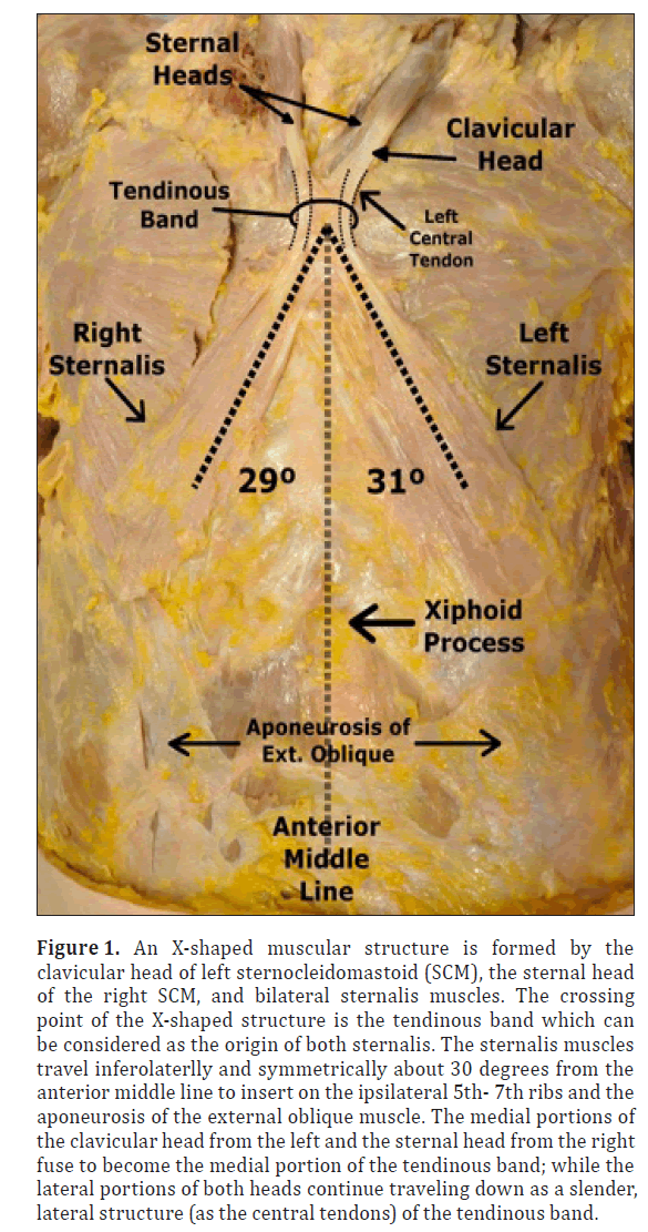
The origins of the sternalis muscles in this case were actually continuations of the clavicular origin (for the left sternalis) and the sternal origin (for the right sternalis) of the ipsilateral sternocleidomastoid (SCM) muscles (Figure 1). On the left side of the anterior chest wall, the left SCM clavicular origin (head) was not attached to the medial aspect of the clavicle. Instead, the left clavicular origin (head) tapered into an unattached flat tendon extension about 5.6 mm wide at the clavicular area. This flattened tendon extension traveled inferomedially 3.3 cm before entering a 24.5 mm x 35.0 mm rectangular tight connective band-like structure, which was named “Tendinous Band” by O’Neil and Folan-Curran (Figure 2) [5]. This band was vertically located near and slightly left of the sternal angle (Figure 1) with the deep aspect of the band attached loosely to the superficial surface of the sternal angle through thin connective tissue. On the right side, the SCM sternal origin did not attach to the manubrium. Instead, it continued inferomedially as a flat tendon about 5.4 mm in width to enter the tendinous band described above. As shown in Figure 1, the medial parts of both the clavicular head of the left SCM and the sternal head of the right SCM fused into the tendinous band at the sternal angle; while the lateral parts of both heads continued as small left and right bundle-like tendons that enter the tendinous band. We referred to these two small bundle-like tendons as “central tendons”, which were slender tendinous connecting structures between the sternalis and the ipsilateral SCM. These bundle-like central tendons were wrapped in the tendinous band but separated by about 13.3 mm. The central tendons, together with remainder of the tendinous band, contributed to the origin of both sternalis muscles at the level of 2nd intercostal space and became fleshy muscle bellies to travel down inferolaterally about 31 degrees (for the left sternalis) or 29 degrees (for the right sternalis) from the body anterior middle line. These fan-shaped muscle bellies traveled along the surface of the sternal portion of the pectoralis major muscle. Each sternalis muscle belly gradually increased in width from 18.2 mm near the 2nd intercostal space to almost 37 mm at their insertions on the ipsilateral 5th – 7th ribs and aponeurosis of the external oblique abdominis muscle (Figure 1).
Figure 1: An X-shaped muscular structure is formed by the clavicular head of left sternocleidomastoid (SCM), the sternal head of the right SCM, and bilateral sternalis muscles. The crossing point of the X-shaped structure is the tendinous band which can be considered as the origin of both sternalis. The sternalis muscles travel inferolaterlly and symmetrically about 30 degrees from the anterior middle line to insert on the ipsilateral 5th- 7th ribs and the aponeurosis of the external oblique muscle. The medial portions of the clavicular head from the left and the sternal head from the right fuse to become the medial portion of the tendinous band; while the lateral portions of both heads continue traveling down as a slender, lateral structure (as the central tendons) of the tendinous band.
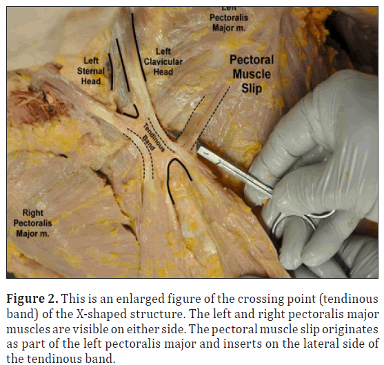
Figure 2: This is an enlarged figure of the crossing point (tendinous band) of the X-shaped structure. The left and right pectoralis major muscles are visible on either side. The pectoral muscle slip originates as part of the left pectoralis major and inserts on the lateral side of the tendinous band.
In general appearance, the shape of the muscular structure from the SCM to the ipsilateral sternalis curved from inferomedial to inferolateral resembling a boomerang (115 degree bend for the left “boomerang” and 127 degree bend for the right). At the bend of each “boomerang” was a central tendon –4 mm wide by approximately 3 cm long– within the tendinous band. The central tendons connected the left SCM clavicular head with the left sternalis and the right SCM sternal head with the right sternalis. Together, both “boomerangs” formed an “X-like” structure across the middle of the anterior chest with each sternalis muscle located almost symmetrically to either side of the anterior middle line at an angle of about 30 degrees respective to the path of the muscle versus the anterior middle line.
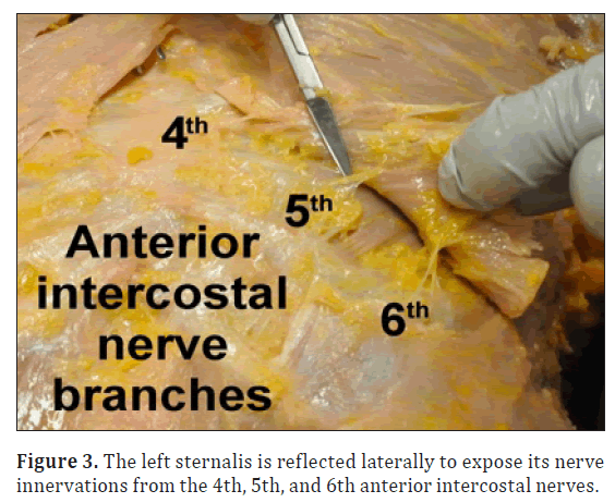
As seen in Figure 2, a pectoral muscle slip was also found laterally connected to the origin of the left sternalis muscle (the tendinous band). The slip was about 9.2 mm wide, horizontally oriented, and medially attached to the lateral edge of the tendinous band. The slip was laterally fused into the left pectoralis major muscle belly. Further, as presented in Figure 3, each of the sternalis muscles was innervated by branches from the, 4th, 5th, and 6th anterior intercostal nerves, which were observed arising from each anterior intercostal space.
Discussion
Although the sternalis muscles described in this case, to some extent, are similar to previous literature reports [1–8], this report demonstrates a few differences. First, one of the sternalis muscles originates from the clavicular head of the SCM muscle, an origin not previously described [2]. Second, the tendinous band named by O’Neill and Folan-Curran [5] is found seemingly suspended over and slightly left of the sternal angle with only a loose connection to the underlying sternal angle. This tendinous band bonds the sternal (right) and clavicular (left) origins of both SCM muscles with the ipsilateral sternalis muscles to form an X-like structure. The crossing point of this structure is held laterally by a small muscle slip originating from the pectoralis major. Third, the sternalis travels inferolaterally and nearly symmetrically on the either side of the sternum at approximately a 30 degree angle to the sternum to insert on the bony ribs of the anterolateral thoracic wall. The sternalis was previously reported almost parallel to the sternum as it traveled inferiorly to its insertion on the sternocostal arch [2] or the costal cartilages [2–5] of the anteromedial thoracic wall.
The embryological origin and nerve innervation of the sternalis muscles have been extensively discussed in two previous reports [2,5]. Based on location and connections with surrounding muscles, we agree with O’Neill and Folan-Curran’s proposal that the sternalis might originate from the hypoaxial muscle masses for the SCM, ventral muscular wall of the thoracoabdominal cavity, or even the remnants of the panniculus carnosus muscle sheet [5]. Similarity of nerve innervation to the sternalis by the anterior intercostal nerves is noticed between the present study and previous published case reports [2,4,5,7]. However, since the existence of bilateral sternalis muscles are very superficial, the nerve innervation from other places such as the medial and/or lateral pectoral nerves may have been destroyed with routine dissection. We can not, therefore, exclude the possibility of other nerve contributions to the sternalis. For a better understanding of this, a more purposeful and accurate dissection may be needed in future cases.
Clinical implications of the sternalis have been described for radiology to avoid misdiagnosis of a malignant lump(s) during mammography [2,3,7,9] and surgery to avoid misjudgment during breast operations like mastectomy [3,6,10]. Functionally, due to the attachment to the lower aspect of the rib cage, the sternalis may act as an accessory muscle to elevate the rib cage during inspiration [4]. Based on the structural observations in this case, no matter which direction the head rotates, the rib cage may be elevated through the “X” like structure. For example, when the head rotates right, the left SCM contracts to pull the left sternalis directly through the central tendon between the two left muscles as well as pull the right sternalis indirectly through the tight connective tissue (“tendinous band”) binding the right and left sternalis central tendons.
References
- Turner WM. On the musculus sternalis. J Anat Physiol. 1867; 1: 246–253.
- Raikos A, Paraskevas GK, Tzika M, Faustmann P, Triaridis S, Kordali P, Kitsoulis P, Brand-Saberi B. Sternalis muscle: an underestimated anterior chest wall anatomical variant. J Cardiothorac Surg. 2011; 6: 73.
- Sarikcioglu L, Demirel BM, Oguz N, Ucar Y. Three sternalis muscles associated with abnormal attachments of the pectoralis major muscle. Anatomy. 2008; 2: 67–71.
- Jeng H, Su SJ. The sternalis muscle: an uncommon anatomical variant among Taiwanese. J Anat. 1998; 193: 287–288.
- O’Neill MN, Folan-Curran J. Case report: bilateral sternalis muscle with a bilateral pectoralis anomaly. J Anat. 1998; 193: 289–292.
- Kale SS, Herrmann G, Kalimuthu R. Sternomastalis: a variant of the sternalis. Ann Plast Surg. 2006; 56: 340–341.
- Mehta V, Arora J, Yadav Y, Suri RK, Rath G. Rectus thoracis bifurcalis: a new variant in the anterior chest wall musculature. Rom J Morphol Embryol. 2010; 51: 799–801.
- Young Lee B, Young Byun J, Hee Kim H, Sook Kim H, Mee Cho S, Hoon Lee K, Sup Song K, Soo Kim B, Mun Lee J. The sternalis muscles: incident and imaging findings on MDCT. J Thorac Imaging. 2006; 21: 179–183.
- Bradley FM, Hoover HC Jr, Hulka CA, Whitman GJ, McCarthy KA, Hall DA, Moore R, Kopans DB. The sternalis muscle: an unusual normal finding seen on mammography. AJR Am J Roentgenol. 1996; 166: 33–36.
- Bailey PM, Tzarnas CD. The sternalis muscle: a normal finding encountered during breast surgery. Plast Reconstr Surg. 1999; 103: 1189–1190.
Hao (Howe) Liu1*, Vic Holmes2, Amy Nordon-Craft1 and Rusty Reeves3
1Department of Physical Therapy, USA
2Department of Physician Assistant Studies, USA
3Department of University of North Texas Health Science Center, Fort Worth, TX, USA
- *Corresponding Author:
- Hao (Howe) Liu, PT, PhD, MD
Physical Therapy, Department University of North Texas Health Science Center, 3500 Camp Bowie Blvd, Fort Worth TX, 76107, USA
Tel: +1 (817) 735-2457
E-mail: howe.liu@unthsc.edu
Date of Received: March 21st, 2012
Date of Accepted: July 6th, 2012
Published Online: October 21st, 2012
© Int J Anat Var (IJAV). 2012; 5: 59–61.
Abstract
During dissection of an anterior chest wall, bilateral appearance of the sternalis muscle (SM) was observed. The clavicular origin of the left sternocleidomastoid (SCM), the sternal origin of the right SCM, and both the left and right SMs together form an X-like structure. The crossing point of this structure is the tendinous band as the common origin of both SMs. Both SMs travel down inferolaterally and symmetrically to insert on ipsilateral 5th – 7th ribs and the aponeurosis of the external oblique muscle. The innervation to the muscle could be traced to the 4th, 5th, and 6th anterior intercostal nerves. Awareness of the location of the sternalis will help medical doctors avoid misdiagnosis during mammography or misjudgment during breast surgery. Because of its superior attachment to the SCM, therapists may need to be aware of that a person with such an anomaly may have an automatic accessory inspiration with head rotation.
-Keywords
sternalis, sternocleidomastoid, pectoralis major
Introduction
The sternalis muscle was first documented by Turner [1] in the 19th century and has been well reported in recent years as a variant muscle in anatomical literature [2,3]. Basically, the sternalis is a long, flat band-like muscle that appears unilaterally or bilaterally on the anterior surface of chest wall. It is usually located superficial to the pectoralis major muscle and lateral to the sternum [2–5]. There are still variances in reported literature regarding the origins, insertions, direction, extra muscle slips, and nerve innervations.
Superiorly, the sternalis has been reported to attach to the sternal origin of the ipsilateral sternocleidomastoid (SCM) muscle [2,3,5,7], to the jugular notch [4], or to the sternal angle [2,6]. In instances of bilateral existence of the sternalis, cases have been described in which superior end of the muscle tendons fuse to become a single tendon attaching to the sternal origin of the SCM [3,4]. The sternalis muscle travels inferiorly in a flat, fan-like [2,5–7] or a bundle-like [3,4] shape immediately lateral and almost parallel to the sternum [2–7]. The muscle may insert on the sternocostal arch [2], the aponeurosis of the external oblique abdominis [2,5–7] and the 6th and 7th costal cartilages [3–5]. An extra muscle slip to the sternalis is not commonly reported and has only been mentioned in two case reports [3,5]. The neural innervation to the muscle has been documented extending primarily from orgthe anterior intercostal nerves (AINs) [2–7]. Three reports documented detailed innervation: single AIN (the 3rd AIN [7], or the 6th AIN [4]) and multiple AINs (all 2nd, 3rd, and 4th AINs [5]) that can provide branches to the ipsilateral sternalis muscle. With this information in mind, we observed that the sternalis muscles discovered during our routine dissection of the anterior chest wall in the gross lab displayed several differences than have been previously reported.
Case Report
During routine gross anatomy dissection, bilateral appearance of the sternalis muscle was discovered in a 100-year-old female cadaver whose cause of death was listed as “natural causes”.
The origins of the sternalis muscles in this case were actually continuations of the clavicular origin (for the left sternalis) and the sternal origin (for the right sternalis) of the ipsilateral sternocleidomastoid (SCM) muscles (Figure 1). On the left side of the anterior chest wall, the left SCM clavicular origin (head) was not attached to the medial aspect of the clavicle. Instead, the left clavicular origin (head) tapered into an unattached flat tendon extension about 5.6 mm wide at the clavicular area. This flattened tendon extension traveled inferomedially 3.3 cm before entering a 24.5 mm x 35.0 mm rectangular tight connective band-like structure, which was named “Tendinous Band” by O’Neil and Folan-Curran (Figure 2) [5]. This band was vertically located near and slightly left of the sternal angle (Figure 1) with the deep aspect of the band attached loosely to the superficial surface of the sternal angle through thin connective tissue. On the right side, the SCM sternal origin did not attach to the manubrium. Instead, it continued inferomedially as a flat tendon about 5.4 mm in width to enter the tendinous band described above. As shown in Figure 1, the medial parts of both the clavicular head of the left SCM and the sternal head of the right SCM fused into the tendinous band at the sternal angle; while the lateral parts of both heads continued as small left and right bundle-like tendons that enter the tendinous band. We referred to these two small bundle-like tendons as “central tendons”, which were slender tendinous connecting structures between the sternalis and the ipsilateral SCM. These bundle-like central tendons were wrapped in the tendinous band but separated by about 13.3 mm. The central tendons, together with remainder of the tendinous band, contributed to the origin of both sternalis muscles at the level of 2nd intercostal space and became fleshy muscle bellies to travel down inferolaterally about 31 degrees (for the left sternalis) or 29 degrees (for the right sternalis) from the body anterior middle line. These fan-shaped muscle bellies traveled along the surface of the sternal portion of the pectoralis major muscle. Each sternalis muscle belly gradually increased in width from 18.2 mm near the 2nd intercostal space to almost 37 mm at their insertions on the ipsilateral 5th – 7th ribs and aponeurosis of the external oblique abdominis muscle (Figure 1).
Figure 1: An X-shaped muscular structure is formed by the clavicular head of left sternocleidomastoid (SCM), the sternal head of the right SCM, and bilateral sternalis muscles. The crossing point of the X-shaped structure is the tendinous band which can be considered as the origin of both sternalis. The sternalis muscles travel inferolaterlly and symmetrically about 30 degrees from the anterior middle line to insert on the ipsilateral 5th- 7th ribs and the aponeurosis of the external oblique muscle. The medial portions of the clavicular head from the left and the sternal head from the right fuse to become the medial portion of the tendinous band; while the lateral portions of both heads continue traveling down as a slender, lateral structure (as the central tendons) of the tendinous band.
Figure 2: This is an enlarged figure of the crossing point (tendinous band) of the X-shaped structure. The left and right pectoralis major muscles are visible on either side. The pectoral muscle slip originates as part of the left pectoralis major and inserts on the lateral side of the tendinous band.
In general appearance, the shape of the muscular structure from the SCM to the ipsilateral sternalis curved from inferomedial to inferolateral resembling a boomerang (115 degree bend for the left “boomerang” and 127 degree bend for the right). At the bend of each “boomerang” was a central tendon –4 mm wide by approximately 3 cm long– within the tendinous band. The central tendons connected the left SCM clavicular head with the left sternalis and the right SCM sternal head with the right sternalis. Together, both “boomerangs” formed an “X-like” structure across the middle of the anterior chest with each sternalis muscle located almost symmetrically to either side of the anterior middle line at an angle of about 30 degrees respective to the path of the muscle versus the anterior middle line.
As seen in Figure 2, a pectoral muscle slip was also found laterally connected to the origin of the left sternalis muscle (the tendinous band). The slip was about 9.2 mm wide, horizontally oriented, and medially attached to the lateral edge of the tendinous band. The slip was laterally fused into the left pectoralis major muscle belly. Further, as presented in Figure 3, each of the sternalis muscles was innervated by branches from the, 4th, 5th, and 6th anterior intercostal nerves, which were observed arising from each anterior intercostal space.
Discussion
Although the sternalis muscles described in this case, to some extent, are similar to previous literature reports [1–8], this report demonstrates a few differences. First, one of the sternalis muscles originates from the clavicular head of the SCM muscle, an origin not previously described [2]. Second, the tendinous band named by O’Neill and Folan-Curran [5] is found seemingly suspended over and slightly left of the sternal angle with only a loose connection to the underlying sternal angle. This tendinous band bonds the sternal (right) and clavicular (left) origins of both SCM muscles with the ipsilateral sternalis muscles to form an X-like structure. The crossing point of this structure is held laterally by a small muscle slip originating from the pectoralis major. Third, the sternalis travels inferolaterally and nearly symmetrically on the either side of the sternum at approximately a 30 degree angle to the sternum to insert on the bony ribs of the anterolateral thoracic wall. The sternalis was previously reported almost parallel to the sternum as it traveled inferiorly to its insertion on the sternocostal arch [2] or the costal cartilages [2–5] of the anteromedial thoracic wall.
The embryological origin and nerve innervation of the sternalis muscles have been extensively discussed in two previous reports [2,5]. Based on location and connections with surrounding muscles, we agree with O’Neill and Folan-Curran’s proposal that the sternalis might originate from the hypoaxial muscle masses for the SCM, ventral muscular wall of the thoracoabdominal cavity, or even the remnants of the panniculus carnosus muscle sheet [5]. Similarity of nerve innervation to the sternalis by the anterior intercostal nerves is noticed between the present study and previous published case reports [2,4,5,7]. However, since the existence of bilateral sternalis muscles are very superficial, the nerve innervation from other places such as the medial and/or lateral pectoral nerves may have been destroyed with routine dissection. We can not, therefore, exclude the possibility of other nerve contributions to the sternalis. For a better understanding of this, a more purposeful and accurate dissection may be needed in future cases.
Clinical implications of the sternalis have been described for radiology to avoid misdiagnosis of a malignant lump(s) during mammography [2,3,7,9] and surgery to avoid misjudgment during breast operations like mastectomy [3,6,10]. Functionally, due to the attachment to the lower aspect of the rib cage, the sternalis may act as an accessory muscle to elevate the rib cage during inspiration [4]. Based on the structural observations in this case, no matter which direction the head rotates, the rib cage may be elevated through the “X” like structure. For example, when the head rotates right, the left SCM contracts to pull the left sternalis directly through the central tendon between the two left muscles as well as pull the right sternalis indirectly through the tight connective tissue (“tendinous band”) binding the right and left sternalis central tendons.
References
- Turner WM. On the musculus sternalis. J Anat Physiol. 1867; 1: 246–253.
- Raikos A, Paraskevas GK, Tzika M, Faustmann P, Triaridis S, Kordali P, Kitsoulis P, Brand-Saberi B. Sternalis muscle: an underestimated anterior chest wall anatomical variant. J Cardiothorac Surg. 2011; 6: 73.
- Sarikcioglu L, Demirel BM, Oguz N, Ucar Y. Three sternalis muscles associated with abnormal attachments of the pectoralis major muscle. Anatomy. 2008; 2: 67–71.
- Jeng H, Su SJ. The sternalis muscle: an uncommon anatomical variant among Taiwanese. J Anat. 1998; 193: 287–288.
- O’Neill MN, Folan-Curran J. Case report: bilateral sternalis muscle with a bilateral pectoralis anomaly. J Anat. 1998; 193: 289–292.
- Kale SS, Herrmann G, Kalimuthu R. Sternomastalis: a variant of the sternalis. Ann Plast Surg. 2006; 56: 340–341.
- Mehta V, Arora J, Yadav Y, Suri RK, Rath G. Rectus thoracis bifurcalis: a new variant in the anterior chest wall musculature. Rom J Morphol Embryol. 2010; 51: 799–801.
- Young Lee B, Young Byun J, Hee Kim H, Sook Kim H, Mee Cho S, Hoon Lee K, Sup Song K, Soo Kim B, Mun Lee J. The sternalis muscles: incident and imaging findings on MDCT. J Thorac Imaging. 2006; 21: 179–183.
- Bradley FM, Hoover HC Jr, Hulka CA, Whitman GJ, McCarthy KA, Hall DA, Moore R, Kopans DB. The sternalis muscle: an unusual normal finding seen on mammography. AJR Am J Roentgenol. 1996; 166: 33–36.
- Bailey PM, Tzarnas CD. The sternalis muscle: a normal finding encountered during breast surgery. Plast Reconstr Surg. 1999; 103: 1189–1190.


