Variation of the veins of the head and neck – external jugular vein and facial vein
Balachandra N*, Padmalatha K, Prakash BS and Ramesh BR
Department of Anatomy, Dr. B.R. Ambedkar Medical College, Bengaluru, Karnataka, India
- *Corresponding Author:
- Dr. Balachandra N
Associate Professor, Department of Anatomy, Dr. B. R. Ambedkar Medical College, Bengaluru, Karnataka, India
Tel: +91 988 6592995
E-mail: suvirincha@gmail.com
Date of Received: June 23rd, 2011
Date of Accepted: July 8th, 2012
Published Online: December 12th, 2012
© Int J Anat Var (IJAV). 2012; 5: 99–101.
[ft_below_content] =>Keywords
external jugular vein, posterior auricular vein, retromandibular vein, common facial vein, internal jugular vein
Introduction
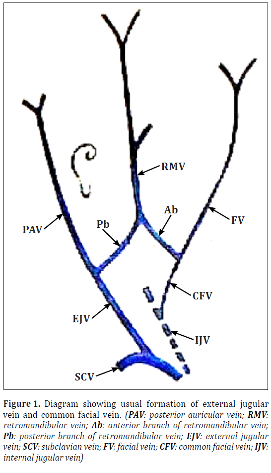
External jugular vein is formed by the union of the posterior auricular vein and the posterior division of the retromandibular vein near the angle of the mandible just below the substance of the parotid gland. It descends from the angle of the mandible obliquely to the middle of clavicle, superficial to the sternocleidomastoid muscle, then to the subclavian triangle where it pierces investing layer of deep cervical fascia to drain in the subclavian vein, lateral or anterior to the anterior scalene muscle. Common facial vein is formed by the union of the anterior division of the retromandibular vein and the facial vein and drains into the internal jugular vein (Figure 1).
Figure 1: Diagram showing usual formation of external jugular vein and common facial vein. (PAV: posterior auricular vein; RMV: retromandibular vein; Ab: anterior branch of retromandibular vein; Pb: posterior branch of retromandibular vein; EJV: external jugular vein; SCV: subclavian vein; FV: facial vein; CFV: common facial vein; IJV: internal jugular vein)
Case Report
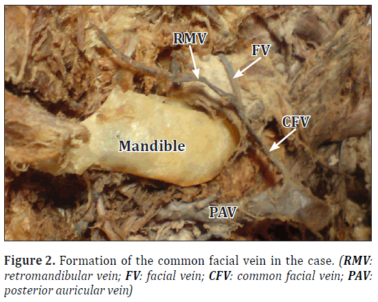
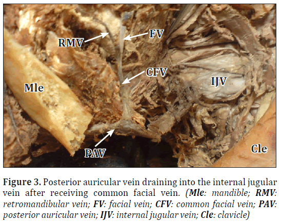
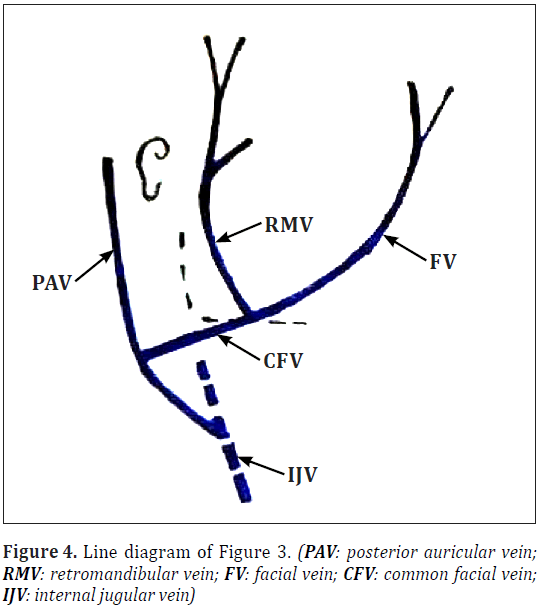
On the right side of a male cadaver aged about fifty years, we found that the external jugular vein was absent (Figure 2). The retromandibular vein was not dividing into anterior and posterior divisions; it was uniting with the facial vein to form the common facial vein (Figure 2). The common facial vein was uniting with the posterior auricular vein and draining into the internal jugular vein (Figures 3,4).
Discussion
Generally, external jugular vein is formed by the union of the posterior auricular vein and the posterior division of the retromandibular vein near the angle of the mandible. It then descends obliquely superficial to the sternocleidomastoid muscle and the subclavian triangle where it traverses the deep fascia to end in the subclavian vein. Hollinshead reported that in 1/3 of cases the external jugular vein drains into the internal jugular vein [1]. Bergman et al. opined that the formation and the termination of the external jugular vein are variable and the usual pattern is difficult to determine [2]. They also reported the absence and duplication of the external jugular vein [2]. Yadav et al. reported termination of the external jugular vein into the internal jugular vein at the level of superior belly of the omohyoid muscle [3]. Singh et al. reported an unusual course of the external jugular vein [4]. Lalwani et al. reported an unusual communication between the external jugular vein of one side, draining into the internal jugular vein of the opposite side, by a venous communication [5]. Rajanigandha et al. have found variant left external jugular vein draining into the right subclavian vein [6].
The facial vein is the continuation of the angular vein which is joined by the anterior division of the retromandibular vein in the neck to form the common facial vein, which ultimately drains into the internal jugular vein. Facial vein usually drains into the internal jugular vein but variations in its drainage have been reported. In the present case, it was draining into the posterior auricular vein instead of the internal jugular vein. In addition, it did not join with the anterior division of the retromandibular vein to form the common facial vein. Choudary et al. observed facial vein draining into the external jugular vein in 4/40 (10%) cases [7]. Gupta et al. found various patterns of facial vein draining into the external jugular vein. They have described two types of drainage. In type I, they have reported Y shaped pattern in 6 cases (37.5%); U shaped pattern in 3 cases (18.7%); tuning fork shape in 2 cases (12.5%); and N shaped pattern in 1 case (6.2%). In type II they have reported, inverted A shaped pattern in 2 cases (12.5%) and step ladder pattern in 2 cases (12.5%) [8]. Pai et al. reported 7.7% of 104 cases of facial vein draining into the external jugular vein [9]. Sumalatha et al. reported the common facial vein draining into external jugular vein [10].
The facial vein is formed by the anastomosis between the primitive maxillary vein and the linguofacial vein. The external jugular vein is developed from a tributary of the cephalic vein from the tissues of the neck and anastomoses secondarily with the anterior facial vein. At this stage the cephalic vein forms a venous ring around the clavicle from which it is connected with the caudal part of the precardinal vein. The deep segment of the venous ring forms the subclavian vein and receives the external jugular vein. The superficial segment of the venous ring dwindles, but may persist in adult life [11]. These variations are due to the persistence of the communication between the primitive linguofacial veins with the secondarily developing external jugular vein after birth. They indicate the retention and regression of the various parts of the veins formed in the rhesus monkey during development [7] or the drainage pattern found in horses, ox and dog where the veins of the face drain into the external jugular vein, the internal jugular vein being either absent or is present as a small vein accompanying the carotid artery [8].
External jugular vein is used as a venous manometer as well as catheterization and also is important medicolegally [3]. Facial veins can be used for carotid endarterectomy and for oral reconstruction [12]. The normally draining common facial vein (into internal jugular vein just above the bifurcation of the common carotid artery) provides a useful landmark for locating the carotid bifurcation. Retromandibular vein is used as a guide to expose the facial nerve branches in superficial parotidectomy and in open reduction of mandibular condylar fractures [13].
Conclusion
Clinical importance of the internal jugular vein lies in the fact that often inspection, auscultation and Doppler-sonographic examination of the jugular veins may give a clue to the diagnosis of cardiac diseases. Dilatation of these veins indicates possible compression of the superior vena cava by an underlying pathology of the mediastinum or the pericardium. Jugular veins are important for any ligations that are performed during radical neck dissection surgeries. These veins are often used for catheterization and also as venous manometers and prior anatomical knowledge is needed before such procedures. Knowledge of unusual communications between external jugular vein and internal jugular veins are of importance to radiologists performing angiographic and sonographic studies. Facial veins can be used for microvascular anastomosis in reconstruction surgeries of the head and neck. They are also used as patches for carotid endarterectomies and for oral reconstruction. Knowledge of variant drainage of the facial vein is important to avoid inappropriate dissection which may cause severe damage.
References
- Hollinshead WH. Anatomy for Surgeons. 3rd Ed., New York, Harper & Row. 1982; 467–469.
- Bergman RA, Afifi AK, Miyauchi R. Compendium of Human Anatomic Variation. Baltimore–Munich, Urban and Schwarzenberg. 1988; 82.
- Yadav S, Ghosh S K, Anand C. Variations of superficial veins of the head and neck. J Anat Soc India. 2000; 49: 61–62.
- Singh G. Variations of jugular veins: phylogenic correlation and clinical implications. http://www.thefreelibrary.com/Variations of jugular veins: phylogenic correlation and clinical...-a0155098584 (accessed July 2012)
- Lalwani R, Rana KK, Das S, Khan RQ. Communication of the external and internal jugular veins: a case report. Int J Morphol. 2006; 24: 721–722.
- Rajanigandha V, Rajalakshmi R, Ranade AV, Pai MM, Prabhu LV, Ashwin K, Jiji PJ. An anomalous left external jugular vein draining into right subclavian vein: a case report. Int J Morphol. 2008; 26: 893–895.
- Choudhry R, Tuli A, Choudhry S. Facial vein terminating in the external jugular vein. An embryologic interpretation. Surg Radiol Anat. 1997; 19: 73–77.
- Gupta V, Tuli A, Choudhry R, Agarwal S, Mangal A. Facial vein draining into external jugular vein in humans: Its variations, phylogenetic retention and clinical relevance. Surg Radiol Anat. 2003; 25: 36–41.
- Pai M, Vadgaonkar R, Prabhu L, Shetty P. The different termination patterns of the facial vein – A cadaveric study. Firat Tip Dergisi. 2008; 13: 32–34.
- D’Silva SS, Pulakunta T, Potu BK. Termination of the facial vein into the external jugular vein: an anatomical variation. J Vasc Bras. 2008; 7: 174–175.
- Williams PL, ed. Gray’s Anatomy. 38th Ed., Edinburgh, Churchill Livingstone. 1999: 327.
- Prakash B, Potu BK. A rare termination of left common facial vein into left subclavian vein: A case report. Int J Morphol. 2007; 25: 555–556.
- Vollala VR, Bolla SR, Pamidi N. Important vascular anomalies of face and neck – a cadaveric study with clinical implications. Firat Tip Dergisi. 2008; 13: 123–126.
Balachandra N*, Padmalatha K, Prakash BS and Ramesh BR
Department of Anatomy, Dr. B.R. Ambedkar Medical College, Bengaluru, Karnataka, India
- *Corresponding Author:
- Dr. Balachandra N
Associate Professor, Department of Anatomy, Dr. B. R. Ambedkar Medical College, Bengaluru, Karnataka, India
Tel: +91 988 6592995
E-mail: suvirincha@gmail.com
Date of Received: June 23rd, 2011
Date of Accepted: July 8th, 2012
Published Online: December 12th, 2012
© Int J Anat Var (IJAV). 2012; 5: 99–101.
Abstract
Knowledge of the variation of the veins of the head and neck, especially the variations of external jugular vein and the facial vein are important as they are used for various clinical procedures. External jugular veins are used for catheterization and as a venous manometer. Facial vein can be used as patches for carotid endarterectomy. Here we describe a variation of the external jugular vein and the facial vein on the right side of a cadaver. The external jugular vein was not seen. The posterior auricular vein was draining into the internal jugular vein. The retromandibular vein had no divisions; it was joining the facial vein to form the common facial vein which in turn was seen to be draining into the posterior auricular vein.
-Keywords
external jugular vein, posterior auricular vein, retromandibular vein, common facial vein, internal jugular vein
Introduction
External jugular vein is formed by the union of the posterior auricular vein and the posterior division of the retromandibular vein near the angle of the mandible just below the substance of the parotid gland. It descends from the angle of the mandible obliquely to the middle of clavicle, superficial to the sternocleidomastoid muscle, then to the subclavian triangle where it pierces investing layer of deep cervical fascia to drain in the subclavian vein, lateral or anterior to the anterior scalene muscle. Common facial vein is formed by the union of the anterior division of the retromandibular vein and the facial vein and drains into the internal jugular vein (Figure 1).
Figure 1: Diagram showing usual formation of external jugular vein and common facial vein. (PAV: posterior auricular vein; RMV: retromandibular vein; Ab: anterior branch of retromandibular vein; Pb: posterior branch of retromandibular vein; EJV: external jugular vein; SCV: subclavian vein; FV: facial vein; CFV: common facial vein; IJV: internal jugular vein)
Case Report
On the right side of a male cadaver aged about fifty years, we found that the external jugular vein was absent (Figure 2). The retromandibular vein was not dividing into anterior and posterior divisions; it was uniting with the facial vein to form the common facial vein (Figure 2). The common facial vein was uniting with the posterior auricular vein and draining into the internal jugular vein (Figures 3,4).
Discussion
Generally, external jugular vein is formed by the union of the posterior auricular vein and the posterior division of the retromandibular vein near the angle of the mandible. It then descends obliquely superficial to the sternocleidomastoid muscle and the subclavian triangle where it traverses the deep fascia to end in the subclavian vein. Hollinshead reported that in 1/3 of cases the external jugular vein drains into the internal jugular vein [1]. Bergman et al. opined that the formation and the termination of the external jugular vein are variable and the usual pattern is difficult to determine [2]. They also reported the absence and duplication of the external jugular vein [2]. Yadav et al. reported termination of the external jugular vein into the internal jugular vein at the level of superior belly of the omohyoid muscle [3]. Singh et al. reported an unusual course of the external jugular vein [4]. Lalwani et al. reported an unusual communication between the external jugular vein of one side, draining into the internal jugular vein of the opposite side, by a venous communication [5]. Rajanigandha et al. have found variant left external jugular vein draining into the right subclavian vein [6].
The facial vein is the continuation of the angular vein which is joined by the anterior division of the retromandibular vein in the neck to form the common facial vein, which ultimately drains into the internal jugular vein. Facial vein usually drains into the internal jugular vein but variations in its drainage have been reported. In the present case, it was draining into the posterior auricular vein instead of the internal jugular vein. In addition, it did not join with the anterior division of the retromandibular vein to form the common facial vein. Choudary et al. observed facial vein draining into the external jugular vein in 4/40 (10%) cases [7]. Gupta et al. found various patterns of facial vein draining into the external jugular vein. They have described two types of drainage. In type I, they have reported Y shaped pattern in 6 cases (37.5%); U shaped pattern in 3 cases (18.7%); tuning fork shape in 2 cases (12.5%); and N shaped pattern in 1 case (6.2%). In type II they have reported, inverted A shaped pattern in 2 cases (12.5%) and step ladder pattern in 2 cases (12.5%) [8]. Pai et al. reported 7.7% of 104 cases of facial vein draining into the external jugular vein [9]. Sumalatha et al. reported the common facial vein draining into external jugular vein [10].
The facial vein is formed by the anastomosis between the primitive maxillary vein and the linguofacial vein. The external jugular vein is developed from a tributary of the cephalic vein from the tissues of the neck and anastomoses secondarily with the anterior facial vein. At this stage the cephalic vein forms a venous ring around the clavicle from which it is connected with the caudal part of the precardinal vein. The deep segment of the venous ring forms the subclavian vein and receives the external jugular vein. The superficial segment of the venous ring dwindles, but may persist in adult life [11]. These variations are due to the persistence of the communication between the primitive linguofacial veins with the secondarily developing external jugular vein after birth. They indicate the retention and regression of the various parts of the veins formed in the rhesus monkey during development [7] or the drainage pattern found in horses, ox and dog where the veins of the face drain into the external jugular vein, the internal jugular vein being either absent or is present as a small vein accompanying the carotid artery [8].
External jugular vein is used as a venous manometer as well as catheterization and also is important medicolegally [3]. Facial veins can be used for carotid endarterectomy and for oral reconstruction [12]. The normally draining common facial vein (into internal jugular vein just above the bifurcation of the common carotid artery) provides a useful landmark for locating the carotid bifurcation. Retromandibular vein is used as a guide to expose the facial nerve branches in superficial parotidectomy and in open reduction of mandibular condylar fractures [13].
Conclusion
Clinical importance of the internal jugular vein lies in the fact that often inspection, auscultation and Doppler-sonographic examination of the jugular veins may give a clue to the diagnosis of cardiac diseases. Dilatation of these veins indicates possible compression of the superior vena cava by an underlying pathology of the mediastinum or the pericardium. Jugular veins are important for any ligations that are performed during radical neck dissection surgeries. These veins are often used for catheterization and also as venous manometers and prior anatomical knowledge is needed before such procedures. Knowledge of unusual communications between external jugular vein and internal jugular veins are of importance to radiologists performing angiographic and sonographic studies. Facial veins can be used for microvascular anastomosis in reconstruction surgeries of the head and neck. They are also used as patches for carotid endarterectomies and for oral reconstruction. Knowledge of variant drainage of the facial vein is important to avoid inappropriate dissection which may cause severe damage.
References
- Hollinshead WH. Anatomy for Surgeons. 3rd Ed., New York, Harper & Row. 1982; 467–469.
- Bergman RA, Afifi AK, Miyauchi R. Compendium of Human Anatomic Variation. Baltimore–Munich, Urban and Schwarzenberg. 1988; 82.
- Yadav S, Ghosh S K, Anand C. Variations of superficial veins of the head and neck. J Anat Soc India. 2000; 49: 61–62.
- Singh G. Variations of jugular veins: phylogenic correlation and clinical implications. http://www.thefreelibrary.com/Variations of jugular veins: phylogenic correlation and clinical...-a0155098584 (accessed July 2012)
- Lalwani R, Rana KK, Das S, Khan RQ. Communication of the external and internal jugular veins: a case report. Int J Morphol. 2006; 24: 721–722.
- Rajanigandha V, Rajalakshmi R, Ranade AV, Pai MM, Prabhu LV, Ashwin K, Jiji PJ. An anomalous left external jugular vein draining into right subclavian vein: a case report. Int J Morphol. 2008; 26: 893–895.
- Choudhry R, Tuli A, Choudhry S. Facial vein terminating in the external jugular vein. An embryologic interpretation. Surg Radiol Anat. 1997; 19: 73–77.
- Gupta V, Tuli A, Choudhry R, Agarwal S, Mangal A. Facial vein draining into external jugular vein in humans: Its variations, phylogenetic retention and clinical relevance. Surg Radiol Anat. 2003; 25: 36–41.
- Pai M, Vadgaonkar R, Prabhu L, Shetty P. The different termination patterns of the facial vein – A cadaveric study. Firat Tip Dergisi. 2008; 13: 32–34.
- D’Silva SS, Pulakunta T, Potu BK. Termination of the facial vein into the external jugular vein: an anatomical variation. J Vasc Bras. 2008; 7: 174–175.
- Williams PL, ed. Gray’s Anatomy. 38th Ed., Edinburgh, Churchill Livingstone. 1999: 327.
- Prakash B, Potu BK. A rare termination of left common facial vein into left subclavian vein: A case report. Int J Morphol. 2007; 25: 555–556.
- Vollala VR, Bolla SR, Pamidi N. Important vascular anomalies of face and neck – a cadaveric study with clinical implications. Firat Tip Dergisi. 2008; 13: 123–126.


