Variations in branching pattern of musculocutaneous nerve with respect to communicating branch between musculocutaneous and median nerve
2 Demonstrator, Dr. D Y Patil Medical College, Hospital & Research Centre, Pimpri, Pune, Dr. D.Y. Patil Vidyapeeth, Pune, India
3 Professor and Head, Department of Anatomy, M.I.M.E.R Medical College, Talegaon, Pune, India
Received: 25-Jun-2018 Accepted Date: Jul 09, 2018; Published: 23-Jul-2018
Citation: Khake SA, Tom DK, Belsare SM. Variations in branching pattern of musculocutaneous nerve with respect to communicating branch between musculocutaneous and median nerve. Int J Anat Var. 2018;11(3):77-80.
This open-access article is distributed under the terms of the Creative Commons Attribution Non-Commercial License (CC BY-NC) (http://creativecommons.org/licenses/by-nc/4.0/), which permits reuse, distribution and reproduction of the article, provided that the original work is properly cited and the reuse is restricted to noncommercial purposes. For commercial reuse, contact reprints@pulsus.com
Abstract
Aim: To study and classify the variations in branching of musculocutaneous nerve when the communicating branch is present between median nerve and musculocutaneous nerve.
Methods: The study was conducted on 60 upper limbs from 30 embalmed cadavers. Stepwise dissection of axilla and arm was carried out and formation of brachial plexus and its branches were studied. Variations from the normal pattern were noted and photographed.
Results: Communicating branch between musculocutaneous nerve and median nerve was observed in 21 of 60 upper limbs specimen studied (36.66%). In these specimens, variations in the branching pattern of musculocutaneous nerve were observed. These variations are described and discussed in the study
Conclusions: Knowledge of the variations involving musculocutaneous nerve when communicating branch between musculocutaneous nerve and median nerve is present is clinically important in the evaluation and diagnosis of unusual presentations of peripheral nerve injuries, and effective management of the upper limb motor deficits caused by them.
Keywords
Musculocutaneous nerve; Median nerve; Nerve to coracobrachialis muscle; Communication; Lateral cord.
Introduction
At the infra-clavicular level, the lateral cord of the brachial plexus bifurcates into the musculocutaneous nerve (MCN), lateral pectoral nerve and the lateral root (LR) of the median nerve (MN) [1]. The musculocutaneous nerve (MCN) pierces the coracobrachialis muscle (CBM) supplies it and descends between biceps muscle (BM) & brachialis muscle (BrM) to supply them, and below the elbow continues as the lateral cutaneous nerve of forearm (LCNF) [2,3].
The median nerve (MN) is formed in axilla by the medial root (MR) of medial cord (MC) and the lateral root (LR) of lateral cord of the brachial plexus. It supplies the flexor muscles of the forearm and thenar muscles [4].
Normally the musculocutaneous nerve and the median nerve pass through the flexor compartment of the arm without having any communicating branch between them [5]. The incidence of communication between musculocutaneous nerve and the median nerve have been reported with wide variability between 2.1% to 63.5% by various authors [6-9]. Communications between the musculocutaneous nerve (MCN) and median nerve (MN) have been observed and classified in different ways, by Le Minor in five types [10], by Venieratos and Anagnostopoulou (1998) in three types [11] and Choi et al. in three types [8]. The possible reason for these communications may be attributed to the stage of embryonic development. It is possible that during embryonic development some nerve fibers which were originally part of the median nerve traverse through the musculocutaneous nerve. In the arm these fibers separate from musculocutaneous nerve to form a communicating branch to join back with the median nerve [7].
Knowledge of this communicating branch between musculocutaneous nerve and median nerve and associated variations in branching pattern of musculocutaneous nerve is relevant to clinicians. It helps in assessment and appropriate management of peripheral nerve lesions causing motor deficits of upper limb. It also helps the surgeon in proper planning of the surgical approach to avoid iatrogenic neurological damage while operating in the region of arm and surgical neck of humerus [12].
Materials and Methods
The study was conducted on 60 upper limbs from 30 cadavers dissected by undergraduate students of M.I.M.E.R Medical College, Talegaon Dabhade, Pune. The cadavers were embalmed with 10 per cent formalin and fixed. Stepwise dissection of axilla and arm was carried out and formation of brachial plexus and its branches were studied. Variations from the normal pattern were noted and photographed.
Observation
We studied the course and branching pattern of the musculocutaneous nerve with special emphasis on specimens having a communicating branch arising from musculocutaneous nerve and joining the median nerve. We also studied the anatomical relationship of this communicating branch with coracobrachialis muscle.
Normally the lateral cord divides into lateral root of median nerve, the musculocutaneous nerve and the lateral pectoral nerve. Median nerve was formed by lateral root of lateral cord and medial root of medial cord of the brachial plexus. In these cases coracobrachialis muscle was supplied by musculocutaneous nerve while piercing it and this normal anatomy was seen in 38 out of 60 upper limb specimens.
In 22 out of 60 upper limb specimens (i.e 36.66% of all specimens) a communicating branch from musculocutaneous nerve to median nerve was observed. In these specimens variations in the branching pattern of musculocutaneous nerve was seen. A separate nerve for coracobrachialis muscle originating from musculocutaneous nerve was also seen in 4 specimens. And in 2 upper limb specimens the nerve to coracobrachialis was given by the communicating branch.
The different variations observed and studied are described below:
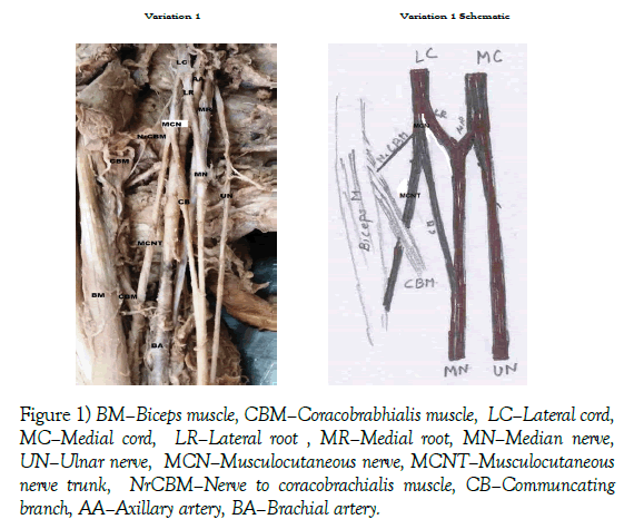
Variation 1: The musculocutaneous nerve divided into two branches before piercing the coracobrachialis muscle. First branch the nerve to coracobrachialis and second the communicating branch. The communicating branch joined the median nerve without piercing the coracobrachialis muscle. The trunk of musculocutaneous nerve pierced the coracobrachialis muscle to supply the biceps brachii and brachialis muscles and continued further as the lateral cutaneous nerve of forearm. This variation was observed unilaterally in one cadaver (1.66%) (Figure 1).
Figure 1) BM–Biceps muscle, CBM–Coracobrabhialis muscle, LC–Lateral cord, MC–Medial cord, LR–Lateral root , MR–Medial root, MN–Median nerve, UN–Ulnar nerve, MCN–Musculocutaneous nerve, MCNT–Musculocutaneous nerve trunk, NrCBM–Nerve to coracobrachialis muscle, CB–Communcating branch, AA–Axillary artery, BA–Brachial artery.
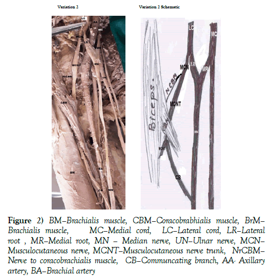
Variation 2: The musculocutaneous nerve gave a branch to coracobrachialis muscle before piercing it. The trunk of the musculocutaneous nerve pierced the coracobrachialis muscle gave muscular branches to supply the biceps brachii and brachialis muscles, a communicating branch which joined the median nerve. After giving the muscular and communicating branches as the musculocutaneous nerve continued as lateral cutaneous nerve of forearm. This variation was observed 3 of 60 upper limb specimens (5%) (Figure 2).
Figure 2) BM–Brachialis muscle, CBM–Coracobrabhialis muscle, BrM– Brachialis muscle, MC–Medial cord, LC–Lateral cord, LR–Lateral root , MR–Medial root, MN – Median nerve, UN–Ulnar nerve, MCN– Musculocutaneous nerve, MCNT–Musculocutaneous nerve trunk, NrCBM– Nerve to coracobrachialis muscle, CB–Communcating branch, AA- Axillary artery, BA–Brachial artery
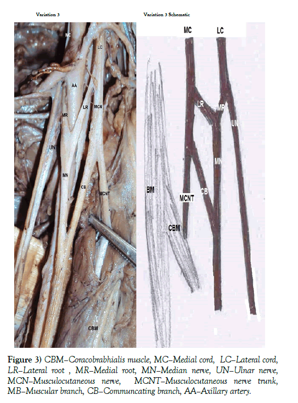
Variation 3: The musculocutaneous nerve gave the communicating branch to the median nerve before piercing the coracobrachialis muscle. The trunk of the musculocutaneous nerve pierced the coracobrachialis muscle supplied it gave muscular branches to the biceps brachii and the brachialis muscles and continued as lateral cutaneous nerve of forearm. This variation was observed 9 of 60 upper limb specimens (15%) (Figure 3).
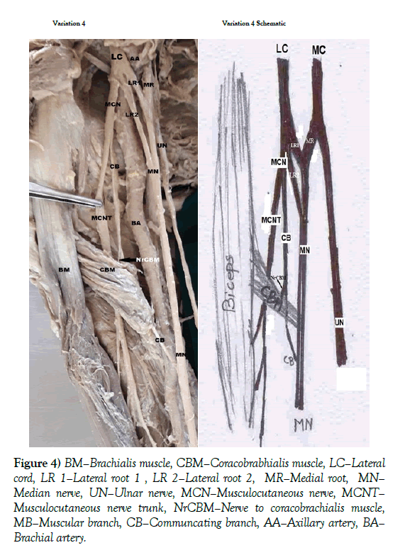
Variation 4: The musculocutaneous nerve gave the communicating branch before piercing the coracobrachialis muscle. The trunk of the musculocutaneous nerve and the communicating branch pierced the coracobrachialis muscle separately. The trunk of the musculocutaneous nerve supplied the biceps brachii and brachialis muscles whereas the coracobrachialis was supplied by the communicating branch. It was also observed that the median nerve was formed by two lateral roots (LR1 and LR2) from lateral cord and one medial root from medial cord of brachial plexus. This variation was observed bilaterally in one cadaver i.e 2 of 60 upper limb specimens (3.33%) (Figure 4).
Figure 4) BM–Brachialis muscle, CBM–Coracobrabhialis muscle, LC–Lateral cord, LR 1–Lateral root 1 , LR 2–Lateral root 2, MR–Medial root, MN– Median nerve, UN–Ulnar nerve, MCN–Musculocutaneous nerve, MCNT– Musculocutaneous nerve trunk, NrCBM–Nerve to coracobrachialis muscle, MB–Muscular branch, CB–Communcating branch, AA–Axillary artery, BA– Brachial artery.
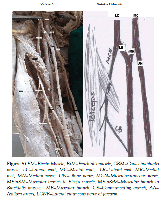
Variation 5: The musculocutaneous nerve pierced coracobrachialis muscle and supplied it. It further gave muscular branches to biceps brachii and brachialis muscle and a communicating nerve which joined the median nerve and continued as lateral cutaneous nerve of forearm. This variation was observed 7 of 60 upper limb specimens (11.66%) (Figure 5).
Figure 5) BM–Biceps Muscle, BrM–Brachialis muscle, CBM–Coracobrabhialis muscle, LC–Lateral cord, MC–Medial cord, LR–Lateral root, MR–Medial root, MN–Median nerve, UN–Ulnar nerve, MCN–Musculocutaneous nerve, MBtoBM–Muscular branch to Biceps muscle, MBtoBrM–Muscular branch to Brachialis muscle, MB–Muscular branch, CB–Communcating branch, AA– Axillary artery, LCNF–Lateral cutaneous nerve of forearm.
Discussion
Communication between musculocutaneous nerve and the median nerve is the most common and frequently observed variation amongst branches of brachial plexus and is reported by many investigators [9-12].
Veinreratos and Anagnostopolou studied 158 upper limb specimens of 79 cadavers and found communications between the musculocutaneous nerve and the median nerve in 44 upper limbs of 22 cadavers. They classified communications between the musculocutaneous nerve and the median nerve in three types based on relation with coracobrachialis muscle.
In type I the communication was proximal to the entrance of the musculocutaneous nerve into the coracobrachialis muscle (18/44 upper limb specimen). In our study we observed the communicating branch was proximal to coracobrachialis muscle and did not pierce it in 10 out of 60 upper limb specimens and we classified them as Variation 1 and Variation 3. Whereas in 2 upper limb specimens (variation 4), though the communicating branch was given proximal to coracobrachialis muscle but it pierced it.
In type II the communication was distal to the coracobrachialis muscle (20/44 upper limb specimen). In our study we observed the communicating branch distal to coracobrachialis muscle 10 out of 60 upper limbs specimens; we classified them Variation 2 and Variation 3. Whereas in 2 upper limb specimens (variation 4), though the communicating branch was given proximal to coracobrachialis muscle but it pierced it.
In type III the nerve as well as the communicating branch did not pierce the muscle (6/44 upper limb specimen) [12], we did not observe this variation in our study.
Choi et al. classified the communications between the musculocutaneous nerve and the median nerve into three types.
Type I the musculocutaneous nerve and median nerve were fused,
Type II one connecting branch between the musculocutaneous nerve and median nerve and
Type III two connecting branches were present between musculocutaneous nerve and median nerve [13].
In our study we observed only one communicating branch in all the 33.66% variations. We have not observed Type I and Type II variations in our study.
Chauhan R et al. studied 400 upper limb specimens and observed one case where the communicating branch joined the median nerve below the insertion of coracobrachialis muscle. The communicating branch was joined by a twig from musculocutaneous nerve at level of insertion of coracobrachialis muscle. The musculocutaneous nerve did not pierce the coracobrachialis muscle [14].
In our study we did not find such kind of variation.
Luis et al conducted the study on 106 fresh frozen upper limbs, they observed communicating branches between musculocutaneous nerve and median nerve in 21/106 (19.8%) upper limbs. It was bilaterally in 10 (47.6%); unilaterally in 11 (52.4%). They classified the variations depending on the origin of communicating nerve from musculocutaneous nerve as follows [8].
Type I communication was observed in 18 cases (17%) , where the communicating branch emerged after musculocutaneous nerve pierced coracobrachialis muscle and connected to the median nerve.
They further classified the type I into four subtypes depending on the emergence of the communicating branch from the musculocutaneous nerve.
Subtype I a communicating branch arose from intramuscular part of musculocutaneous nerve in 2 cases (11.1%).
Subtype I b from the proximal segment of musculocutaneous nerve before the branch to biceps muscle in 2 cases (11.1%).
Subtype I c from segment of the musculocutaneous nerve between the emergence of the branches to biceps muscle and brachialis muscle in 8 specimens (44.5%).
Subtype I d in 6 cases (33.3%) the communicating branch arose from the branch to brachialis muscle.
Type II the communicating branch was found from median nerve to musculocutaneous nerve in 3 specimens (2.8%). We have not observed such type of variations.
Priti C et al. studied 60 upper limb specimens. They observed that the communicating branch was given before the musculocutaneous nerve pierced the coracobrachialis muscle in 6 upper limb specimens [15].We observed similar finding in 10 of 60 upper limbs studied.
The communication between the musculocutaneous nerve and median nerve have been reported, discussed and classified by many authors. These variations may lead to unusual presentations of neuropathies. The musculocutaneous nerve injury proximal to communicating branch may lead to unexpected presentation of weakness of forearm and thenar muscles. This may lead to difficulty in diagnosis and treatment [16,17].
Conclusion
In our study, in variation 2 and variation 3 we observed that the nerve to coracobrachialis muscle was given as a separate branch from the musculocutaneous nerve before piercing it. And in variation 4 the nerve to coracobrachialis muscle was a branch from communicating nerve. These findings are not yet reported in literature.
Existence of the musculocutaneous nerve and median nerve communication in the arm may be the cause of unusual presentation of peripheral neuropathies. Knowledge of these communications is of great significance, especially when considering the physical examination, diagnosis, prognosis and surgical treatment of upper limb motor disorders caused by peripheral nerve injuries. This may be of value in correct surgical planning and approaches of axilla and arm, in the peripheral nerve surgery, especially in nerve transfers techniques or muscle grafts from arm.
REFERENCES
- Kosugi K, Shibata S, Yamashita H. Supernumerary head of biceps brachii and branching pattern of the musculocutaneous nerve in Japanese. Surg Radiol Anat. 1992;14:175-85.
- Kaus M, Wotowicz Z. Communicating branch between the musculocutaneous and median nerves in human. Folia Morphol (Warsz). 1995;54:273-7.
- Maeda S, Kawai K, Koizumi M, et al. Morphological study of the communication between the musculocutaneous and median nerves. Anat Sci Int. 2009;84:34-40.
- David J, Harold E. Gray’s Anatomy. In: Pectoral girdle, shoulder region and axilla. New York Churchill Livingstone Elsevier. 2008;791-822.
- Hollinshead WH. Functional anatomy of the limbs and back. 1976;P:134-40.
- Mehta V, Yadav Y, Arora J, et al. A new variant in the brachium musculature with reinforced innervation from a median-musculocutaneous nerve communication. Morphologie. 2009;93:63-6.
- Uysal II, Karabulut AK, Buyukmumcu M, et al. The course and variations of the branches of the musculocutaneous nerve in human fetuses. Clin Anat. 2009;22:337-45.
- Choi D, Rodriguez-Niedenfuhr M, Vazquez T, et al. Patterns of connections between the musculocutaneous and median nerves in the axilla and arm. Clin Anat. 2002;15:11-7.
- Guerri-Guttenberg RA, Ingolotti M. Classifying musculocutaneous nerve variations. Clin Anat. 2009;22:671-83.
- Le Minor JM. A rare variant of the median and musculocutaneous nerves in man. Archieves Anatomy Histology Embryology. 1992;73:33-42.
- Venieratos D, Anangnostopoulou S. Classification of communications between the musculocutaneous and median nerves. Clin Anat. 1998;11:327- 31.
- Luis EB, Pedro LF, Edna RB. Communication between the musculocutaneous and median nerves in the arm: an anatomical study and clinical implications. Revista Brasileria De Ortopedia. 2015;50:567-72.
- Kosugi K, Mortia T, Yamashita H. Branching pattern of the musculocutaneous nerve. 1. Cases possessing normal biceps brachii. Jikeakai Med J. 1986;33:63- 71.
- Williams PL, Bannister LH, Berry MM, et al. Gray’s Anatomy In: Nervous system. Churchill Livingston, Edinburgh. 1995;pp:1266-74.
- Chauhan R, Roy TS. Communication between the median and musculocutaneous nerve: a case report. J Anat Soc India. 2002;51:72-5.
- Priti C, Gurudeep K, Rajan S, et al. Communication between musculocutaneous and median nerve different types and their incidence in north Indian population. Indian J clini practice. 2013;24:364-71.
- Sunderland S. Nerves and Nerve Injury In: The Median Nerve: Anatomical and Physiological features. Churchill Livingstone. 1978;pp:672-7.


