Variations in origin and course of the Dorsalis pedis artery - A case study
Chun Chung Cheung*, Martin Keogh and Abduelmenem Alashkham
School of Biomedical Sciences, University of Edinburgh, Edinburgh, UK
- *Corresponding Author:
- Chun Chung Cheung
School of Biomedical Sciences
University of Edinburgh, Edinburgh, UK
Tel: +44 (0) 131 650 5103
E-mail: s1670701@sms.ed.ac.uk
Published Online : 01 February 2017
Citation: Cheung CC, Keogh M, Alashkham A. Variations in origin and course of the Dorsalis pedis artery: A case study. Int J Anat Var. 2017;10(1):001-3.
Copyright: © This open-access article is distributed under the terms of the Creative Commons Attribution Non-Commercial License (CC BY-NC) (http://creativecommons.org/licenses/by-nc/4.0/), which permits reuse, distribution and reproduction of the article, provided that the original work is properly cited and the reuse is restricted to noncommercial purposes. For commercial reuse, contact reprints@pulsus.com
[ft_below_content] =>Keywords
Dorsalis pedis; Anterior tibial artery; Peroneal artery; Aberrant; Pulse
Introduction
The development of the arterial system is complex and it is not uncommon to observe anatomical variations [1,2,3]. The dorsalis pedis artery is also known as the dorsal artery of the foot and is the major arterial supply to the forefoot. It is a continuation of the anterior tibial artery at the talocrural joint just distal to the inferior extensor retinaculum. It runs to the space between the first and second metatarsals and divides into the first dorsal metatarsal artery and the deep plantar artery, which contributes to the deep plantar arch. The anterior tibial artery is one of the two direct branches of the popliteal artery, arising at the popliteal fossa at the posterior aspect of the knee joint. Immediately after the bifurcation of the popliteal artery, the anterior tibial artery runs to the anterior compartment of the leg through an aperture in the proximal part of the interosseous membrane. It descends anterior to the membrane and becomes the dorsalis pedis artery as described above. The posterior tibial artery, the other direct branch of the popliteal artery, supplies the posterior compartment of the leg and the foot. It immediately gives off the peroneal artery. The posterior tibial artery terminates by dividing into the medial and lateral plantar arteries which supply the sole of the foot deep to the flexor retinaculum. The peroneal artery descends obliquely towards the fibula and terminates at the lateral malleolus [4,5]. The dorsalis pedis pulse is commonly evaluated in a physical examination and a weakened or absent pulse could be a result of arterial diseases [6]. Therefore, aspects of the clinical and surgical implications relating to systemic and regional examinations are considered. This paper describes the unusual origin of the dorsalis pedis artery and the associated aberrant arterial anatomy documented by photographic images in a female cadaver.
Case Report
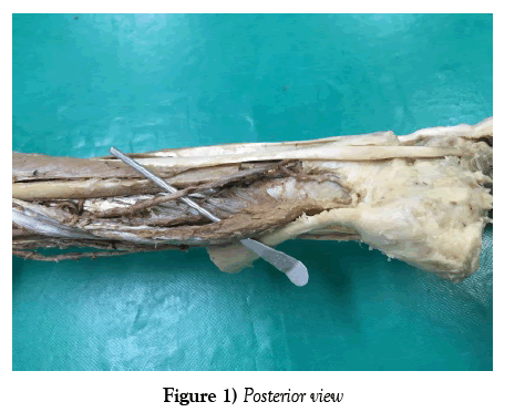
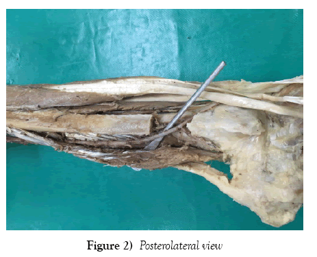
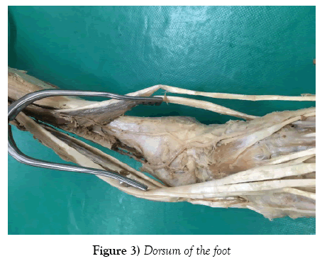
A variation of the origin of the dorsalis pedis artery was identified while performing dissection of the right lower limb of an 86 years old formaldehyde fixed female cadaver (Figure 1) with carcinoma of the distal third of the transverse colon as the certified cause of death. The usual branching of the popliteal artery into anterior and posterior tibial arteries was observed at the posterior aspect of the knee joint just inferior to the inferior border of popliteus. The anterior tibial artery (with a diameter of 2.43mm at its origin) pierced through the interosseous membrane, entered the anterior compartment of the leg and descended deep to extensor digitorum longus. However, the anterior tibial artery was tapering off and terminated 6.3cm proximal to the talocrural joint (with a diameter of 0.16mm at its termination. The posterior tibial artery followed its usual course and gave rise to the peroneal artery 5cm distal to the inferior border of popliteus. The peroneal artery (with a diameter of 3.80mm at its origin) descended inferolaterally between tibialis posterior and flexor hallucis longus parallel to posterior tibial artery to supply the lateral compartment of the leg. It gave rise to muscular branches supplying peroneus longus and brevis, nutrient branch to the fibula, communicating branches with the posterior tibial, calcaneal branches to the calcaneus and a perforating 6.3cm proximal to the talocrural joint. This perforating branch branch (with a diameter of 2.24mm) pierced the interosseous membrane and entering the anterior compartment of the leg. Then, it passed deep to the flexor retinaculum continued as the dorsalis pedis artery (with a diameter of 2.28mm) which descended in between the tendons of the extensor hallucis longus medially and extensor digitorum longus laterally.
Discussion
In this case, we have described the dorsalis pedis artery origin being from the peroneal (fibular) artery and not directly from the anterior tibial artery running a tortuous course to its terminal continuance supplying the dorsum of the foot as previously described in the literature by Lippert and Pabst [1] as occurring in 6% of specimens. While core curriculum reference texts such as Grays [4] and Grants [5] provide acknowledgement of variations in vascular branching and distribution in the leg scant recourse is given to clinical or surgical implications, or the associated developmental structure(s) [7] (Figure 1).
The arteries of the lower limb arise from two sources, the primary limb bud artery (sciatic artery) and the femoral artery. With regression of the sciatic the middle and distal segments persist forming the popliteal and peroneal arteries. The anterior tibial arises from the popliteal artery. The popliteal artery and the early distal femoral artery anastomose and form the posterior tibial. In conclusion the arterial variations in the lower limb can be explained by combinations of persistent primitive arterial segments, abnormal fusions and segmental hypoplasia or absence [2,3] (Figure 2).
Previous authors [8] point out that the identification and examination of the pulse from the dorsalis pedis artery in association with the posterior tibial and the popliteal arteries are important in assessing peripheral vascular disease, hypertension, diabetic arteriopaths; and for monitoring of peripheral integrity distal to fracture fixation stabilisation and suggest that while clinical education teaches how to locate and examine there is no guidance or developed protocol when there is difficulty in locating the dorsalis pedis pulse [9,10]. Aberrant arteries are often associated with further developmental variation [7] relating to the embryonic axial artery development [2,3] and need to be born in mind. With the percentage score sizes [11] of the artery lumen and length in regular and atypical branching origins of the artery changes in pulse wave formation, velocity and volume may lead to clinical misinterpretation [10]. The surgical implication for skin tissue flaps [8] being viable from the dorsum of the foot are dependent upon dorsalis pedis anchoring in plastic and burns procedures and have previously been identified as important considerations for surgeons (Figure 3).
In Orthopaedics however these authors believe that this case study illustrates the need to comprehend how external fixation (especially casting and bracing) and internal fixation can lead to impaired healing of associated tissues in cases of uni and bilateral malleoli, fibular or tibial shaft fractures. Interestingly trauma to the anterior talocrural joint structures will not produce the same degree of vascular sequelae in cases where the origin is not from the anterior tibial artery in front of the mid joint. The more oblique path of the artery in this case study may very well compound hyper-pronation syndromes and cases of pes cavus particularly in diabetic peripheral neuropathy and arthropathy; in addition to being vulnerable during ankle arthroscopy. It is theorised that a peroneal (fibular) origin could make the dorsalis pedis artery more at risk from lateral and posterior compartment syndromes as opposed to anterior [12].
Arterial puncture sites include the dorsalis pedis artery in the infant, child and adult with a successful outcome is uncommon and probably reflects aberrant anatomy and inadequate anatomical knowledge.
Peripheral vascular resistance is determined by vascular musculature tone [11] and blood vessel diameter and length therefore dimensional changes associated with the arteries as described are co morbidity factors in the ever-increasing rate and prevalence of cardiovascular and circulatory disease (Figure 4). In previous large study [6] of 53 limbs dissected, a normal branching pattern was seen in 56% of dorsalis pedis arteries, with variable origins in 8% and branching variations in 16%. There was an absence or a duplication in 2%. In this particular account described in this paper shows a more unusual triad of variation and in particular highlights the extended course and associated attrition of arterial caliber and gives supporting evidence to the need for a high index of clinical suspicion assessing the vascular integrity of the ankle and foot; and for anatomical educators to be alert to unusual findings [3].
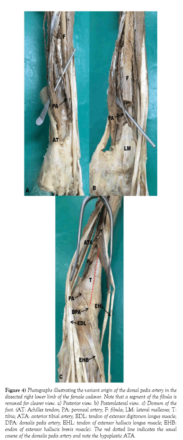
Figure 4: Photographs illustrating the variant origin of the dorsal pedis artery in the dissected right lower limb of the female cadaver. Note that a segment of the fibula is removed for clearer view. a) Posterior view. b) Posterolateral view. c) Dorsum of the foot. (AT: Achilles tendon; PA: peroneal artery; F: fibula; LM: lateral malleous; T: tibia; ATA: anterior tibial artery; EDL: tendon of extensor digitorum longus muscle; DPA: dorsalis pedis artery; EHL: tendon of extensor hallucis longus muscle; EHB: endon of extensor hallucis brevis muscle). The red dotted line indicates the usual course of the dorsalis pedis artery and note the hypoplastic ATA.
References
- Lippert H, Pabst R. Arterial variations in man: classification and frequency. Munich, J.F. Bergman Verlag. 1995; 63.
- Sahin B, Bilgic S. Two rare arterial variations of the deep femoral artery in the new born. Surg Radial Anat. 1998; 20:233-35.
- Atanasova M, Georgiev GP, Jelev L. Intriguing variations of the tibial arteries and their clinical implications. International Journal of Anatomic Variations. 2011; 4: 45-47.
- Standring S. Gray’s Anatomy the Anatomical Basis Of Clinical Practice. 41st Ed, London, Elsevier. 2016; 1405-15.
- Detton A J. Grants Dissector. 16th Ed, Philadelphia, Wolters Kluwer. 2015; 210.
- Vijayalakshmi S, Gunapriya R, Varsha S. Anatomical study of Dorsalis pedis artery and its clinical correlations. Journal of Clinical and Diagnostic Reseach. 2011; 5(2): 287-90.
- Taser,f., Shafiq, Q., Ebraheim, N.A. et al. Enlarged perforating branch of peroneal artery and extra crural fascia in close relationship with the tibiofibular syndemossi. Surg Radiol Anat. 2006; 28: 108.
- Clemenza J W, Rogers S, Magennis P. Pre operative evaluation of the lower extremity prior to microvascular free fibular flap harvest. Annals of the Royal College of Surgeons of England. March 2000; 82(2): 122-27.
- Thomas A, Kimber C, Bramwell D, Jaarsma R. Improving clinical examination in acute tibial fractures by enhancing visual cues; the case for always “cutting back” a tibial backslab and marking the dorsalis pedis. International Journal of Orthopaedic and Trauma Nursing. 2016; 22: 36-43.
- Glyn M, Drake W M. Hutchinson’s Clinical Methods An Integrated Approach to Clinical Practice. 23d Ed. London. Elsevier. 2012.
- Su H W, Lin TH, Hsu P C, et al. Association of bilateral ankle pulse wave velocity difference with peripheral vascular disease and left ventricular mass index. Catapano A, Ed. PLoS ONE. 2014; 9:e88331.
- Bellezza P A, Elliott E, Conlee T, Clements J R. Aplastic posterior tibial artery in presence of trimalleolar ankle fracture dislocation resulting in below knee amputation. The Journal of Foot and Ankle Surgery. 2016; 10:1053.
Chun Chung Cheung*, Martin Keogh and Abduelmenem Alashkham
School of Biomedical Sciences, University of Edinburgh, Edinburgh, UK
- *Corresponding Author:
- Chun Chung Cheung
School of Biomedical Sciences
University of Edinburgh, Edinburgh, UK
Tel: +44 (0) 131 650 5103
E-mail: s1670701@sms.ed.ac.uk
Published Online : 01 February 2017
Citation: Cheung CC, Keogh M, Alashkham A. Variations in origin and course of the Dorsalis pedis artery: A case study. Int J Anat Var. 2017;10(1):001-3.
Copyright: © This open-access article is distributed under the terms of the Creative Commons Attribution Non-Commercial License (CC BY-NC) (http://creativecommons.org/licenses/by-nc/4.0/), which permits reuse, distribution and reproduction of the article, provided that the original work is properly cited and the reuse is restricted to noncommercial purposes. For commercial reuse, contact reprints@pulsus.com
Abstract
The application of anatomical knowledge of the arterial system is important to clinical practitioners requiring access to available sites for investigations, procedures or clinical examination. The increasing reliance upon high-tech diagnostics and at the other spectral end devolved clinical examination skills to professions supplementary to medicine may fail to take into account any anatomical basis of changes in peripheral pulses particularly when they are notoriously difficult to identify reliably such as the dorsalis pedis. Dissection for anatomical study of a female cadaver identified aberrant right dorsalis pedis originating from the peroneal artery as a continuance of the perforating branch as opposed to the usual anterior tibial artery that in this case was tapering off. The other structures of the anterior, posterior and lateral compartments were observed to follow the same course as described in standard anatomy textbooks.
-Keywords
Dorsalis pedis; Anterior tibial artery; Peroneal artery; Aberrant; Pulse
Introduction
The development of the arterial system is complex and it is not uncommon to observe anatomical variations [1,2,3]. The dorsalis pedis artery is also known as the dorsal artery of the foot and is the major arterial supply to the forefoot. It is a continuation of the anterior tibial artery at the talocrural joint just distal to the inferior extensor retinaculum. It runs to the space between the first and second metatarsals and divides into the first dorsal metatarsal artery and the deep plantar artery, which contributes to the deep plantar arch. The anterior tibial artery is one of the two direct branches of the popliteal artery, arising at the popliteal fossa at the posterior aspect of the knee joint. Immediately after the bifurcation of the popliteal artery, the anterior tibial artery runs to the anterior compartment of the leg through an aperture in the proximal part of the interosseous membrane. It descends anterior to the membrane and becomes the dorsalis pedis artery as described above. The posterior tibial artery, the other direct branch of the popliteal artery, supplies the posterior compartment of the leg and the foot. It immediately gives off the peroneal artery. The posterior tibial artery terminates by dividing into the medial and lateral plantar arteries which supply the sole of the foot deep to the flexor retinaculum. The peroneal artery descends obliquely towards the fibula and terminates at the lateral malleolus [4,5]. The dorsalis pedis pulse is commonly evaluated in a physical examination and a weakened or absent pulse could be a result of arterial diseases [6]. Therefore, aspects of the clinical and surgical implications relating to systemic and regional examinations are considered. This paper describes the unusual origin of the dorsalis pedis artery and the associated aberrant arterial anatomy documented by photographic images in a female cadaver.
Case Report
A variation of the origin of the dorsalis pedis artery was identified while performing dissection of the right lower limb of an 86 years old formaldehyde fixed female cadaver (Figure 1) with carcinoma of the distal third of the transverse colon as the certified cause of death. The usual branching of the popliteal artery into anterior and posterior tibial arteries was observed at the posterior aspect of the knee joint just inferior to the inferior border of popliteus. The anterior tibial artery (with a diameter of 2.43mm at its origin) pierced through the interosseous membrane, entered the anterior compartment of the leg and descended deep to extensor digitorum longus. However, the anterior tibial artery was tapering off and terminated 6.3cm proximal to the talocrural joint (with a diameter of 0.16mm at its termination. The posterior tibial artery followed its usual course and gave rise to the peroneal artery 5cm distal to the inferior border of popliteus. The peroneal artery (with a diameter of 3.80mm at its origin) descended inferolaterally between tibialis posterior and flexor hallucis longus parallel to posterior tibial artery to supply the lateral compartment of the leg. It gave rise to muscular branches supplying peroneus longus and brevis, nutrient branch to the fibula, communicating branches with the posterior tibial, calcaneal branches to the calcaneus and a perforating 6.3cm proximal to the talocrural joint. This perforating branch branch (with a diameter of 2.24mm) pierced the interosseous membrane and entering the anterior compartment of the leg. Then, it passed deep to the flexor retinaculum continued as the dorsalis pedis artery (with a diameter of 2.28mm) which descended in between the tendons of the extensor hallucis longus medially and extensor digitorum longus laterally.
Discussion
In this case, we have described the dorsalis pedis artery origin being from the peroneal (fibular) artery and not directly from the anterior tibial artery running a tortuous course to its terminal continuance supplying the dorsum of the foot as previously described in the literature by Lippert and Pabst [1] as occurring in 6% of specimens. While core curriculum reference texts such as Grays [4] and Grants [5] provide acknowledgement of variations in vascular branching and distribution in the leg scant recourse is given to clinical or surgical implications, or the associated developmental structure(s) [7] (Figure 1).
The arteries of the lower limb arise from two sources, the primary limb bud artery (sciatic artery) and the femoral artery. With regression of the sciatic the middle and distal segments persist forming the popliteal and peroneal arteries. The anterior tibial arises from the popliteal artery. The popliteal artery and the early distal femoral artery anastomose and form the posterior tibial. In conclusion the arterial variations in the lower limb can be explained by combinations of persistent primitive arterial segments, abnormal fusions and segmental hypoplasia or absence [2,3] (Figure 2).
Previous authors [8] point out that the identification and examination of the pulse from the dorsalis pedis artery in association with the posterior tibial and the popliteal arteries are important in assessing peripheral vascular disease, hypertension, diabetic arteriopaths; and for monitoring of peripheral integrity distal to fracture fixation stabilisation and suggest that while clinical education teaches how to locate and examine there is no guidance or developed protocol when there is difficulty in locating the dorsalis pedis pulse [9,10]. Aberrant arteries are often associated with further developmental variation [7] relating to the embryonic axial artery development [2,3] and need to be born in mind. With the percentage score sizes [11] of the artery lumen and length in regular and atypical branching origins of the artery changes in pulse wave formation, velocity and volume may lead to clinical misinterpretation [10]. The surgical implication for skin tissue flaps [8] being viable from the dorsum of the foot are dependent upon dorsalis pedis anchoring in plastic and burns procedures and have previously been identified as important considerations for surgeons (Figure 3).
In Orthopaedics however these authors believe that this case study illustrates the need to comprehend how external fixation (especially casting and bracing) and internal fixation can lead to impaired healing of associated tissues in cases of uni and bilateral malleoli, fibular or tibial shaft fractures. Interestingly trauma to the anterior talocrural joint structures will not produce the same degree of vascular sequelae in cases where the origin is not from the anterior tibial artery in front of the mid joint. The more oblique path of the artery in this case study may very well compound hyper-pronation syndromes and cases of pes cavus particularly in diabetic peripheral neuropathy and arthropathy; in addition to being vulnerable during ankle arthroscopy. It is theorised that a peroneal (fibular) origin could make the dorsalis pedis artery more at risk from lateral and posterior compartment syndromes as opposed to anterior [12].
Arterial puncture sites include the dorsalis pedis artery in the infant, child and adult with a successful outcome is uncommon and probably reflects aberrant anatomy and inadequate anatomical knowledge.
Peripheral vascular resistance is determined by vascular musculature tone [11] and blood vessel diameter and length therefore dimensional changes associated with the arteries as described are co morbidity factors in the ever-increasing rate and prevalence of cardiovascular and circulatory disease (Figure 4). In previous large study [6] of 53 limbs dissected, a normal branching pattern was seen in 56% of dorsalis pedis arteries, with variable origins in 8% and branching variations in 16%. There was an absence or a duplication in 2%. In this particular account described in this paper shows a more unusual triad of variation and in particular highlights the extended course and associated attrition of arterial caliber and gives supporting evidence to the need for a high index of clinical suspicion assessing the vascular integrity of the ankle and foot; and for anatomical educators to be alert to unusual findings [3].
Figure 4: Photographs illustrating the variant origin of the dorsal pedis artery in the dissected right lower limb of the female cadaver. Note that a segment of the fibula is removed for clearer view. a) Posterior view. b) Posterolateral view. c) Dorsum of the foot. (AT: Achilles tendon; PA: peroneal artery; F: fibula; LM: lateral malleous; T: tibia; ATA: anterior tibial artery; EDL: tendon of extensor digitorum longus muscle; DPA: dorsalis pedis artery; EHL: tendon of extensor hallucis longus muscle; EHB: endon of extensor hallucis brevis muscle). The red dotted line indicates the usual course of the dorsalis pedis artery and note the hypoplastic ATA.
References
- Lippert H, Pabst R. Arterial variations in man: classification and frequency. Munich, J.F. Bergman Verlag. 1995; 63.
- Sahin B, Bilgic S. Two rare arterial variations of the deep femoral artery in the new born. Surg Radial Anat. 1998; 20:233-35.
- Atanasova M, Georgiev GP, Jelev L. Intriguing variations of the tibial arteries and their clinical implications. International Journal of Anatomic Variations. 2011; 4: 45-47.
- Standring S. Gray’s Anatomy the Anatomical Basis Of Clinical Practice. 41st Ed, London, Elsevier. 2016; 1405-15.
- Detton A J. Grants Dissector. 16th Ed, Philadelphia, Wolters Kluwer. 2015; 210.
- Vijayalakshmi S, Gunapriya R, Varsha S. Anatomical study of Dorsalis pedis artery and its clinical correlations. Journal of Clinical and Diagnostic Reseach. 2011; 5(2): 287-90.
- Taser,f., Shafiq, Q., Ebraheim, N.A. et al. Enlarged perforating branch of peroneal artery and extra crural fascia in close relationship with the tibiofibular syndemossi. Surg Radiol Anat. 2006; 28: 108.
- Clemenza J W, Rogers S, Magennis P. Pre operative evaluation of the lower extremity prior to microvascular free fibular flap harvest. Annals of the Royal College of Surgeons of England. March 2000; 82(2): 122-27.
- Thomas A, Kimber C, Bramwell D, Jaarsma R. Improving clinical examination in acute tibial fractures by enhancing visual cues; the case for always “cutting back” a tibial backslab and marking the dorsalis pedis. International Journal of Orthopaedic and Trauma Nursing. 2016; 22: 36-43.
- Glyn M, Drake W M. Hutchinson’s Clinical Methods An Integrated Approach to Clinical Practice. 23d Ed. London. Elsevier. 2012.
- Su H W, Lin TH, Hsu P C, et al. Association of bilateral ankle pulse wave velocity difference with peripheral vascular disease and left ventricular mass index. Catapano A, Ed. PLoS ONE. 2014; 9:e88331.
- Bellezza P A, Elliott E, Conlee T, Clements J R. Aplastic posterior tibial artery in presence of trimalleolar ankle fracture dislocation resulting in below knee amputation. The Journal of Foot and Ankle Surgery. 2016; 10:1053.


