Variations in the branching pattern of axillary artery with high origin of radial artery
Syed Rehan Daimi*, Abu Ubaida Siddiqui and Rajendra Namdeo Wabale
Department of Anatomy, Rural Medical College, PIMS, Ahmednagar, Maharashtra, India
- *Corresponding Author:
- Syed Rehan Daimi, MD
Assistant Professor Department of Anatomy, Rural Medical College, PIMS, Loni 413736 Ahmednagar, Maharashtra, India
Tel: +91 9970441463
E-mail: daimi_dr@yahoo.com
Date of Received: October 9th, 2009
Date of Accepted: April 7th, 2010
Published Online: May 17th, 2010
© Int J Anat Var (IJAV). 2010; 3: 76–77.
[ft_below_content] =>Keywords
axillary artery, lateral thoracic artery, thoracoacromial artery, subscapular artery, radial artery
Introduction
Accurate knowledge of the normal and variant anatomy of the axillary region is a prerequisite for correct diagnosis of the underlying pathology. The subsequent clinical procedures are of utmost significance for the vascular radiologists, surgeons and clinical anatomists. The vascular variations of the region should be well known [1]. The course and branching pattern of the axillary artery vary with race, sex and ethnic groups [1,2].
Axillary artery is the continuation of subclavian artery from the outer border of the first rib and continues as the brachial artery at the inferior border of teres major. Pectoralis minor muscle divides the artery in three parts, as the first part (proximal), second part (posterior) and third part (distal) to the muscle. As classically described in anatomical texts, axillary artery gives six branches; first part giving rise to superior thoracic, second part to thoracoacromial and lateral thoracic and the third part to subscapular, anterior circumflex humeral and posterior circumflex humeral arteries [3].
Various authors have described the variations in the branching pattern of the axillary artery [1,2,4–9]. During routine dissection of the upper limb, we came across a unique branching pattern, which has not yet been described in the literature. The variant observed is not only rare but seems to be relevant and significant for understanding the formation of the arteries of the arm.
Case Report
The described arterial variation was found in the right upper limb of a cadaver, 70-year-old Indian male, during routine dissection in the Department of Anatomy, Rural Medical College, Loni. The history of the individual and the cause of death are not known. The topographic details of the arteries were examined by casual dissection and the variations were recorded and photographed.
Several variations were observed, namely:
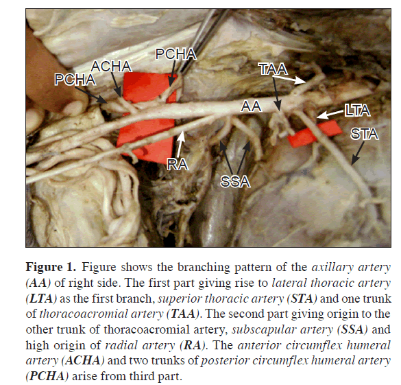
1) Lateral thoracic artery emerged as the first branch of the axillary artery (instead of the superior thoracic artery, as classically described), arising from the first part of the axillary artery. It crossed the superior thoracic artery and passed laterally.
2) Two trunks of thoracacromial artery were observed. One took origin from the superior aspect of the first part of the axillary artery and the other one from the anterior aspect of the second part of the axillary artery. Both divided into 3-4 terminal branches.
3) Subscapular artery arose from the second part of the axillary artery (instead from the third part) and immediately after origin, divided in two branches. One branch ran downwards, after giving off a small circumflex scapular artery, and continued as thoracodorsal artery, while the other branch supplied the subscapularis muscle.
4) High origin of the radial artery was another interesting feature observed, which arose from the second part of the axillary artery. The course of the artery was superficial, running downwards and laterally, crossing the brachial artery and median nerve from medial to lateral side in the arm, without giving any branch.
Figure 1: Figure shows the branching pattern of the axillary artery (AA) of right side. The first part giving rise to lateral thoracic artery (LTA) as the first branch, superior thoracic artery (STA) and one trunk of thoracoacromial artery (TAA). The second part giving origin to the other trunk of thoracoacromial artery, subscapular artery (SSA) and high origin of radial artery (RA). The anterior circumflex humeral artery (ACHA) and two trunks of posterior circumflex humeral artery (PCHA) arise from third part.
5) Two trunks of the posterior circumflex humeral arteries were observed, arising from the third part of the axillary artery. Both were situated on a plane posterior to the origin of anterior circumflex humeral artery. The diameter of both the posterior circumflex humeral arteries was larger as compared to the diameter of anterior circumflex humeral artery. One artery continued laterally together with axillary nerve and appeared in the quadrangular space. Other artery passed medially piercing the teres minor muscle and appeared on the dorsal surface of scapula.
Discussion
It is not uncommon to encounter variations of the axillary artery. Many authors have described variants in the course and branching pattern and the number of branches [1,2,4]. Patnaik et al., quoted that De Garis and Swartley (1928) in a study of 512 axillary arteries found 5-11 branches, the commonest number being eight [4], whereas the textbooks description gives the named branches as six [3]. We found eight branches, amongst those, additional branches were one extra branch each of thoracoacromial and posterior circumflex humeral artery.
Patnaik et al. described lateral thoracic artery arising from second part of axillary artery in 92% of the limbs and in 6% directly from first part [4]. In the present case, it was arising from the first part, and also emerging as the first branch of the axillary artery.
Yang et al. studied 304 Korean cadavers and described high origin of radial artery from axillary artery in 2.3% of cases. This was the most frequent variation found in the western populations as quoted by them [2]. Baeza AR found the radial artery arising from the axillary artery in 16 cases out of 150 cadavers [5].
Most frequently described variant is that, the axillary artery dividing into 2 trunks; one continuing as the brachial artery and the other trunk giving rest of the branches [6–9]. But two thoracoacromial trunks and two posterior circumflex humeral arteries and an early division of the subscapular artery; which are concurrently present in our case; is not yet described in the available literature.
Arterial anomalies in the upper limb are due to defects in embryonic development of the vascular plexus of upper limb bud. This may be due to arrest at any stage of development of vessels followed by regression, retention or reappearance, thus leading to variations in the arterial origin and course of major upper limb vessels [10].
The high origin and superficial course of the radial artery, as we reported, may be hazardous and vulnerable to injury during venepuncture and surgical procedures. On the other hand, its superficial course makes arterial grafting and cardiac catheterization easier. Therefore knowledge of both normal and variant anatomy of the region is a must for accurate diagnosis and treatment.
References
- Saeed M, Rufai AA, Elsayed SE, Sadiq MS. Variations in subclavian-axillary arterial system. Saudi Med J. 2002; 23: 206–212.
- Yang HJ, Gil YC, Jung WS, Lee HY. Variations of the superficial brachial artery in Korean cadavers. J Korean Med Sci. 2008; 23: 884–887.
- Susan Standring ed. Gray’s Anatomy. 40th Ed., London, Churchill Livingstone. 2008; 815–817.
- Patnaik VVG, Kalsey G, Singla RK. Branching pattern of axillary artery – a morphological study. J Anat Soc India. 2000; 49: 127–132.
- Rodriguez-Baeza A, Nebot J, Ferreira B, Reina F, Perez J, Sanudo JR, Roig M. An anatomical study and ontogenetic explanation of 23 cases with variations in the main pattern of the human brachio-antebrachial arteries. J. Anat. 1995; 187: 473–479.
- Patnaik VVG, Kalse G, Singla RK. Bifurcation of axillary artery in its 3rd part – a case report. J Anat Soc India. 2001; 50: 166–169.
- Bhat KM, Gowda S, Potu BK, Rao MS. A unique branching pattern of the axillary artery in a South Indian male cadaver. Bratisl Lek Listy. 2008; 109: 587–589.
- Tan CB, Tan CK. An unusual course and relations of the human axillary artery. Singapore Med J. 1994; 35: 263–264.
- Saralaya V, Joy T, Madhyastha S, Vadgaonkar R, Saralaya S. Abnormal branching of the axillary artery: subscapular common trunk. A case report. Int J Morphol. 2008; 26: 963–966.
- Hamilton WJ, Mossman HW, eds. Cardiovascular system. In: Human Embryology. 4th Ed., Baltimore, Williams and Wilkins. 1972; 271–290.
Syed Rehan Daimi*, Abu Ubaida Siddiqui and Rajendra Namdeo Wabale
Department of Anatomy, Rural Medical College, PIMS, Ahmednagar, Maharashtra, India
- *Corresponding Author:
- Syed Rehan Daimi, MD
Assistant Professor Department of Anatomy, Rural Medical College, PIMS, Loni 413736 Ahmednagar, Maharashtra, India
Tel: +91 9970441463
E-mail: daimi_dr@yahoo.com
Date of Received: October 9th, 2009
Date of Accepted: April 7th, 2010
Published Online: May 17th, 2010
© Int J Anat Var (IJAV). 2010; 3: 76–77.
Abstract
An unusual unilateral variation in the branching pattern of axillary artery was observed in the right upper limb of a 70-year-old male cadaver. The axillary artery had eight branches. Lateral thoracic artery was the first branch arising from first part of the axillary artery, two thoracoacromial arteries; one from the first part and other from the second part, two posterior circumflex humeral arteries arose from the third part. There was high origin of radial artery from the second part of axillary artery. Early division of the subscapular artery was also observed. The normal and variant anatomy of this region has pragmatic importance for surgeons and anatomists for accurate diagnosis and surgical procedures. Literature is replete with reports related to variants of branching pattern of the axillary artery. We report a case showing a hitherto unknown and unreported unique branching pattern of the axillary artery.
-Keywords
axillary artery, lateral thoracic artery, thoracoacromial artery, subscapular artery, radial artery
Introduction
Accurate knowledge of the normal and variant anatomy of the axillary region is a prerequisite for correct diagnosis of the underlying pathology. The subsequent clinical procedures are of utmost significance for the vascular radiologists, surgeons and clinical anatomists. The vascular variations of the region should be well known [1]. The course and branching pattern of the axillary artery vary with race, sex and ethnic groups [1,2].
Axillary artery is the continuation of subclavian artery from the outer border of the first rib and continues as the brachial artery at the inferior border of teres major. Pectoralis minor muscle divides the artery in three parts, as the first part (proximal), second part (posterior) and third part (distal) to the muscle. As classically described in anatomical texts, axillary artery gives six branches; first part giving rise to superior thoracic, second part to thoracoacromial and lateral thoracic and the third part to subscapular, anterior circumflex humeral and posterior circumflex humeral arteries [3].
Various authors have described the variations in the branching pattern of the axillary artery [1,2,4–9]. During routine dissection of the upper limb, we came across a unique branching pattern, which has not yet been described in the literature. The variant observed is not only rare but seems to be relevant and significant for understanding the formation of the arteries of the arm.
Case Report
The described arterial variation was found in the right upper limb of a cadaver, 70-year-old Indian male, during routine dissection in the Department of Anatomy, Rural Medical College, Loni. The history of the individual and the cause of death are not known. The topographic details of the arteries were examined by casual dissection and the variations were recorded and photographed.
Several variations were observed, namely:
1) Lateral thoracic artery emerged as the first branch of the axillary artery (instead of the superior thoracic artery, as classically described), arising from the first part of the axillary artery. It crossed the superior thoracic artery and passed laterally.
2) Two trunks of thoracacromial artery were observed. One took origin from the superior aspect of the first part of the axillary artery and the other one from the anterior aspect of the second part of the axillary artery. Both divided into 3-4 terminal branches.
3) Subscapular artery arose from the second part of the axillary artery (instead from the third part) and immediately after origin, divided in two branches. One branch ran downwards, after giving off a small circumflex scapular artery, and continued as thoracodorsal artery, while the other branch supplied the subscapularis muscle.
4) High origin of the radial artery was another interesting feature observed, which arose from the second part of the axillary artery. The course of the artery was superficial, running downwards and laterally, crossing the brachial artery and median nerve from medial to lateral side in the arm, without giving any branch.
Figure 1: Figure shows the branching pattern of the axillary artery (AA) of right side. The first part giving rise to lateral thoracic artery (LTA) as the first branch, superior thoracic artery (STA) and one trunk of thoracoacromial artery (TAA). The second part giving origin to the other trunk of thoracoacromial artery, subscapular artery (SSA) and high origin of radial artery (RA). The anterior circumflex humeral artery (ACHA) and two trunks of posterior circumflex humeral artery (PCHA) arise from third part.
5) Two trunks of the posterior circumflex humeral arteries were observed, arising from the third part of the axillary artery. Both were situated on a plane posterior to the origin of anterior circumflex humeral artery. The diameter of both the posterior circumflex humeral arteries was larger as compared to the diameter of anterior circumflex humeral artery. One artery continued laterally together with axillary nerve and appeared in the quadrangular space. Other artery passed medially piercing the teres minor muscle and appeared on the dorsal surface of scapula.
Discussion
It is not uncommon to encounter variations of the axillary artery. Many authors have described variants in the course and branching pattern and the number of branches [1,2,4]. Patnaik et al., quoted that De Garis and Swartley (1928) in a study of 512 axillary arteries found 5-11 branches, the commonest number being eight [4], whereas the textbooks description gives the named branches as six [3]. We found eight branches, amongst those, additional branches were one extra branch each of thoracoacromial and posterior circumflex humeral artery.
Patnaik et al. described lateral thoracic artery arising from second part of axillary artery in 92% of the limbs and in 6% directly from first part [4]. In the present case, it was arising from the first part, and also emerging as the first branch of the axillary artery.
Yang et al. studied 304 Korean cadavers and described high origin of radial artery from axillary artery in 2.3% of cases. This was the most frequent variation found in the western populations as quoted by them [2]. Baeza AR found the radial artery arising from the axillary artery in 16 cases out of 150 cadavers [5].
Most frequently described variant is that, the axillary artery dividing into 2 trunks; one continuing as the brachial artery and the other trunk giving rest of the branches [6–9]. But two thoracoacromial trunks and two posterior circumflex humeral arteries and an early division of the subscapular artery; which are concurrently present in our case; is not yet described in the available literature.
Arterial anomalies in the upper limb are due to defects in embryonic development of the vascular plexus of upper limb bud. This may be due to arrest at any stage of development of vessels followed by regression, retention or reappearance, thus leading to variations in the arterial origin and course of major upper limb vessels [10].
The high origin and superficial course of the radial artery, as we reported, may be hazardous and vulnerable to injury during venepuncture and surgical procedures. On the other hand, its superficial course makes arterial grafting and cardiac catheterization easier. Therefore knowledge of both normal and variant anatomy of the region is a must for accurate diagnosis and treatment.
References
- Saeed M, Rufai AA, Elsayed SE, Sadiq MS. Variations in subclavian-axillary arterial system. Saudi Med J. 2002; 23: 206–212.
- Yang HJ, Gil YC, Jung WS, Lee HY. Variations of the superficial brachial artery in Korean cadavers. J Korean Med Sci. 2008; 23: 884–887.
- Susan Standring ed. Gray’s Anatomy. 40th Ed., London, Churchill Livingstone. 2008; 815–817.
- Patnaik VVG, Kalsey G, Singla RK. Branching pattern of axillary artery – a morphological study. J Anat Soc India. 2000; 49: 127–132.
- Rodriguez-Baeza A, Nebot J, Ferreira B, Reina F, Perez J, Sanudo JR, Roig M. An anatomical study and ontogenetic explanation of 23 cases with variations in the main pattern of the human brachio-antebrachial arteries. J. Anat. 1995; 187: 473–479.
- Patnaik VVG, Kalse G, Singla RK. Bifurcation of axillary artery in its 3rd part – a case report. J Anat Soc India. 2001; 50: 166–169.
- Bhat KM, Gowda S, Potu BK, Rao MS. A unique branching pattern of the axillary artery in a South Indian male cadaver. Bratisl Lek Listy. 2008; 109: 587–589.
- Tan CB, Tan CK. An unusual course and relations of the human axillary artery. Singapore Med J. 1994; 35: 263–264.
- Saralaya V, Joy T, Madhyastha S, Vadgaonkar R, Saralaya S. Abnormal branching of the axillary artery: subscapular common trunk. A case report. Int J Morphol. 2008; 26: 963–966.
- Hamilton WJ, Mossman HW, eds. Cardiovascular system. In: Human Embryology. 4th Ed., Baltimore, Williams and Wilkins. 1972; 271–290.


