Varicosities affecting the lower limb veins consequent to a unique variant drainage pattern of the small saphenous vein
Received: 10-May-2018 Accepted Date: May 24, 2018; Published: 06-Jun-2018, DOI: 10.37532/1308-4038.18.11.63
Citation: Rice SM, Schipani ES, Santos AMR, et al. Varicosities affecting the lower limb veins consequent to a unique variant drainage pattern of the small saphenous vein. Int J Anat Var. 2018;11(2):63-65.
This open-access article is distributed under the terms of the Creative Commons Attribution Non-Commercial License (CC BY-NC) (http://creativecommons.org/licenses/by-nc/4.0/), which permits reuse, distribution and reproduction of the article, provided that the original work is properly cited and the reuse is restricted to noncommercial purposes. For commercial reuse, contact reprints@pulsus.com
Abstract
We report varicosities affecting the right deep femoral vein and the right femoral vein consequent to a unique unilateral variation in the course of the small saphenous vein. The right small saphenous vein, instead of draining into the popliteal vein at the popliteal fossa, expanded considerably in girth and thickness and continued as the deep femoral vein. The deep femoral vein then pierced the adductor magnus muscle, appeared in the anterior compartment, and joined the femoral vein. Four centimeters distal to this junction, a two-centimeter long varicosity in the deep femoral vein was noted. There was also a one-centimeter long varicosity on the femoral vein at its point of attachment to the deep femoral vein. The abnormal course of the small saphenous vein has several clinical implications, including pathogenesis and treatment of varicose veins, planning of coronary artery bypass grafting, and pathogenesis of venous thrombosis and pulmonary embolism.
Keywords
Small saphenous vein; Varicose veins; Variant venous drainage; Defective morphogenesis
Introduction
Typically, the small (short) saphenous vein (SSV) originates from the dorsal venous arch on the foot, proximal to the calcaneal tuberosity. It courses posterior to the lateral malleolus, ascends on the posterior leg to the popliteal fossa, accompanies the sural nerve, and drains into the popliteal vein.
The presence of variable drainage in the small saphenous vein in the general population could be as high as 47.2% [1]. Previous studies have described variable termination of the small saphenous vein into the great saphenous vein [2], into the femoral vein [3], and into a femoropopliteal (“Giacomini”) vein that courses in the fat tissue of the popliteal fossa and terminates at varying positions [4-6].
We report a unilateral variant drainage of the small saphenous vein into the deep femoral vein and the clinical implications thereof.
Case Report
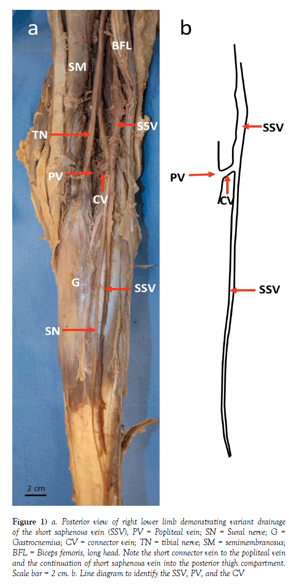
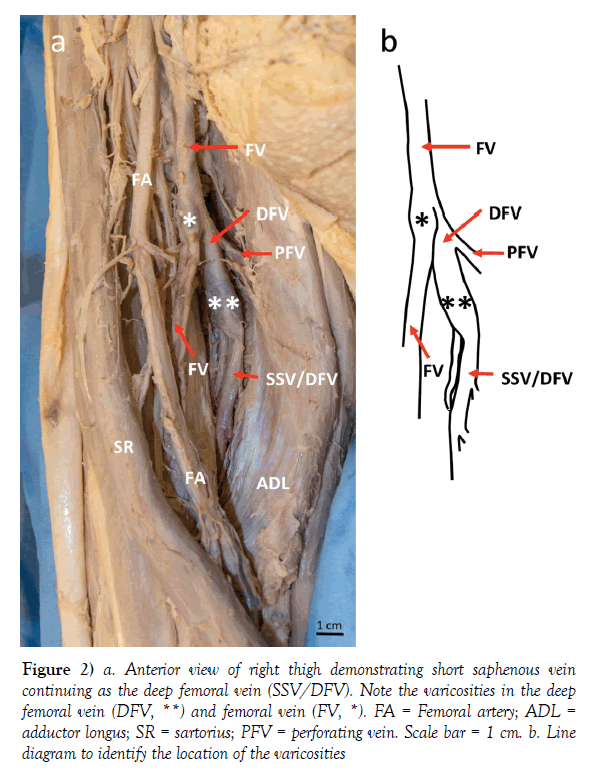
The present case of variation of the small saphenous vein (SSV) was observed during routine dissection of the right lower limb in an 87-year-old female cadaver; cause of death was reported as cerebral hemorrhage. The variation was detected only in the right lower limb. The course of the right SSV originated at the lateral side of the dorsal venous arch of the foot and traveled up the posterior leg towards the popliteal fossa. The thickness of this segment of the SSV increased gradually from 0.2 cm (most distal part at origin) to 0.6 cm (in the popliteal fossa). Upon reaching the popliteal fossa, there was a communicating vein between the SSV and the popliteal vein (PV). Instead of terminating within the PV, however, the SSV continued up as the deep femoral vein (DFV), diving deeper into the thigh and widening in girth (Figure 1). The thickness of this segment of the SSV/DFV was 0.8 cm. The DFV passed deep to the common tendon of biceps femoris and along the underside of the short head of biceps femoris muscle. In the thigh, several perforating veins, as is typical, drained into the DFV. In the proximal thigh, the DFV pierced through the adductor magnus to enter the anterior compartment of the thigh. Viewed anteriorly, the DFV joined the femoral vein (FV), as is typical (Figure 2). At this intersection, the FV was noted to narrow down significantly as it coursed distally down the thigh alongside the femoral artery. The FV was 1.2 cm thick proximal to and only 0.6 cm thick distal to it’s joining with the DFV. Approximately 4 cm distal to this joining, a 2 cm long varicosity of the DFV was noted. There was also a 1 cm long varicosity on the FV at its point of attachment to the DFV. The great saphenous vein normally drained to the FV. The left small saphenous vein had a normal drainage pattern and varicosities were not noted in any of the left lower limb veins.
Figure 1) a. Posterior view of right lower limb demonstrating variant drainage of the short saphenous vein (SSV), PV = Popliteal vein; SN = Sural nerve; G = Gastrocnemius; CV = connector vein; TN = tibial nerve; SM = semimembranosus; BFL = Biceps femoris, long head. Note the short connector vein to the popliteal vein and the continuation of short saphenous vein into the posterior thigh compartment. Scale bar = 2 cm. b. Line diagram to identify the SSV, PV, and the CV
Figure 2) a. Anterior view of right thigh demonstrating short saphenous vein continuing as the deep femoral vein (SSV/DFV). Note the varicosities in the deep femoral vein (DFV, **) and femoral vein (FV, *). FA = Femoral artery; ADL = adductor longus; SR = sartorius; PFV = perforating vein. Scale bar = 1 cm. b. Line diagram to identify the location of the varicosities
Discussion
Variations in the drainage pattern of the lower limb, particularly of superficial veins, are not uncommon. These have both developmental and clinical implications.
During embryonic development, sensory nerves regulate the pattern of vasculogenesis by secretion of vascular endothelial growth factor [7,8]. The sural nerve directs the development of the SSV in the leg; axial extension of the SSV is directed by the axial or the ‘greater sciatic’ nerve [9]. The embryological basis of a SSV and the thigh extension of the vein might be regarded as a unique venous channel, wholly axial throughout its course, formed by the SSV proper in the leg and by a persistent and functional sciatic (ischiatic) vein, which usually disappears, satellite of the ischiatic nerve, in the thigh [10]. Knowledge of embryological origin is important for understanding the basis of venous variations, particularly those that develop unilaterally.
Understanding the course of the SSV in patients is important in several clinical scenarios, including coronary artery bypass grafts [11,12] and treatment of varicose veins [13]. Thus, the knowledge of this particular variation is of relevance to vascular and plastic surgeons, particularly with use of pre-operative ultrasound. Furthermore, it is of particular interest that a superficial vein that drains the fascia of the lower limb drains into a vein typically responsible for draining the deep structures of the thigh. This has clinical importance for ergonomics of drainage: previous cases have reported varicosities as a result of anomalous SSV drainage, which has implications for venous return, including the potential for venous congestion [3]. For clinicians, it is important to note that the deep femoral vein is often used by intravenous drug users (IVDU) as an injection site; variations in drainage and potential for varicosities have clinical implications for this population. Variations in the deep femoral vein also have implications for nerve growth [9], deep vein thrombosis [14], and pulmonary embolism [15].
Conclusion
It is important to be aware of variant anatomic entities for their morphogenetic and clinical implications. We predict that the reported case of variant drainage in the SSV was consequent to defective morphogenesis. It contributed to the development of the varicosities noted in the right deep femoral vein and the right femoral vein. The presence of such varicosities in the vicinity of a variation in the path of a superficial vein is noteworthy for a wide range of health care providers, most specifically for vascular surgeons.
Acknowledgement
We wish to thank those who donate their bodies for the advancement of education and research, and the donor families that have made these donations possible. We would also like to thank Amanda Collins, Director of Anatomical Services at the University of Massachusetts Medical School, for her support of this project. Finally, we are grateful to Charlene Baron, Biomedical Photographer and Digital Imaging Specialist at the University of Massachusetts Medical School, for her help with photography related to the project.
REFERENCES
- de Oliveira A, Vidal EA, Franca GJ, et al. Anatomic variation study of small saphenous vein termination using color Doppler ultrasound. J Vasc Br. 2004;3:223-30.
- Abhinitha P, Mohandas Rao KG, Satheesha Nayak B, et al. Anomalous termination of small (short) saphenous vein associated with its abnormal course in the thigh: A case report. OA Case Reports. 2013;2:63.
- Shetty PD, Souza, MR, Nayak SB. An Unusual Course and Termination of Small Saphenous Vein: A Case Report. J Clin Diagn Res. 2016;10:AD01-2.
- Delis KT, Knaggs AL, Khodabakhsh P. Prevalence, anatomic patterns, valvular competence, and clinical significance of the Giacomini vein. J Vasc Surg. 2004; 40:1174-83.
- Prakash Kumari J, Nishanth Reddy N, Kalyani Rao P, et al. A review of literature along with a cadaveric study of the prevalence of the Giacomini vein (the thigh extension of the small saphenous vein) in the Indian population. Rom J Morphol Embryol. 2008;49:537-9.
- Natsis K, Paraskevas G, Lazaridis N, et al. Giacomini vein: thigh extension of the small saphenous vein-report of two cases and review of the literature. Hippokratia. 2015;19: 263-5.
- Mukouyama YS, Shin D, Britsch S, et al. Sensory nerves determine the pattern of arterial differentiation and blood vessels branching in the skin. Cell. 2002;109:693-705.
- Risau W. Mechanisms of angiogenesis. Nature. 1997;386:671-74.
- Uhl JF, Gillot C, Chahim M. Anatomical variations of the femoral vein. J Vasc Surg. 2010;52:714-9.
- Barberini F, Cavallini A, Caggiati A. The thigh extension of the small saphenous vein: a hypothesis about its significance, based on morphological, embryological and anatomo-comparative reports. Ital J Anat Embryol. 2006;111:187-98.
- Brandt CP, Greene GC, Maggart ML, et al. Endoscopic vein harvest of the lesser saphenous vein in the supine position: a unique approach to an old problem. Interact Cardiovasc Thorac Surg. 2012;16:1-4.
- Sarwar U, Chetty G, Sarkar P. The short saphenous vein: A viable alternative conduit for coronary artery bypass grafts harvested using a novel technical approach. J Surg Tech Case Rep. 2012;4:61-3.
- Winterborn RJ, Campbell WB, Heather BP, et al. The management of short saphenous varicose veins: a survey of the members of the vascular surgical society of Great Britain and Ireland. Eur J Vasc Endovasc Surg. 2004;28:400-3.
- Quinlan DJ, Alikhan R, Gishen P. Variations in lower limb venous anatomy: implications for US diagnosis of deep vein thrombosis. Radiol. 2003;228:443-8.
- Federman J, Anderson ST, Rosengarten DS, et al. Pulmonary embolism secondary to anomalies of deep venous system of the leg. Br Heart J. 1977;39:547-55.


