Alteration in the serum levels of interleukin-6 and interleukin-15 in the patients of knee osteoarthritis
2 Department of Biochemistry, Pt. BD Sharma PGIMS University, Haryana, India
3 Department of Orthopaedics, Pt. BD Sharma PGIMS University, Haryana, India
Received: 02-Mar-2023, Manuscript No. PULJCGG-23-6207; Editor assigned: 07-Mar-2023, Pre QC No. PULJCGG-23-6207; Reviewed: 21-Mar-2023 QC No. PULJCGG-23-6207; Revised: 30-Mar-2023, Manuscript No. PULJCGG-23-6207; Published: 27-Apr-2023, DOI: 10.37532/puljcgg.23.6(1).1-4
Citation: Chauhan A, Bala M, Rohilla R, et al. Alteration in the serum levels of interleukin-6 and interleukin-15 in the patients of knee osteoarthritis. J Clin Genet Genom 2023; 6(1):1-4.
This open-access article is distributed under the terms of the Creative Commons Attribution Non-Commercial License (CC BY-NC) (http://creativecommons.org/licenses/by-nc/4.0/), which permits reuse, distribution and reproduction of the article, provided that the original work is properly cited and the reuse is restricted to noncommercial purposes. For commercial reuse, contact reprints@pulsus.com
Abstract
Background: Osteoarthritis is the most common type of arthritis affecting mostly the weight bearing joints. Pro-inflammatory cytokines including IL-6 and IL-15 are predominately secreted by macrophages and other cells and they are involved in up regulation of inflammatory response. IL-6 and IL-15 may induce expression of proteolytic enzymes which cause cartilage damage and in turn osteoarthritis. This study is designed to evaluate the serum levels of IL-6 and IL-15 in the patients of knee OA and their possible role in its pathogenesis.
Aim: To evaluate the role of IL-6 and IL-15 in the patients of knee osteoarthritis.
Materials and methods: The present study was conducted in the department of biochemistry in collaboration with department of orthopedics from May 2018 till July 2020. Forty patients with knee osteoarthritis in the age group of 30-60 years and forty healthy controls were enrolled for the study. After taking written consent, five mL of venous blood from the patients and controls was taken from antecubital vein in a red capped vaccutainer under aseptic conditions. Estimation of IL-6 and IL-15 were done by the Enzyme-Linked Immunosorbent Assay kit (ELISA kit). The routine biochemistry tests were done on the same day by standard autoanalyzer kit method.
Results: The mean serum levels of IL-6 and IL-15 were found higher in cases as compared to the controls (p<0.001).
Conclusion: The serum levels of IL-6 and IL-15 found higher in the patients which may cause cartilage damage and in turn osteoarthritis.
Keywords
Osteoarthritis; Interleukins; Cartilage; Cytokines; Matrix Metalloproteinases (MMPs); Inflammation
Introduction
Osteoarthritis is a degenerative joint disorder of the weight bearing joints like knee and hip. It is the most common cause of joint paint. OA is due to the progressive cellular and changes in all tissues of the joint. Ageing is the most common cause of the OA. It affects joint cartilage, subchondral bone as well as synovium. The ageing cells alter mitochondrial function due to the oxidative stress which lead to the cell death. In ageing cells, there is down regulation of the Transforming Growth Factor-β (TGF-β) and chondrocytes instead of matrix synthesis shift towards catabolic side and Matrix Metalloproteinases (MMPs) expression. Obesity is another etiological factor in the development of OA. It is the most promising problem in recent years due to the modification in the life style of people. Adipokines are adipose tissues derived cytokines which are released by adipocytes in obese persons. In obese person, the levels of pro-inflammatory cytokines are found elevated. This lead to NB-kb pathway activation which ultimately lead to initiation of catabolic process in the chondrocytes and ECM degradation. Sport injury is another etiological factor in the development of OA, especially in young population. It is the most common cause of OA in young and middle aged population [1].
In recent years, researchers have demonstrated the role of inflammation in the development and progression of knee OA. Inflammation may be caused by cytokine-induced Mitogen Activated Protein kinase (MAP), NF-kB activation and oxidative phosphorylation. It involves the entire joint including cartilage, synovium and subchondral bone. It is well established that the low grade inflammation found in OA lead to further diseases development and progression. Although it is yet not clear that at what extent inflammation can cause cartilage demage. It plays a central role in diverse host defense mechanisms such as the immune response, hematopoiesis and acute phase reactions. IL-6 and IL-15 are lymphokine produced by antigen or mitogenic activated T-cells, fibroblasts, macrophages and other cells that serve as a differentiation factor for B-cells and thymocytes which stimulates immunoglobulin production [2].
Cytokines are low molecular weight proteins and their molecules carry same polypeptide fold with four α-helical bundles. They are produced from TH1 cells, TH2 cells, TH17 cells or TReg cells. They can be classified as: Chemokines i.e these cytokines are chemotactic for inflammatory cells, lymphokines i.e these cytokines are produced by T-cells and are responsible for immune response, pro-inflammatory cytokines i.e responsible for inflammation and ant-inflammatory i.e cytokines which oppose the inflammation. Cytokines bind with their receptors and produce cellular and humoral response, initiation of inflammatory response, control of hematopoiesis, cellular proliferation and differentiation. Cytokines often produce in cascade and acts in paracrine (Acts on the cells which produce them), autocrine (Acts on nearby cells) or endocrine manner (Acts on cells at distance after being carried in blood or tissue fluids) [3].
Pro-inflammatory cytokines are responsible for the inflammation and includes: IL-1, IL-6, IL-8, IL-12, IL-15, IL-17, IL-18, Tumor Necrosis Factor-α (TNF-α), Interferon-γ (IFN-γ), Oncostatin M (OSM), Granulocyte- Macrophage Colony-Stimulating Factor (GM-CSF) and Macrophage Colony- Stimulating Factor (M-CSF). IL-6 is a glycoprotein consisting of 184 amino acid residues. IL-6 exerts its biological activities through two molecules: IL-6R (also known as IL-6Rα, gp80 or CD126) and gp130 (also referred to as IL-6Rβ or CD130). It is most potent proinflammatory cytokine. Proinflammatory cytokines stimulate both cellular and humoral inflammatory response. Anti-inflammatory cytokines oppose the inflammation and includes: IL-4, IL-10, IL-11, IL-13 and Tumor Necrosis Factor-β (TNF-β). They inhibit the pro-inflammatory cytokines production [4].
The most common method used to diagnose the knee OA is radiography. Since OA may take years to develop, it is an attractive idea to develop the noval biomarkers which can predict the diseases before the radiological changes appears. So this study is aimed to establish the role of IL-6 and IL-15 in the patients of knee osteoarthritis [5].
Materials and Methods
The present study (case-control study) was conducted in the department of biochemistry in collaboration with department of orthopedics from May 2018 till June 2021. The present study was approved and ethical clearance was taken from the institutional ethical committee (Approval number- IEC/Th/18/BioChem/02). Written consents were taken from all patients and controls. The participation in our study was voluntary and no benefits or compensation were given to the participants [6].
Study population
Forty patients (33 female and 07 male) with knee osteoarthritis in the age group of 30-60 years visiting at orthopedics OPD were enrolled for the study. Forty healthy volunteers (33 female and 07 male) {Hospital workers} of the same age group were taken as control. Informed written consent has been taken from patients as well as controls. All volunteers fulfilled inclusion and exclusion criteria. Patients of knee osteoarthritis suggestive of Diabetes, atherosclerosis, depression, systemic lupus erythematous, any malignancy and other chronic diseases were excluded from this study [7].
Sample collection
After getting the written consent from cases and controls, detailed history was taken and recorded in their respective performa. Five mL of fasting venous blood from cases as well as controls were drawn from antecubital vein in a red capped vaccutainer under aseptic conditions after an informed written consent. Serum was separated by centrifugation at 2000 rpm for 10 minutes after clotting. Samples of serum for the estimation of IL-6 and IL-15 were stored at -70°C until processing. Estimation of IL-6 and IL-15 were done by the Enzyme-Linked Immunosorbent Assay kit (ELISA kit). ELISA kit (Boster Biological Technology Co. Ltd.) was used to quantitatively measure serum concentrations of Interleukin-6 (IL-6) and IL-15 [8].
Statistical analysis
The data was compiled and analysed by unpaired t-test using SPSS 20 version. Correlations between serum levels of IL-6 and IL-15 were done by Spearman’s rank correlation coefficients. P values less than 0.05 (2-tailed) were considered significant and P values less than 0.001 (2-tailed) were considered highly significant [9].
Results
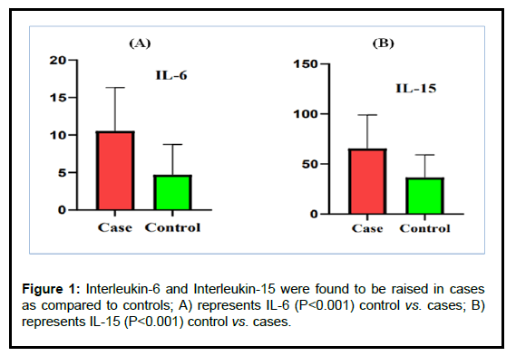
The baseline clinical data of cases and controls are presented in Table 1. Mean age of cases (47 ± 9.22 years) and controls (44.15 ± 8.02 years) were compared which were found statistically not significant (p<0.05). The BMI of cases (26.28 ± 3.81) and controls (22.69 ± 2.53) were compared and found statistically not significant (p<0.05). In our study, male and females were equally distributed in cases and controls. So, the cases and controls were similar with regard to age, proportion of gender [10]. The biochemical parameters were also analysed in cases and controls which are presented in Table 1. The mean serum levels of calcium in cases and controls were (Mean ± SD) 9.56 ± 1.03 (mg/dL) and 10.5 ± 0.86 (mg/dL) respectively which were found statistically highly significant (p<0.001). The mean serum levels of vitamin D in cases and controls were (Mean ± SD) 23.48 ± 18.69 (ng/ mL) and 20.68 ± 14.79 (ng/mL) respectively which were found statistically not significant (p>0.05). The mean serum levels of phosphorus in cases and controls were (Mean ± SD) 3.51 ± 0.60 (mg/ dL) and 3.79 ± 0.76 (mg/dL) respectively which were found statistically not significant (p>0.05). The mean serum levels of ALP in cases and controls were (Mean ± SD) 61.95 ± 20.58 (IU/L) and 66.38 ± 21.43 (IU/L) respectively which were also found statistically not significant (p>0.05). The levels of IL-6 and IL-15 were analysed in cases and control groups which is presented in Table 1 and Figure 1 [11].
| Parameter | Cases (n=40) (Mean ± SD) | Controls (n=40) (Mean ± SD) | Range | 95% CI | P value |
|---|---|---|---|---|---|
| Age (years) | 47 ± 9.22 | 44.18 ± 8.02 | 31-60 | 44.05-49.94 | P<0.05 |
| BMI (kg/m2) | 26.28 ± 3.81 | 22.69 ± 2.53 | 19-28.5 | 18.52-27.3 | P<0.05 |
| Serum calcium (mg/dL) | 9.56 ± 1.03 | 10.5 ± 0.86 | 6.1-11.3 | 9.23-9.88 | P<0.001 |
| Serum vitamin D (ng/mL) | 23.48 ± 18.69 | 20.68 ± 14.79 | 10-100 | 17.50-29.46 | P>0.05 |
| (ALP) (IU/L) | 61.95 ± 20.58 | 66.38 ± 21.43 | 29-98 | 55.37-68.53 | P>0.05 |
| Phosphorus (mg/dL) | 3.51 ± 0.60 | 3.79 ± 0.76 | 2.2-5.0 | 3.32-3.70 | P>0.05 |
| Serum interleukin-6 (pg/mL) | 10.54 ± 5.80 | 4.69 ± 4.07 | 3.63-24.94 | 8.68-12.40 | P<0.001 |
| Serum interleukin-15 (pg/mL) | 64.87 ± 34.39 | 36.48 ± 22.77 | 4.01-153.9 | 53.87-75.87 | P<0.001 |
Table 1: Serum levels of calcium, vitamin D, Alkaline Phosphatase (ALP), phosphorus, interleukin-6 and interleukin-15 in cases and controls.
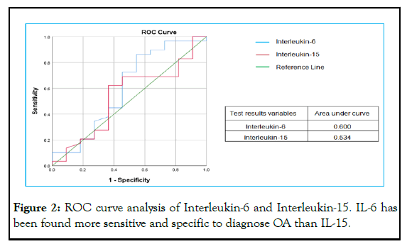
The levels of IL-6 and IL-15 were found significantly elevated in cases as compared to controls. The mean serum levels of IL-6 in cases and controls were (Mean ± SD) 10.54 ± 5.80 (pg/mL) and 4.69 ± 4.07 (pg/mL) respectively which were found statistically highly significant (p<0.001). The mean serum levels of IL-15 in cases and controls were 64.87 ± 34.39 (pg/mL) and 36.48 ± 22.77 (pg/mL) which were also found statistically highly significant (p<0.001) [12]. A ROC analysis to determine the predictive power of cytokine revealed that in this analysis, Area under the Curve (AUC) of IL-6 was best compared to IL-15. IL-6 is found more sensitive and specific than IL-15 (Table 2 and Figure 2) [13].
| Parameters | Correlation coefficient (r) | P value |
|---|---|---|
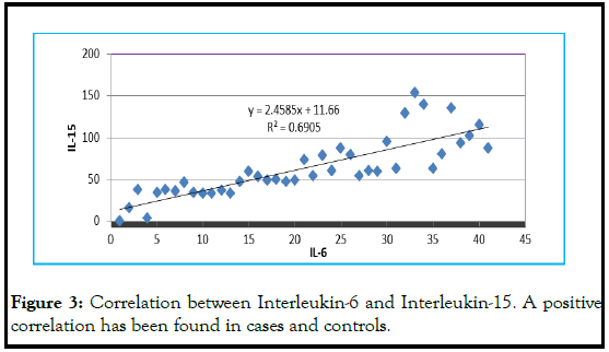
| Serum IL-6 and IL-15 | (+) 0.69 | P<0.05 |
Table 2: Correlation coefficient of serum interleukin-6 and interleukin-15.
Discussion
The present study explored the serum concentration of IL-6 and IL-15 in patients of knee OA and healthy controls. IL-6 and IL-15 are a proinflammatory cytokines which are responsible for the communication among cells of immune system [14]. The increased levels of proinflammatory cytokines are related to development and progression of OA due to upregulation of metalloproteinase gene expression, stimulation of reactive oxygen species production, alteration of chondrocyte metabolism and increased osteoclastic bone reabsorption. IL-6 and IL-15 are produced by T cells, B cells, monocytes, fibroblasts as well as osteoblasts from subchondral bone, osteophytes of OA under mechanical loading and adipocytes of the infrapatellar fat pad [15]. Our findings suggests that higher serum levels of IL-6 and IL-15 may cause knee OA by intensifying the collagen chain destruction. Radiographic evaluation of cases has been done along with clinical sign and symptoms [16]. The severity of radiographic finding with higher levels of IL-6 and IL-15 cannot be rule out. We also demonstrated a statistically significant positive association between serum levels of IL-6 and IL-15 in the patients. Both IL-6 and IL-15 was founder higher simultaneously and their high levels in the serum could be a possible cause of damage to the knee cartilage [17]. These observations support the hypothesis that IL-6 and IL-15 may be good marker in the diagnosis of OA. Our findings are in accordance with study performed. In their study, IL-6 and IL-10 has been analysed in the patients of knee OA but they did not analyse other biochemical parameters to establish the alteration in routine biochemical parameters in cases as compared to healthy controls. A ROC curve has been generated to find the sensitivity and specificity of IL-6 and IL-15 (Table 3 and Figure 3) [18].
| Cases | Positive | Negative |
|---|---|---|
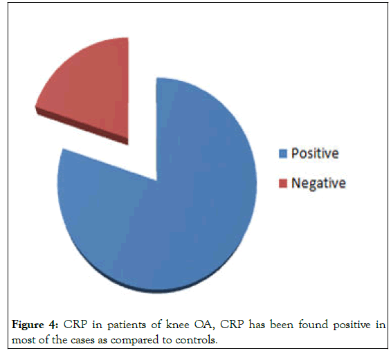
| 40 | 24 | 6 |
Table 3: C-Reactive Protein (CRP) in the patients with knee osteoarthritis.
It has been found that IL-6 is more sensitive and specific than IL-15 in OA. IL-6 acts as a major pro-inflammatory mediator for the induction of the acute phase response, leading to a wide range of local and systemic changes, so can damage the knee cartilage aggressively than IL-15. The CRP is a pentameric protein which is synthesized in liver. We also found that CRP is positive in most of the patients as compared to controls. The serum levels of CRP rises in response to the inflammation. In our study the CRP levels were found higher in 90% of cases and so significantly associated with the knee osteoarthritis. One of the major causes for the higher levels of CRP is that its production is stimulated by IL-6 and IL-15 in the patients of knee osteoarthritis. Although estimation of IL-6 and IL-15 are not currently in practice to diagnose the OA, but its inflammatory role in the pathogenesis cannot be rule out. Evaluation of IL-6 and IL-15 can be done along with the established diagnostic tool for confirmation of the diseases. The studies of these biomarkers on larger scale should be done to establish cut off levels to diagnose the OA (Figure 4) [19].
To summarize, our results indicate that IL-6 and IL-15 can play a role in the early stages of OA development as it can be detected with a high prevalence and at elevated concentrations in serum of OA patients. IL-6 and IL-15 can represent a relevant, independent biomarker candidate for prediction of the development of RA.
Conclusion
Osteoarthritis is thought to be degenerative disease of cartilage but accumulating evidences indicates that inflammation has a critical role in its pathogenesis. In our study, the serum levels of IL-6 and IL-15 were found higher as compared to controls. Quantification of serum levels of IL-6 and IL-15 could be a useful as a complementary diagnostic and prognostic test for knee OA. Improved knowledge of the new biomarkers will permit a better means for the assessment of the effects of new therapeutic interventions in OA. As inflammation appears much earlier before the symptoms appears, so the invention of novel biomarkers is better idea for earlier detection of knee OA.
Funding
No funding, grants or support has been provided by any organization for this study.
Competing Interest
The authors don’t have any financial or non-financial interests to disclose.
Authors Contributions
• Design of research-Aman Chauhan, Manju Bala, Rajesh Rohilla.
• Data collection and drafting the work-Aman Chauhan, Rooma Devi.
• Final approval of the version to be published-Manju Bala, Rajesh Rohilla, Aman Chauhan, Rooma Devi.
Ethics Approval
The ethical approval has been taken from the institutional ethical committee of Pt. BD Sharma, university of health science, Rohtak, Haryana, India (Approval number- IEC/Th/18/BioChem/02).
Consent to Participate
Written informed consent has been taken from patients as well as healthy controls to participate in the present study.
Consent to Publish
Informed consent has been taken from all the participants in this study to participate as well as to get it published. However, no personal data of an individual in any form like image, personal information or videos has been used in this manuscript.
Availability of Data and Materials
All data generated or analysed during this study are included in this manuscript.
References
- Pal CP, Singh P, Chaturvedi S, et al. Epidemiology of knee osteoarthritis in India and related factors. Indian J Orthopaed. 2016;50:518-22.
[Crossref] [Google Scholar] [PubMed]
- Loeser RF. Ageing and osteoarthritis. Curr Opin Rheumatol. 2019;91:123-59.
- Chen DI, Shen J, Zhao W, et al. Osteoarthritis: Toward a comprehensive understanding of pathological mechanism. Bone Res. 2017;5(1):1-3.
[Crossref] [Google Scholar] [PubMed]
- Chauhan A, Bala M, Rohilla R. Various treatment aspects of arthritis (Allopathic and Ayurvedic) IJSR. 2021;10:85-89.
- Nedoszytko B, Sokolowska-Wojdyło M, Ruckemann-Dziurdzinska K, et al. Chemokines and cytokines network in the pathogenesis of the inflammatory skin diseases: Atopic dermatitis, psoriasis and skin mastocytosis. Adv Dermatol Allergol/Postępy Dermatol Alergol. 2014;31(2):84-91.
- Lozano R, Naghavi M, Foreman K, et al. Global and regional mortality from 235 causes of death for 20 age groups in 1990 and 2010: A systematic analysis for the global burden of disease study. Lancet. 2012;380:2095-128.
- Ling SM, Patel DD, Garnero P, et al. Serum protein signatures detect early radiographic osteoarthritis. Osteoarthritis Cartilage. 2009;17(1):43-8.
[Crossref] [Google Scholar] [PubMed]
- Li YP, Wei XC, Zhou JM, et al. The age-related changes in cartilage and osteoarthritis. Biomed Res Int. 2013;20:916-30.
- Sokolove J, Lepus CM. Role of inflammation in the pathogenesis of osteoarthritis: Latest findings and interpretations. Ther Adv Musculoskelet Dis. 2013;5(2):77-94.
[Crossref] [Google Scholar] [PubMed]
- Qiu GX. Osteoarthritis: Diagnosis and treatment of osteoarthritis. Orthop Surg. 2010;2:01-06.
- Bay-Jensen AC, Reker D, Kjelgaard-Petersen CF, et al. Osteoarthritis year in review 2015: Soluble biomarkers and the BIPED criteria. Osteoarthritis Cartilage. 2016;24(1):9-20.
[Crossref] [Google Scholar] [PubMed]
- Shimura Y, Kurosawa H, Sugawara Y, et al. The factors associated with pain severity in patients with knee osteoarthritis vary according to the radiographic disease severity: A cross-sectional study. Osteoarthritis Cartilage. 2013;21(9):1179-84.
[Crossref] [Google Scholar] [PubMed]
- Malathi R, Kothari S, Chattopadhyay A, et al. Raised serum IL-6 and CRP in radiographic knee osteoarthritis in eastern India. JMSCR. 2013;5:21687-92.
- Azim S, Nicholson J, Rebecchi MJ, et al. Interleukin-6 and leptin levels are associated with preoperative pain severity in patients with osteoarthritis but not with acute pain after total knee arthroplasty. Knee. 2018;25(1):25-33.
[Crossref] [Google Scholar] [PubMed]
- Loeser RF, Goldring SR, Scanzello CR, et al. Osteoarthritis: A disease of the joint as an organ. Arthritis Rheumatism. 2012;64(6):1697.
[Crossref] [Google Scholar] [PubMed]
- Felson DT. Osteoarthritis of the knee. N Engl J Med. 2006;354:841-48.
- Imamura M, Ezquerro F, Marcon Alfieri F, et al. Serum levels of proinflammatory cytokines in painful knee osteoarthritis and sensitization. Int J Inflam. 2015.
- Dhagestani HN, Kraus VB. Inflamatory biomarkers in osteoarthritis. Osteoarthr Cartil. 2016;23:1890-96.
- Sproston NRS, Ashworth JJ. Role of C-reactive protein at sites of inflammation and infection. Front Immunol. 2018;9:754.
[Crossref] [Google Scholar] [PubMed]


