Incomplete Unilateral Ureter Duplication a Case Report
2 Georgetown University School of Medicine, Georgetown University Medical Center, Washington, DC, USA
3 Baylor College of Medicine, School of Medicine, Houston, TX, USA
4 College of Natural Sciences, University of Texas at Austin, Austin, TX, USA
5 Department of Physiology and Anatomy, University of North Texas Health Science Center, Fort Worth, TX, USA
Received: 01-Dec-2023, Manuscript No. ijav-23-6876; Editor assigned: 04-Dec-2023, Pre QC No. ijav-23-6876; Reviewed: 21-Dec-2023 QC No. ijav-23-6876; Revised: 25-Dec-2023, Manuscript No. ijav-23-6876 (R); Published: 30-Dec-2023, DOI: 10.37532/1308-4038.16(12).331
Citation: Ballout B. Incomplete Unilateral Ureter Duplication a Case ReportReport. Int J Anat Var. 2023;16(12): 442-444
This open-access article is distributed under the terms of the Creative Commons Attribution Non-Commercial License (CC BY-NC) (http://creativecommons.org/licenses/by-nc/4.0/), which permits reuse, distribution and reproduction of the article, provided that the original work is properly cited and the reuse is restricted to noncommercial purposes. For commercial reuse, contact reprints@pulsus.com
Abstract
The ureter is a muscular tube that carries urine from the collecting system of the kidney to the bladder for collection and, ultimately, excretion. Of mesodermal origin, the ureter arises from reciprocal induction between the metanephric blastema and the ureteric bud. Deviations from the normal intricate developmental process can lead to congenital anatomical alterations. During a routine dissection of a female cadaver, an incomplete unilateral ureteral duplication was discovered prompting further evaluation of the rare anatomical alterations’ incidence, manifestations, and clinical implications. The discovered ureter originated from two anatomically independent collecting systems from the same kidney that later converged along their course, forming one relatively thicker ureter that feeds into the bladder. Realizing such an anatomical alteration’s overall incidence and presence in patients is essential for healthcare workers as its diagnosis would guide treatment and reduce errors in radiologic studies, bedside procedures, and invasive surgeries.
Keywords
Kidney; Incomplete duplicated ureter; Duplex ureter; Renal pelvis
INTRODUCTION
Ureteral duplications are rare congenital variations in the kidney and urinary tract (CAKUT) spectrum that present with a multitude of clinical expressions and treatments. These various anatomical alterations, although often asymptomatic, can pose potential complications, such as vesicoureteral reflux and hydronephrosis. Duplex systems exhibit diverse phenotypes, with various classifications proposed to categorize these pathological formations [1]. Incomplete duplication involves two poles of a duplex kidney sharing the same ureteral orifice of the bladder. On the other hand, complete duplications usually involve an anatomical variation of the upper pole and a normal lower pole of the kidney, often originating from an ectopic ureter that emerges anteriorly to the position of the normal ureter. Moreover, inverted Y-ureteral duplication and rare conditions such as H-shaped ureters or multiplex ureters further exemplify the diverse range of anatomical alterations encountered in duplex kidney formations [1].
Additionally, the growth of the ureteric bud and its interaction with the metanephric mesoderm play pivotal roles in nephron formation, essential for the development of the ureter, calyces, and collecting tubules [2]. Disruptions in these developmental processes can result in congenital anatomical alterations, underscoring the critical interdependence between the ureteric bud and metanephric mesenchyme in kidney development [2].
The aim of this case report is to provide a comprehensive description and analysis of an incomplete ureteral duplication. By exploring this specific presentation, the report aims to contribute to the understanding of the anatomy, embryological formation, clinical manifestations, and potential treatments of this condition.
CASE REPORT
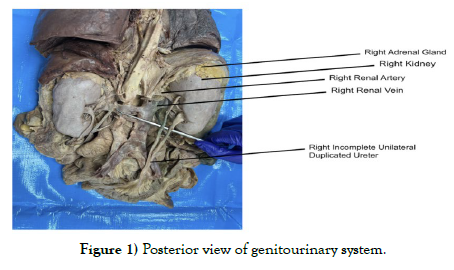
A case of an incomplete unilateral ureteral duplication presented during a standard dissection of a middle-aged female cadaver. The cadaver was received through the Willed Body Program with signed informed consent form from the donor. The method of dissection for this cadaver used a technique in which the entire gastrointestinal and genitourinary systems from tongue to anus were removed from the body as one complete entity. This technique allowed us to dissect and reflect the organs, vasculature, and nerves in a unique perspective while keeping all organs in anatomical position without distortion. The dissection began with an anterior sagittal cut and reflection from the chin all the way down to the pelvis. Once the anterior aspect of the cadaver was retracted, we were able to begin dissecting the organs out of the body. Upon detachment of all the organs from the anterior, lateral, and posterior aspects of the inner cavity walls, we were able to remove all the organs. This then continued with an entire dissection of the anterior aspect of the gastrointestinal tract including landmarks, spaces, and vasculature. Upon completion of an anterior dissection of the entire gastrointestinal system, we turned over the specimen to begin dissection of the posterior aspect. We cleaned and identified all the structures and landmarks. The right kidney weighed 145 grams. 3D measurements were taken of the kidney, as well as to note for any size anatomical deviations. The superior to inferior aspect of the kidney was 11.5cm in length, the medial to lateral aspect of kidney was 6.5 cm, and the anterior to posterior aspect of the kidney was 3 cm. The external surface of the kidney appeared to be normal. The kidneys were beanshaped with a brown-tan color. The external surface had a soft smooth texture with no excessive lobulation. There was a green bile stain on the kidney due to its location on the right side of the abdomen in close proximity to the gallbladder. The bile also slightly stained the unilateral incomplete duplicated ureter of the right kidney giving it a light green appearance midway through the descending part of the ureter. The renal artery, renal vein, and ureter all identified bilaterally. However, upon further cleaning and dissection of the right kidney, we found an additional ureter coming out of the right kidney superior to the originally identified ureter (Figure 1).
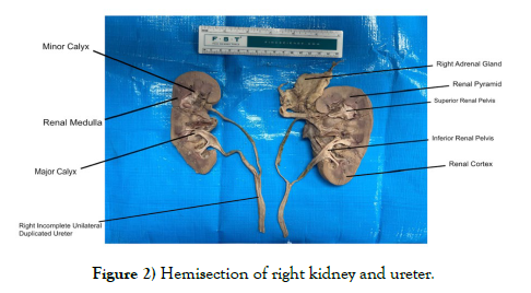
These two ureters would go on to descend and merge into one large ureter anterior to the psoas muscle. This ureter attached to the right kidney was roughly 17cm long from the kidney all the way down to the site it was dissected at. The rest of the ureter was attached to the bladder with a length of about 6.5cm. This gave the ureter a rough total length of around 23.5 cm. The incomplete unilateral ureter duplication length came out to be around 23.5cm (±2.33). The average ureter length is 23.8cm (±2.18) for men and 23.2cm (±2.44) for women [3]. Given this study where there was a positive correlation found between the donor’s size and ureteral length, we can see that the incomplete unilateral ureteral duplication is sitting right at the general average ureter length and slightly higher than women’s average ureter length. Once the external data needed were collected, we proceeded with a hemi-section of the right kidney and ureters with a sagittal cut to note the interior of this kidney. Upon hemi-section, we found that each ureter originated from separate renal pelvis. The superior ureter attachment had a superior renal pelvis of around 0.7 cm., while the inferior ureter attachment had an inferior renal pelvis of around 1 cm. There was about an average of 3 cm. separating the superior renal pelvis from the inferior renal pelvis within the right kidney. The renal parenchyma between each pelvis was unremarkable. Furthermore, the superior renal pelvis had a total of 5 renal pyramids (2 renal pyramids per major calyx, except 1 renal pyramid for most inferior pyramid on superior renal pelvis). The inferior renal pelvis had a total of 4 renal pyramids (2 minor calyces per 1 major calyx; 3 major calyces for the entire pelvis) (Figure 2).
DISCUSSION
Prenatal anatomical alterations of the genitourinary system make up 20%- 30% of all congenital anatomical alterations and, therefore, must not be overlooked in a clinical or surgical setting [4]. Specifically in our case, cadaveric studies have shown that the prevalence of an incomplete ureteral duplication amongst the general population can vary and may reach 6.25% [5]. The incidence of such an anatomical alteration is shown to be two times more common in females than in males, as in this case [5]. It is important to understand the embryological formation of the anatomical variations in order to understand the complexity of the anatomy and its relationship to the surrounding structures. The development of the ureteric bud and its interaction with the metanephric mesoderm play a critical role in nephron formation. These reciprocal interactions are vital for continual branching of the ureteric bud, leading to the development of the ureter, calyces, and collecting tubules [2]. Disruptions in these developmental processes often result in congenital anatomical alterations, underscoring the importance of the interdependence between the ureteric bud and metanephric mesenchyme during kidney formation [2]. Key signaling pathways such as GDNF–RET and FGF, along with transcription factors in the metanephric mesenchyme, regulate ureteric bud budding. Dysregulation of these pathways and their negative regulators can lead to various kidney and urinary tract defects, including renal agenesis, duplex kidneys, and ureteric bud anatomical alterations, emphasizing the delicate balance required for proper kidney and urinary tract formation [2]. Exploring the formation of ureteral duplications offers insight into the complex molecular interactions governing kidney development, thereby enriching our understanding of these anatomical alterations.
Oftentimes, adults with an incomplete unilateral ureteral duplication may never know of it as it often does not impose any clinical significance. And if found, its diagnosis would be merely incidental [6]. Nevertheless, having such an anatomical alteration does incur a higher risk of developing clinical syndromes. It has been consistently shown that patients an incomplete ureteral duplication were at an increased risk of developing of ureteroureteric reflux, and hydronephrosis, recurrent UTIs, and pyelonephritis [4-6].
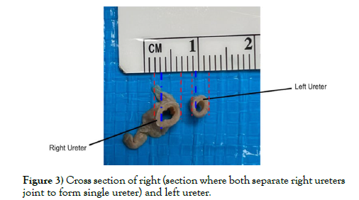
Moreover, patients with the anatomical alterations have a higher likelihood of developing kidney stones [7]. Under normal circumstances, a typical ureter encounters anatomic constrictions along its course, such as the pelviureteric and the ureterovesical junctions that have shown to be common sites where kidney stones may lodge. The meeting point of the duplicated ureter was also a constricted point along the ureter and could be an additional point prone to harboring a stone. This donor also demonstrated a thickened merged ureter distal to the duplication which could impose problems (Figure 3).
Ureteral thickening was shown to be a radiographic sign of urothelial neoplasm [8]. As such, in cases where ureter thickness was to be used as a marker for a neoplasm, it must also be proven not to be a duplication of the collecting system.
In children, such rare anatomical alteration may cause more severe effects. The presence of duplication increases the risk of recurrent urinary tract infections. Therefore, children with frequent recurrent UTIs, an incomplete duplicated ureter must be prioritized on the differential diagnosis and be ruled out as part of the patient work up.
Unintentional mischaracterization of the duplicated ureter during invasive procedures may lead to unforeseen complications. Cases of incidental discoveries during surgery of duplicated ureters have been reported in the literature [9,10]. Surgeons and radiologists familiarizing themselves with the possibility and presentation of such an anatomical alteration would reduce the risk of iatrogenic injury to the ureter or surrounding structures. CT scans, bedside ultrasound, venous urography, among other techniques, may be used to find diagnoses in case of clinical suspicion.
ACKNOWLEDGMENTS
The authors would like to thank the Willed Body Program at UNTHSC and the donors and their families for their contribution to advancing medicine and anatomical knowledge.
REFERENCES
- Kozlov VM, A Schedl. Duplex kidney formation: developmental mechanisms and genetic predisposition. F1000Res, 2020; 9: F1000.
- Nagalakshmi VK, Yu J. The ureteric bud epithelium: morphogenesis and roles in metanephric kidney patterning. Mol Reprod Dev, 2015; 82(3):151-66.
- Mansouri A. Is the ureteral length associated with the patient's size. Prog Urol. 2019; 29(2): 127-132.
- Roy M. Anatomical variations of ureter in central India: A cadaveric study. Journal of Datta Meghe Institute of Medical Sciences University. 2017; 12(4): 277-279.
- Nagpal H, Chauhan R. Unilateral duplex collecting system with incomplete duplication of ureter a case report. International Journal of Research in Medical Sciences. 2017; 5(5):2254.
- Bisset GS, Strife JL. The duplex collecting system in girls with urinary tract infection: prevalence and significance. AJR Am J Roentgenol. 1987; 148(3): 497-500.
- Xiao N. Obstruction of bifid ureter by two calculi: A case report. Medicine. 2018; 97(30): E11474.
- Xu AD. Significance of Upper Urinary Tract Urothelial Thickening and Filling Defect Seen on MDCT Urography in Patients With a History of Urothelial Neoplasms. American Journal of Roentgenology. 2010; 195(4): 959-965.
- J Kelly, Moghul FA, Pisani V. Double ureter at repair of obstetric fistula. International Journal of Gynecology & Obstetrics. 2008; 102(1): 77-78.
- Varlatzidou A. Complete unilateral ureteral duplication encountered during intersphincteric resection for low rectal cancer. Journal of Surgical Case Reports. 2018; 2018(10).
Indexed at, Google Scholar, Crossref
Indexed at, Google Scholar, Crossref
Indexed at, Google Scholar, Crossref
Indexed at, Google Scholar, Crossref
Indexed at, Google Scholar, Crossref
Indexed at, Google Scholar, Crossref
Indexed at, Google Scholar, Crossref


