
Page 22
Volume 03
Spine 2019
October 16-17, 2019
Journal of Neurology and Clinical Neuroscience
October 16-17, 2019 | Rome, Italy
SPINE AND SPINAL DISORDERS
5
th
World Congress on
J Neurol Clin Neurosci, Volume 03
Spinal Metastases of two different grade Oligodendrogliomas: A case report and
review of literature
Andreina Martinez Amado
1
, Maria Gabriela Sanchez Paez
2
and
Carlos Enrique Maiguel Carrizosa
2
1
Nueva Granada Military University, Colombia,
2
Foscal Clinic, Colombia
O
ligodendrogliomas (OGD) are glial tumors, together with mixed oligoastrocytoma constituting 5-20% of all gliomas,
which occur predominantly in younger populations and are managed with surgery and chemotherapy with good long-term
prognosis after treatment and additionally present with low rates of metastases. We present the case of a 46-year-old patient with
intracranial right frontal subcortical OGD [World Health Organisation (WHO) grade II] managed at the Neurosurgery Department
in Foscal Clinic, Floridablanca, Colombia. Two years after brain surgery the patient presents with neurological symptomatology
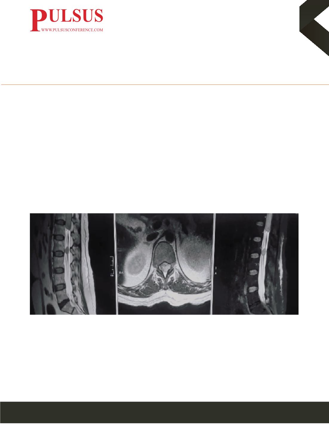
suggestive of Spinal Cord Compression and is found to have a neoplastic lesion with extramedullary compressive strength on
the conus medullary and wrapping all of the roots with the final report of pathology and immunohistochemistry indicating: OGD
(WHO grade III), this lesion was the only one found, the brain studies shows any residual tumor or recurrence in the primary
tumor site.
Biography
Andreina Martinez is a General Physician from Colombia, with deep interest in Neurosurgery and Spine surgery and huge passion for
clinical research. She finished her Medicine Program in Military University – Bogota with honors for her commitment and dedication to
research and have the opportunity of being part to the neurosurgery team at Shaio Clinic, Military Hospital, FOSCAL - Bucaramanga.
She has exhibited remarkable enthusiasm and superb medical understanding in the research area. Her ability to grasp both the medical
concepts as well as the clinical implication presented to her was exceptional.
e
:
u0401380@gmail.comFigure 1. Spine MRI that shows intra-axial and expansive intramedullary lesion at the level of medullary cone and vertebral bodies T12 and L1


