


 7 / 10

Page 26
Volume 03
Spine 2019
October 16-17, 2019
Journal of Neurology and Clinical Neuroscience
October 16-17, 2019 | Rome, Italy
SPINE AND SPINAL DISORDERS
5
th
World Congress on
J Neurol Clin Neurosci, Volume 03
CT study of surgical anatomy of Hepatic Veins – Application in Liver Transplantation
Surgery
Alka Bhingardeo
AIIMS Telangana, India
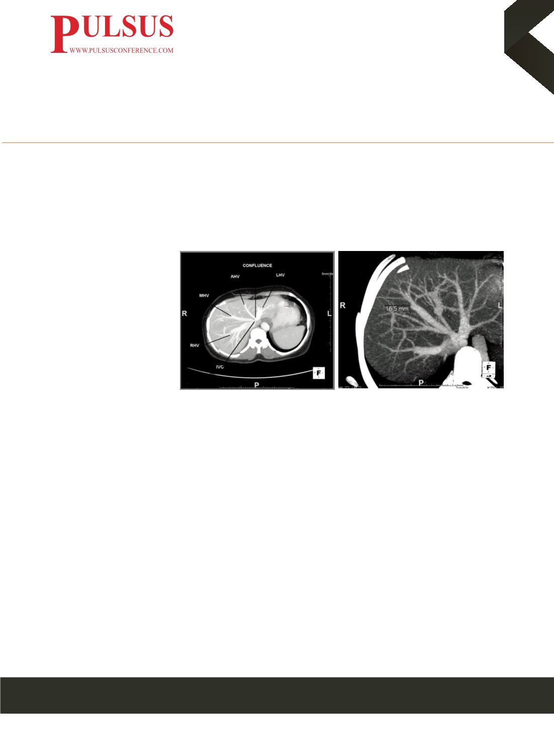
Statement of Problem
: Living-
Donor Liver Transplantation
(LDLT) is a surgical option for
patients who are deteriorating
clinically while awaiting cadaveric
donor liver. In the right lobe
transplantation, the hepatectomy
line passes approximately 1 cm to
the right side of the middle hepatic
vein as a standard procedure. In
LDLT, anatomy of hepatic venous
system is important, not only to
carry out complex reconstructions,
but also to avoid graft congestion after liver transplant in the graft recipient. Hence pre-operative evaluation of venous drainage
and awareness of probable complications is a pre-requisite for transplantation surgeries.
Methodology
: We conducted a retrospective multi-slice spiral CT study of hepatic veins, whereby, we studied 100 abdominal
CT scans which were reported as normal. We studied the length, the number of branches of hepatic veins and measured the
distance of their peripheral-most branch from the nearest hepatic surface. We have also seen for the presence of accessory
hepatic veins.
Findings
: We observed that right hepatic vein was longest (mean length-131.26mm) hepatic vein followed by middle (mean
length-122.62mm) and last of all, the left (mean length- 93.15mm) hepatic vein. Most of the right (46%) and middle (45%)
hepatic veins were visualized up to third order while most of the left (42%) hepatic veins were having less branches and were
visualized up to second order. Most of the right (45) and left (49) hepatic veins were 10-15mm from the hepatic surface while
most of the middle hepatic vein was in the range of 15-20mm. We found Accessory hepatic veins in 18% of cases.
Conclusion and significance
: Hence in liver transplantation, pre-operative evaluation of venous architecture of liver of donor
and recipient is necessary for reconstruction anastomoses and to avoid major hemorrhage during surgery.
Biography
Alka Bhingardeo, currently working as Assistant Professor, at AIIMS Telangana, has passion for research. Her study of CT study
of venous drainage of liver has described variations in draining pattern of hepatic veins which can alter surgical approach in living
live donor liver transplantation and prevent graft rejection and hemorrhage. The information of probable variations in hepatic veins
necessitates transplantation surgeon to have preoperative evaluation of hepatic veins of donor and recipients for successful liver
transplantation.
e
:
dr.alkabhingardeo@gmail.com