
Page 21
Volume 2
July 24-25, 2019 | Rome, Italy
World Hematology 2019 & Nursing Care 2019
July 24-25, 2019
Journal of Blood Disorders and Treatment
47
th
WORLD CONGRESS ON NURSING CARE
11
th
WORLD HEMATOLOGY AND ONCOLOGY CONGRESS
&
Myelodysplastic disorder as the main performance of peritoneal primary sclerosing
epithelioid fibrosarcoma on 18F-FDG PET/CT
Ping Sui, Shujie Song, Dengjun Sun
Yantai Yuhuangding Hospital of Qingdao University, China
Background
: Sclerosing epithelioid fibrosarcoma (SEF) is a rare soft
tissue tumour. Primary SEF in peritoneal is exceedingly rare and has not
be reported before.
Case presentation
: A 67-year-old male patient was presented with
progressive elevated white blood cells for 1 week in his routine physical
examination. Abdominal CT examination revealed peritoneal multiple
space-occupying lesions. Images of 18F-FDG PET/CT showed elevated
18F-FDG uptake in the peritoneal multiple mass. In addition, his cervical,
thoracic and lumbar vertebra presented with wide range of high metabolism
signs, but no bond damage manifestation. Histopathological examination
of the peritoneal lesion and bone marrow cytology and morphology
confirmed the diagnosis of peritoneal primary sclerosing epithelioid
fibrosarcoma accompanied with leukemoid reaction.
Conclusion
: Here we describe a rare case of SEF arising from peritoneal,
an unusual origin and location for such a relatively rare lesion. Besides, the
atypical clinical manifestations and the typical imaging of this patient will
provide guiding significance in diagnosing this disease.
J Blood Disord Treat, Volume 2
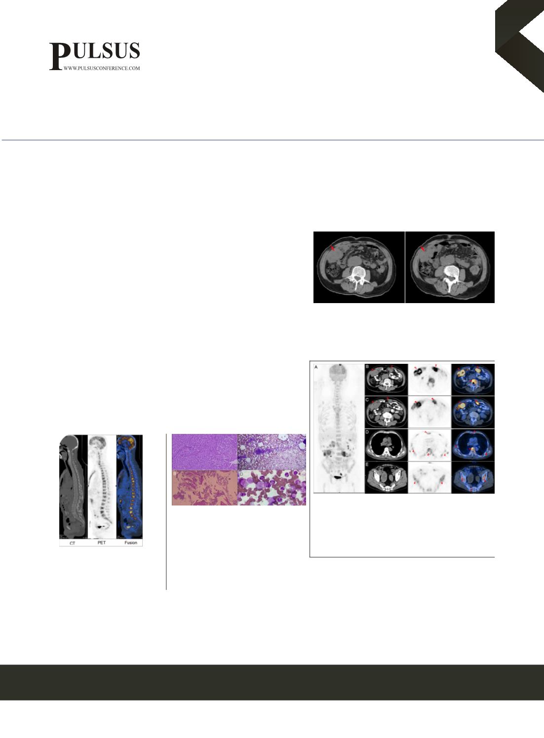
Figure 1
A 67-year-old man was found abnormal peripheral blood
leukocyte count in the Lab test, which fluctuated between 37720/μL and
78570/μL. Abdominal Pelvic CT showed peritoneum multiple occupying
lesions in the parietal peritoneum, the largest of which was 6.0cm×4.0
cm(Red arrow). His physical examination was unimpressive and there
are no significant findings in the Gastroscopy and Colonoscopy, and
18F-FDG PET/CT was performed for further evaluation.
Figure 2
Images of 18F-FDG PET/CT scan were acquired 1 hour after
intravenous injection of 10 mCi of 18F-FDG with a blood glucose level of
76mg/dL(A). The images showed mutiple peritoneal mass with soft tissue
density and had an elevated FDG uptake with SUVmax of 8.5(B,C ). In
addition, the cervical, thoracic and lumbar vertebra presented with wide
range of high metabolism signs with increased FDG uptake (SUVmax,
4.8) (B,D,E),but no bone destruction presentation.
Figure 3
18F-FDG PET/CT sagittal image
showed that the cervical, thoracic and
lumbar vertebra presented with wide range
of high metabolism signs with increased FDG
uptake, but no bone destruction presentation.
Figure 4
Histopathological examination revealed
a neoplasm composed of spindle epithelioid cells,
Immunohistochemistry indicated: Vimentin(+), CK(+),
SMA(-), ALK(-), Desmin(-), CD34(-), CD3(-), CD117(-
), MPO(-), DOG-1(-), CD20(-), CD68(+), Ki-67 index
30%-40%.The diagnosis of this patient was peritoneal
primary sclerosing epithelioid fibrosarcoma(A).
Hyperplasia of bone marrow and neutrophils were shown in the bone marrow pathology( B) and bone marrow morphology test(C,D),
but no abnormal cells infiltration was found. Alkaline phosphatase detection for NAP was 271 points, NAP was 100% positive. The
gene test of BCR/ABL and JAK2V617 were negative. The patient was finally diagnosed as peritoneal primary sclerosing epithelioid
fibrosarcoma accompanied with leukemoid reaction.
Biography
Ping Sui received her medical postgraduate degree in a famous medical college five years ago in china and has passed the standardized
training examination for resident physicians successfully. She has her expertise in comprehensive and targeted cancer therapy. Currently she
is working as a professional oncologist in the Affiliated Yantai Yuhuangding Hospital of Qingdao University, Shandong, China.
suipingsuiying@163.com


